Definition
Cricoid cartilage is part of the larynx (just below the pharynx), and sits on a level plane just below the thyroid gland and thyroid cartilage. It is a circular piece of hyaline cartilage which wraps completely around the trachea, providing a place of attachment for various ligaments, muscles and other cartilage.
Overview
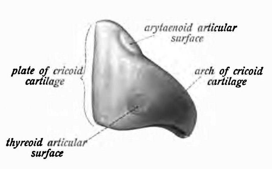
While the majority of cartilaginous rings that surround the trachea are semi-circular, the first ring – the cricoid cartilage ring – is a complete circle and features a narrow arch anterior and a broad lamina posterior to the airway. This form is often likened to a signet ring. In the image below, the broadest side (the cricoid plate) lies in front of the esophagus and the arch faces the front.
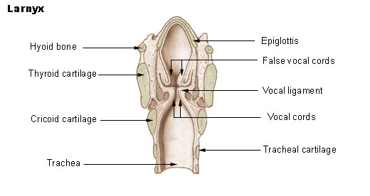
The larynx is a complicated structure with three functions. It contains a valve that protects the airway from debris, especially when swallowing (the epiglottis), it provides structure to the otherwise soft tissue of the airway, and it contains the apparatus for vocalization. The diagram below shows the position of the cricoid cartilage immediately below the vocal chords and in touching distance of the thyroid cartilage.
Housed within this part of the anatomy, just under the feature known as the Adam’s apple (thyroid cartilage), cricoid cartilage is more than a strengthening buttress for the trachea. It is attached to the thyroid and arytenoid cartilages via synovial joints, and is so strong it can support a series of ligaments and muscles. Vocal folds are singularly attached to a small piece of arytenoid cartilage, which is itself attached to the cricoid cartilage by way of a ball and socket joint. The thyroid cartilage is also attached to the cricoid cartilage.
Cricoid cartilage provides an attachment point for three muscles: the lateral cricoarytenoid, posterior cricoarytenoid, and cricothyroid muscles. All of these play a role in opening, closing and elongating the vocal chords, and are essential for voice pitch and quality.
In total, nine cartilages can be found in the larynx. These are either singular (cricoid, thyroid, epiglottis) or in pairs (arytenoids, comiculates, cuneiforms) and, together with the hyoid bone, are described as the ‘visceral skeleton’ of this region of the neck.
Cricoid Cartilage Function
Cricoid cartilage function is to provide attachments for the cricothyroid, posterior cricoarytenoid, and lateral cricoarytenoid muscles, cartilages, and ligaments involved in voice pitch and quality.
In addition, cricoid cartilage supports the soft connective tissue of the trachea together with other semi-circular cartilage bands that run along its entire length.
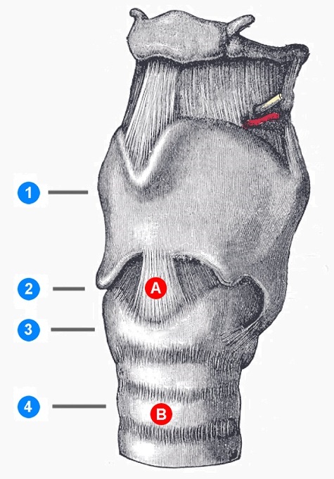
Cricoid cartilage is also used as an anatomical landmark for surgical cricothyrotomy (temporary) and tracheostomy (definitive) procedures that provide a viable airway in emergency settings. The diagram below shows the thyroid cartilage (1), cricothyroid ligament (2), cricoid cartilage (3), and trachea (4). Points A and B relate to cricothyrotomy and tracheostomy incision points respectively.
Cricoid pressure (Sellick’s maneuver) is occasionally implemented during intubation when there is a risk of the stomach contents rising up through the esophagus and into the trachea (and lungs) due to a paralyzed epiglottis. Just before a patient falls asleep, light pressure is placed on the cricoid in order to flatten the soft tube of the esophagus behind it and, in theory, prevent the stomach contents from entering the airway. As the cricoid ring is a complete circle with a broader plate facing onto the esophagus, such pressure will, in principle, close off the esophagus which is not protected by any cartilaginous structures. This is a controversial maneuver, but still used during rapid-sequence induction of anesthesia (RSI). Extremely rarely, cricoid cartilage can fracture under pressure.
How is Cricoid Cartilage Structured?
All cricoid cartilage is hyaline cartilage and its structure is distinguished by type II collagen and chondroitin sulfate within its extracellular matrix. This type of cartilage is durable, and it experiences very little friction due to its smooth, glass-like surface. Cricoid cartilage is surrounded by a perichondrial membrane.
Cricoid cartilage grows in a pattern of circumferential growth (cell proliferation) and interstitial growth (extracellular matrix production) but, as a cartilaginous tissue without an immediate blood supply, this process is slow.
Cricoid cartilage, as all cartilage, is aneural and avascular. However, the density of blood vessels in its immediate vicinity provides nutrients and oxygen to the perichondrium, from which these then pass via diffusion into the cartilage tissue. While cartilage tissue itself contains no nerves, the larynx is significantly innervated, as are the muscles attached to the cricoid cartilage.
In adult patients under general anesthetic where the placement of an endotracheal tubeendotracheal tube is required, the narrowest part of the trachea through which to pass the tube is between the vocal chords. In older children and adults, the trachea remains approximately the same diameter along its length. In babies and very young children however, the trachea is distinctly funnel-shaped, and the narrowest location is not at the vocal chords, but the site of the cricoid cartilage itself.
This image below shows a video-laryngoscope image of an already placed endotracheal tube fixed between the open (relaxed) vocal chords of an adult. In a young child, the comparative narrowness of the part of the trachea surrounded by the cricoid cartilage makes tube placement more complicated. Note the relaxed epiglottis at the top of the screen.

As with all joints, cricoarytenoid and cricothyroid joints can be affected by osteoarthritis. This leads to cracking or fissure-forming in the cartilage tissue, loosening of collagen structures, and loss of proteoglycans. Cricoid cartilage degeneration leads to decreased vocal quality and changes in pitch.
Quiz