Definition
An adipocyte cell is a brown, beige, pink, or white fat cell that stores triglyceride droplets, has a secretory function, and/or helps to convert lipids into energy. Fat tissue is primarily composed of adipocytes. An adipocyte can secrete hormones and other effector chemicals that play important roles in metabolism control. Because of its association with metabolic disorders, adipocyte tissue is undergoing extensive studies to pinpoint its exact role.

What is an Adipocyte?
An adipocyte, fat cell, or lipocyte is the primary cell found in brown, beige, and white adipose tissue. These tissues have varying functions – brown and beige adipocyte cells produce energy; white adipocytes store energy in the form of lipid droplets.
Adipose tissue is full of adipocytes but also features non-adipose cells such as neurons and immune cells. Adipose fat is a complex, high-functioning organ rather than ‘simple’ connective tissue.
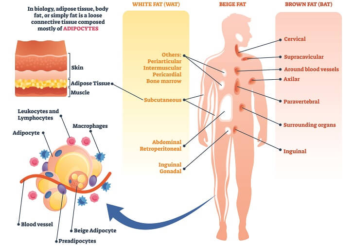
A white adipocyte cell contains a lipid body surrounded by a ring of cytoplasm. The lipid body expands to flatten the nucleus, pushing it toward the cell membrane.
While the number of fat cells in the body is relatively stable in people of a healthy weight, these cells grow or shrink in size. However, the fatter the person, the higher the white lipocyte count and the more likely they are to produce more fat cells.
Brown adipocytes have higher quantities of cytoplasm with small lipid droplets scattered within. The absence of a larger lipid globule in the center means the nucleus is not flat. Large numbers of mitochondria populate the cytoplasm, further indicating the energy-producing role of brown fat cells.
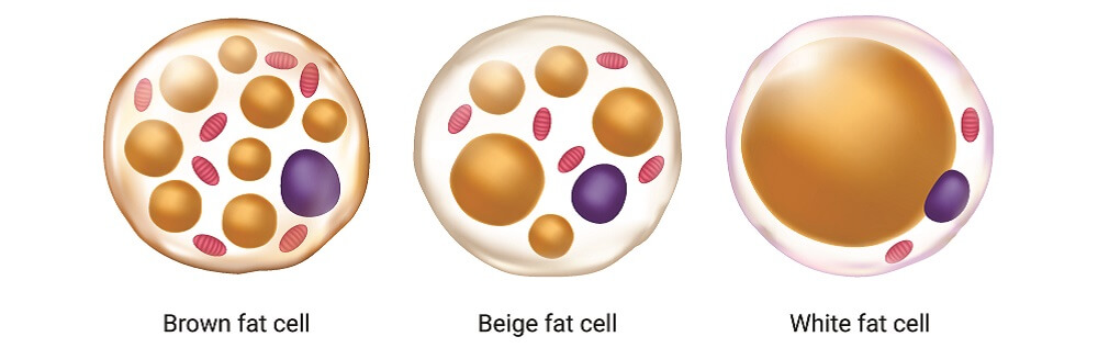
The beige lipocyte is a mix of white and brown adipocyte structure with mid-sized lipid droplets and a median quantity of mitochondria and cytoplasm compared to white and brown cells.
Pink adipocytes contain milk vesicles rather than lipid bodies as well as a much larger Golgi apparatus and endoplasmic reticulum. These cells originate in subcutaneous mammary tissue during pregnancy and are differentiated white lipocytes. They look like a cross between a fat cell and an epithelial cell; so far they have only been described in rodents.
Red and yellow adipocytes are found in red and yellow bone marrow respectively. Bone marrow adipocytes (BMAs) help to regulate blood cell production, also known as hematopoiesis.
Yellow BMAs are large and less responsive to the environment; red BMAs – in the marrow where blood stem cells are found – have a much higher capacity for communication.
All adipose cells contain mitochondria, a nucleus containing the nucleolus, Golgi apparatus, rough and smooth endoplasmic reticulum, a very strong cell membrane, and vacuoles.
Adipocyte Function
When looking at adipocyte function, you need to look at white, pink, beige, bone marrow, and brown cells separately. However, all adipocyte types release chemical signaling molecules called adipocytokines.
Adipocytokines are proinflammatory molecules; dysregulated production of fat cell secretions – as observed in the obese – contributes to chronic inflammatory disorders. Furthermore, levels of these chemicals rise over time as we gain weight and the body responds to them less efficiently. High levels of fat tissue are responsible for insulin resistance, for example.
The most common adipocytokines produced in all fat cell types are:
- Leptin: essential for the acute inflammatory response and T-cell proliferation;
- Adipsin: stimulates insulin secretion in the presence of hyperglycemic signals;
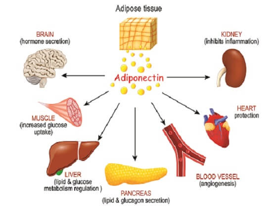
- Adiponectin: modulates glucose levels, fatty acid metabolism, and adipocyte differentiation;
- Omentin: crucial roles in insulin sensitivity, angiogenesis, and vasodilation.
All fat cell types are partially responsible for metabolic and immune pathway functioning. They also define the link between obesity and diabetes, cardiovascular disease, susceptibility to infection, and metabolic dysfunction.
White Adipocyte Function
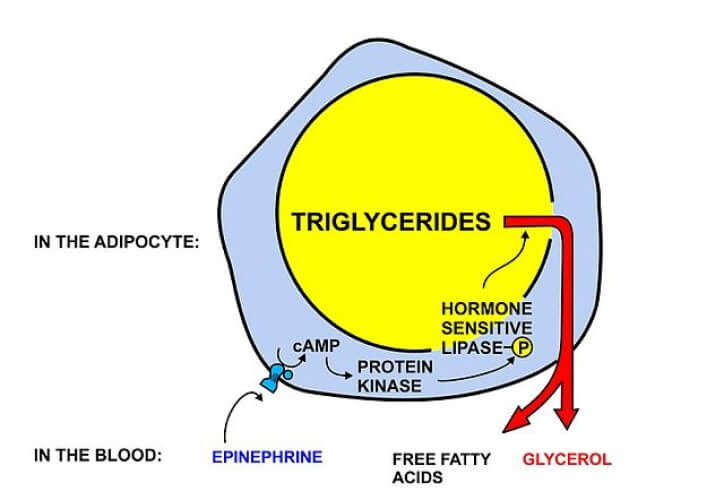
White adipocytes are fat storage cells and the most prolific cell type in white and beige fat tissue. Triglyceride droplets are broken down and released during periods of fasting to provide the basic ingredients for lipolysis – the conversion of fats into fuel.
The fed state that triggers insulin release and the fasting state that triggers catecholamine release control how much fat is released from white fat cells.
Brown Adipocyte Function
Brown adipocytes produce energy in the form of heat – thermogenesis. Nonshivering thermogenesis occurs under the control of norepinephrine and the hypothalamus; it is not possible without the presence of brown fat.
Young babies, who cannot shiver to produce internal heat, have high percentages of brown fat. It is possible to stimulate brown adipocyte differentiation by living in permanently cold environments.
Brown adipocytes produce uncoupling protein-1 because they express the gene that codes for this protein’s synthesis. This is not the case in white, beige, or pink fat cells. It is this protein that converts fatty acids into heat energy – the primary function of brown fat tissue.
Recent discoveries have shown how brown fat tissue is also important for a healthy immune system and protects against metabolic disorders such as diabetes and nonalcoholic fatty liver disease. The lower the percentage of brown fat in the body, the higher the risk of these metabolic disorders.
Beige Adipocyte Function
While it used to be thought that beige fat contained the same types of cell as brown and white tissues, it now appears that beige adipocytes are a separate entity; they do not express the gene for uncoupling protein 1 (UPC1); the beige adipocyte responds primarily to a hormone called irisin.
Irisin is released by working skeletal muscle. This offers us a link between a sedentary lifestyle and increased risk of cardiovascular disease and diabetes. Irisin stimulates fat ‘browning’ – increasing the numbers of brown adipocyte populations in beige and brown fat depots.

Beige adipocytes function in a similar way to brown cells – they produce heat energy from lipid fuel, but are UCP1 negative. While researchers are unsure why, some suggest these are brown cells in a semi-dormant state that could – under longer-term stimulation – increase brown adipose tissue levels.
Bone Marrow Adipocyte Function
Yellow and red lipocyte populations of their corresponding bone marrow locations are a recent topic of study; low or high proportions of fat cells in the bone marrow are associated with insufficient and excess blood cell production respectively.
Blood cell cancers suppress bone marrow adipocyte production; interestingly, solid tumors in other tissues often communicate with cancer-associated adipocytes (CCAs).
Pink Adipocyte Function
The pink adipocyte has only been described in rodents; white, beige, and brown adipocytes can differentiate into pink cells that do not seem to derive from stem cells.
Pink lipocytes have a glandular function; populations increase during pregnancy and lactation. As mammalian milk is lipid rich, this takes advantage of triglyceride droplets in the immediate vicinity.

While scientists are still looking for the equivalent cell in humans, they do agree as to adipocyte plasticity. Furthermore, mammary gland adipocyte populations are linked to obesity and breast cancer.
Adipocyte Differentiation
Adipogenesis – the production of fat cells – begins in the stem cell.
Mesenchymal stem cells produce various cell types and can also regenerate their own populations. When stimulated they divide to produce preadipocytes. Stimulation is regulated by growth factors, the tissue type in which the stem cell is found, hormones, and competition from chemicals that regulate other cell populations.

A preadipocyte has already differentiated but is immature. White preadipocytes become white lipocytes; brown preadipocytes form brown lipocytes.
Adipocytes also transdifferentiate. A differentiated fat cell can divide to become a different type. Most pink cells were originally white; brown adipocytes form via brown preadipocyte division or white adipocyte transdifferentiation.
Adipocyte Dysfunction
Adipocyte dysfunction has various causes. Genetic mutations might alter populations or change how fat cells respond to chemical signals.
Impaired adipogenesis reduces the number of fat cells produced, causing existing white cells to expand in size. This expansion is called adipocyte hypertrophy.
The larger the adipocyte, the higher the degree of inflammation and the greater the risk for insulin resistance, cancer, and obesity.

Every adipocyte must be in contact with a capillary; lipocyte hypertrophy and higher cell proliferation rates stimulate new blood vessel formation (angiogenesis); however, the blood volume does not significantly increase.
This means lower oxygen and nutrient transport to all tissues. To make up for this, the heart rate increases; cardiac output is usually higher in the obese.
Anti-adipocyte antibodies have been successfully used to reduce fat in livestock, producing leaner meats. This is a form of adipocyte immunization. Monoclonal antibodies may one day immunize against obesity. Recent phase II clinical trials of bimagrumab administered intravenously reduced up to 20.5% of body fat in obese subjects with type two diabetes in 48 weeks.