Definition
Articular cartilage is found only in diarthroidal joints (synovial joints), and is comprised of hyaline cartilage – a particularly smooth type of cartilage which allows for easy articulation, increased weight distribution, and shock absorption.
Articular Cartilage Explained
Articular cartilage’s role is often better understood in patients in which it has degenerated (usually due to the ageing process or intensive sports). Inflammation and friction where the surfaces of two bones rub against each other cause pain and limited mobility. In the lower limbs, the weight-bearing properties of articular cartilage are unable to properly distribute weight and the shock-absorbant characteristics of cartilage are lost. Any pain is not caused by damage to the cartilage itself, as cartilaginous connective tissues are aneural (without a nerve network). It is caused by the nerves surrounding the bony layers which are no longer protected by the cushioning effect of cartilage.
Articular cartilage is usually found in layers of between 2 and 4 mm thick. As with all types of cartilage, the absence of blood vessels and lymph vessels creates a very slow metabolic environment. Chondrocyte proliferation and apoptosis (death) exists at much lower rates than in noncartilagenous connective tissue. Low levels of oxygen mean chondrocytes primarily depend upon anaerobic metabolism. Nutrients are provided directly from the synovial fluid and not from the perichondrium, which is absent in articular cartilage.
Articular Cartilage Function
Articular cartilage function is based upon its composition of hyaline cartilage, which is practically frictionless due to the glass-like surface and ability to self-lubricate via lubricating glycoproteins within the extracellular matrix. When articulation is smooth, less stress is exercised on the cartilage surface and the tissue is more resistant to wear, in the same way oil added to a squeaky door hinge prevents the erosion of the touching surfaces.
The structure of articular cartilage into three zones with different characteristics allows for an efficient, load-bearing surface which distributes compressive forces generated during diarthroidal joint loading and diarthroidal joint motion. Wrong movement in load bearing cartilage, for example at a joint between the long bones, is the reason why the knee (between the femur and tibia) is the location of frequent articular cartilage injury. The knee joint can undergo damage through excessive rotation; a common football injury is the dreaded meniscus tear.
A further function of articular cartilage is the ability for that part of the anatomy to move on one or more planes. The joint range of movement depends on the specific type of diarthroidal joint.
Where Is Articular Cartilage Found
Articular cartilage locations are found throughout the body. The term ‘articular cartilage’ does not refer to the type of cartilage structure, but to its location. Cartilaginous joints (growth plates, the symphysis, the spine, and the ribs) have very little movement and no synovial membrane. Fibrous joints (skull sutures, dental sockets, and other immovable joints) have no movement and similarly lack synovial membrane and fluid. It is therefore correct to say that articular cartilage occurs only in the presence of a synovium.
All diarthroidal joints, in which articular cartilage is found, have certain characteristics. These joints are the meeting points of two bones. They allow for movement in at least one axis. To protect the smooth hyaline surface, all diarthroidal joints are covered by a synovial membrane filled with synovial fluid. They are categorized according to structure and type of motion. There are six types of synovial joint.
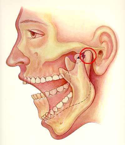
Planar (Gliding) Joints
Planar joints do not rotate, but allow two, relatively flat bone surfaces to glide across one another. Examples of planar joints are the carpals of the hand and the tarsals of the foot, and in the temporomandibular joint as shown below.
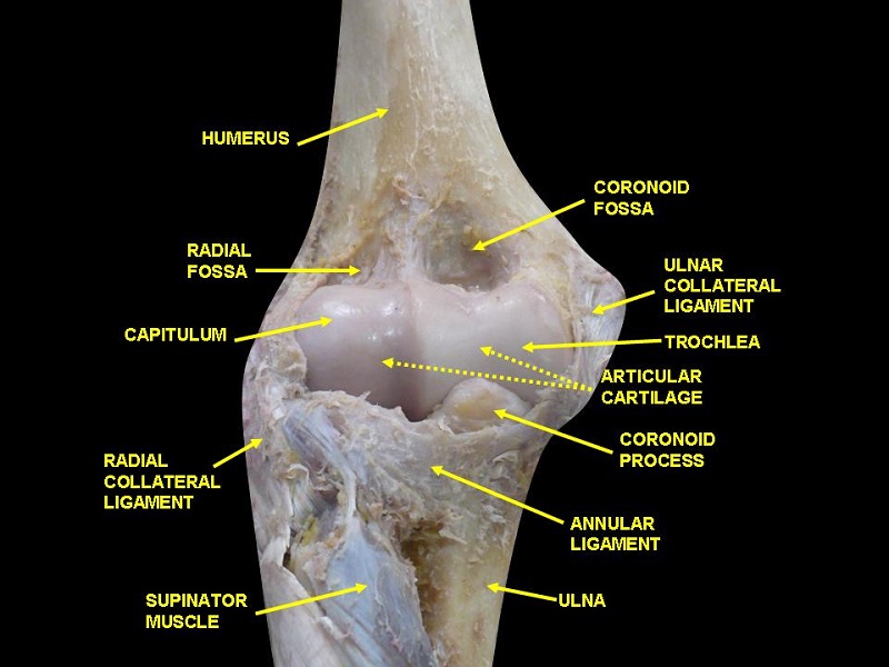
Hinge Joints
The two bone surfaces are of different shapes. One is rounded, the other hollowed. During motion, one bone remains in place and the other moves. This type of joint has a very limited range of motion. The elbow is the best example of a simple hinge joint. The knee, with a wider range of motion, is considered to be a modified hinge joint.
Note the shiny hyaline layer in the humeroulnar joint of the elbow below.
Pivot Joints
Also called rotary or trochoid joints, pivot joints have a circular range of motion on a single axis due to one bone surface having a ring-like shape. Examples are the proximal radioulnar joint, and the joint between the first and second cervical vertebrae. The latter is pictured below, showing the projection of the dens from the second cervical vertebra (C2), or axis, into the arch of the first cervical vertebra (C1), or atlas.

Condyloid (Ellipsoidal) Joints
With the ability to move along two axes (up and down, side to side), ellipse-shaped bone surfaces – one concave and one convex – are found primarily in the hand and wrist, and the foot. Rotation is not possible in condyloid joints.

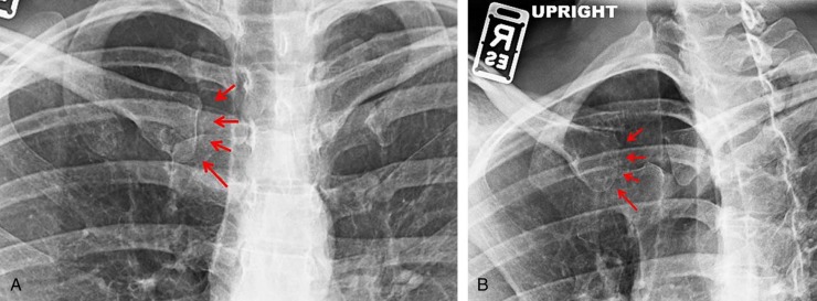
Saddle Joints
Saddle joints have a slightly higher range of non-rotary movement than condyloid joints. The best-known example of the saddle joint is the first metacarpal joint between thumb and wrist. A lesser-known example is the sternoclavicular joint. The x-ray featured below shows the healthy, undamaged sternoclavicular joint of a young male on the left-hand side, and the same joint after trauma and subsequent osteoarthritis to the right.
Ball and Socket Joints
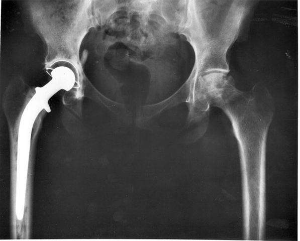
In ball and socket joints, one bone surface is almost spherical in form and the other distinctly and deeply concave, providing a connection which provides maximum range of movement with a lower risk of dislocation. Ball and socket joints are found in the hips and shoulders.
One of the most common orthopedic surgeries in elderly populations, a total hip replacement replaces hip joints where arthritis has worn away the articular cartilage, leading to loss of motion and pain. The image below shows the characteristic ball and socket shape of the prosthetic hip joint to the left, and an arthritic natural hip joint to the right.
Differences in Articular Cartilage Compared to Other Cartilage Types
Articular cartilage does not have a perichondrium, and is composed of four different layers: superficial, transitional (mid), deep (radial), and calcified layers, or zones. A ‘tidemark’ distinguishes between the non-calcified and calcified layers. The extracellular matrix of these four layers are further split into three regions called the pericellular region (immediately surrounding the chondrocyte), the territorial region (protective, collagen-rich area), and the interterritorial region (largest region with high amounts of collagen and proteoglycans, and structurally important).
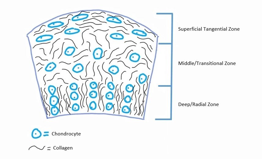
Superficial or Tangential Layer
In the superficial layer – the top layer of articular cartilage – chondrocytes are quite flat in shape. At this level there are fewer proteoglycans and higher numbers of organized collagen fibrils. This layer protects the deeper layers from sheer stresses, but is very thin. It is also in direct contact with the synovial fluid of the joint capsule.
Transitional or Middle Layer
Chondrocytes are rounder in this second layer. Here, proteoglycans are more prevalent than in any of the other layers, while collagen fibrils are relatively disorganized in comparison to the layer above. This layer acts as a bridge between the superficial and deep zones, and is the thickest layer. It can absorb compressive forces, but not to the same degree as the underlying radial zone.
Deep or Radial Layer
Here, the chondrocytes begin to form columns along an axis of collagen fibrils. This structure is at right angles to the underlying bone – an arrangement that provides the most resistance to compressive forces. The first three layers of articular cartilage are shown in the simple diagram below.
Calcified Layer
Distinguished by the ‘tidemark’ between deep and calcified layers, this layer provides the connection between bone and cartilage through partial mineralization. It acts as a transition between cartilage and the underlying subchondral bone, allowing strong adhesion of the two different tissue types. Very few chondrocytes are found in this layer.
If a portion of ‘wet’ articular cartilage with all of its layers was separated into the most important individual elements, water would provide 65–80% of its weight, type II collagen fibrils would account for 10–20% (along with very small percentages of other collagen types), and 10–15% would be made up primarily of Aggrecan, but also other proteoglycans. Chondrocytes only provide approximately 5% of the wet weight of articular cartilage, and lubricating glycoproteins (such as the aptly named lubricin) and noncollagenous proteins are represented in even lower amounts.
Chondrons
Another difference in articular cartilage is the chondron – the collective name for one or more chondrocytes and the immediate surrounding matrix known as the pericellular matrix or PCM. It is believed, although research is still in its infancy, that the PCM enables cellular signaling, with cartilage homeostasis as a result.
Quiz