Foot Definition
The foot is a part of vertebrate anatomy which serves the purpose of supporting the animal’s weight and allowing for locomotion on land. In humans, the foot is one of the most complex structures in the body. It is made up of over 100 moving parts – bones, muscles, tendons, and ligaments designed to allow the foot to balance the body’s weight on just two legs and support such diverse actions as running, jumping, climbing, and walking.
Because they are so complicated, human feet can be especially prone to injury. Strains, sprains, tendonitis, torn ligaments, broken bones, fallen arches, bunions, corns, and plantar warts can all occur. Here we will talk more about the anatomy of the human foot and its many moving parts.
The complexity of the human foot may stem from the fact that it evolved from hand-like, grasping feet like those we see in apes today. Our ancient ancestors were tree-dwellers, and needed to be able to hang onto branches tightly with all four limbs. This caused them to evolve extraordinarily intricate hands and feet, which were capable of grasping, rotating, and gripping with dexterity that engineers are still trying to replicate in fields like robotics today.
Scientists are not sure why our ancestors eventually developed to walk upright, which caused the “fingers” of our feet to fuse and create a flat surface for walking on. It may have been because our ancestors began living on treeless grasslands, where standing tall to be able to see over the grass was more important than climbing. It could also have been because, as we began using tools, the ability to walk on two feet while using our hands to carry items became important.
Feet are present in other species too; especially mammals, birds, reptiles, and amphibians. Invertebrates such as mollusks and insects may have “feet” that they use to walk or move, but these are not complex bony structures like those found in vertebrates.
Here we will discuss the anatomy of the human foot, and some things that can go wrong to cause injuries or disorders.
These descriptions are meant for informational purposes only. You should always see a doctor if foot injury is suspected, as prompt and proper treatment can make for a faster, easier recovery! It is especially important to see a doctor if a suspected foot injury involves numbness, bleeding, or inability to move the foot, as these may be signs of serious complications.
Proper diagnosis and treatment takes a trained professional; improper diagnosis and treatment may lead to longer-lasting problems!
Foot Anatomy
The foot contains 26 bones, 33 joints, and over 100 tendons, muscles, and ligaments. This may sound like overkill for a flat structure that supports your weight, but you may not realize how much work your foot does!
The foot is responsible for balancing the body’s weight on two legs – a feat which modern roboticists are still trying to replicate. This requires strong, subtle muscles which can keep the foot standing firm even as we move our body’s weight around at different positions and angles.
The many bones work together to allow to allow this fine, delicate movement by subtly shifting inside the foot. They also allow us to perform intricate actions such as standing, climbing, and “grasping” at the ground with our feet on moving or uneven surfaces.
Here we will discuss the most important parts of the anatomy of the foot, and some injuries and disorders that can occur when these parts are damaged.
Of note, here we will make general statements about how different foot injuries and disorders may be treated by doctors. This is not a substitute for medical advice.
See a doctor about any suspected foot injury or disorder, as prompt diagnosis and treatment can make for a faster, easier recovery, while improper treatment may lead to long-term damage.
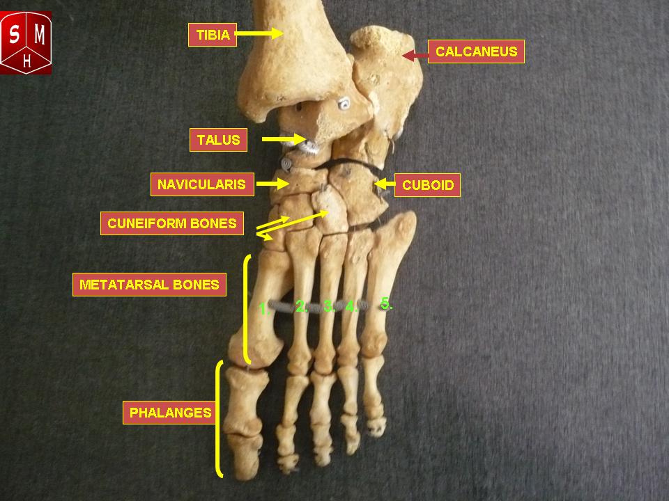
Foot Bones
There are 26 bones in the foot. These include:
- The phalanges, which are the bones in your toes
- The metatarsals, which run through the flat part of your foot
- The cuneiform bones, the navicularis, and the cuboid, all of which function to give your foot a solid yet somewhat flexible foundation
- The calcaneus, which is the bone in your heel
- The talus, which is the bone in your ankle
- The talus connects to the tibia, which is the main bone in your lower leg
While you may not notice these bones in action every day, you’ll notice quickly if something is wrong with one of them. These bones allow your feet to execute the delicate shifts which enable you to keep your balance while walking, running, jumping, climbing, dancing, and playing sports!
Injury to a bone in the foot often results in a sharp or throbbing pain, especially when you move in a way that causes your weight or a nearby muscle to put pressure on the bone.
The most common broken bones in the foot are broken toes, which may occur after hitting a toe on a hard or sharp surface while walking, running, swimming, or playing sports.
Broken bones in the foot usually call for rest, ice, compression, and elevation to reduce any swelling. It is helpful to remember the acronym “RICE” for Rest, Ice, Compression, and Elevation. This combination of at-home treatments is a good first-line response for many leg and foot injuries.
Supportive wraps or protective casts may be used to reduce pain and keep bones properly aligned. Sometimes, crutches or other means of keeping weight off the foot entirely might be prescribed. In rare cases where a bone breaks into two or more pieces and these pieces become misaligned, surgery may be required to move the pieces back into alignment so they can heal.
Physical therapy may also be suggested to help regain healthy use of the muscles after the injury.
Another possible problem with bones in the foot is the problem of bunions, or bone spurs.
Bone spurs occur when extra bone growth occurs, usually near the end or joint of a bone. This can be caused by chronic irritation of the joint, such as rubbing against another bone or joint. The most common types of bone spurs in feet occur in the big toe, and these are called “bunions.”
Bunions and bone spurs can cause significant pain. Internally, they can rub against other bones, muscles, and nerves beneath the skin. Externally, they can change the shape of the foot, resulting in pain and discomfort from wearing normal shoes.
Mild bunions can be treated by wearing more comfortable shoes or shoe inserts, taking over-the-counter anti-inflammatory medications, applying rest, ice, compression, and elevation, and taping, or splinting the affected area. All of these measures might reduce swelling and prevent the bunion from causing pain.
If pain is not relieved by these activities, surgery may be required to remove some of the bunion tissue.
The risk for bunions is increased if you wear tight, narrow shoes, which may force bones to rub against each other. The risk is also increased if you have arthritis or a history of injuries to the foot.
Foot Ligaments
Ligaments are bands of very strong, flexible tissue that perform the important job of connecting bones together. Ligaments are very strong and difficult to injure, but ligament injuries can be serious when they do occur. This is because ligaments do not receive much blood flow like bones and muscles, so they are slow to repair themselves.
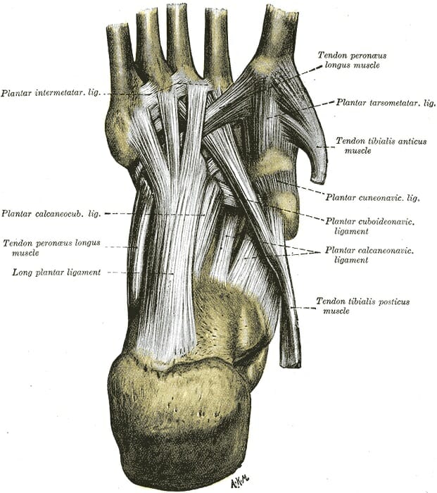
There are a lot of bones in the foot, so you might guess correctly that there are a lot of ligaments. In fact there are so many ligaments that we need three different diagrams to show them all to you!
This diagram shows the sole of the foot. You can see the toes on the top and the heel on the bottom, while the arch and sole of the foot are made up of a thick web of ligaments holding the bones together:
This diagram shows the “medial aspect” of the foot. This term comes from the terms “medial,” meaning “center,” or “in the middle,” and “aspect,” meaning “face.” In other words, this is the “face” that the foot shows to the center of the body. It is the side of the foot that faces inward.
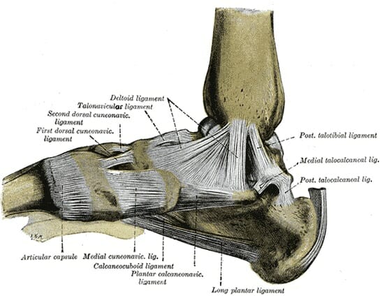
This diagram shows the heel on the right, while the toes reach off the screen to the left.
Here you can see that the ankle is also a thick web of ligaments, where the tibia is connected to the bones of the ankle and the core of the foot. You can also see the bands of ligaments where the metatarsals and phalanges are connected to each other.
Lastly, this diagram shows the “lateral aspect” of the foot, with “lateral” meaning “to the side.” This is the view of the foot from the side of the body, then; the view of the part of the foot that faces outward.
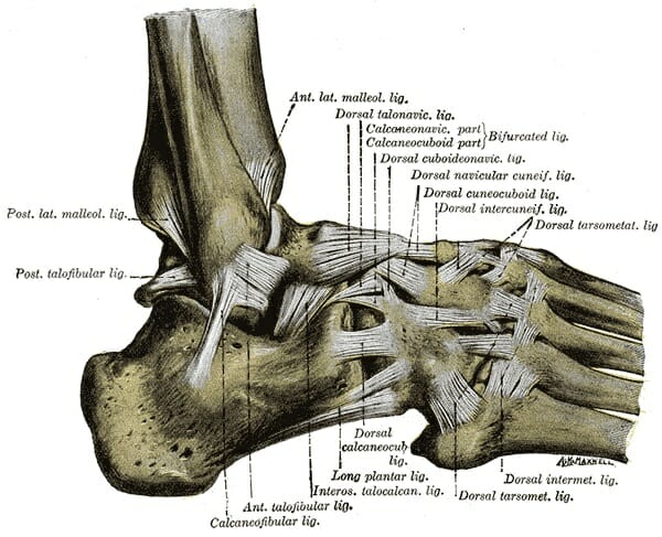
On the left side of the image, above the heel, you can see the delicate leg bone called the fibula. The fibula is smaller than the tibia and runs alongside it. Having two separate bones instead of one connecting the foot to the leg gives the foot and leg extra balance and maneuverability.
You can also see the thick web of ligaments on the top of the foot, where the bones of the foot’s core are connected on the top side.
Now you can begin to see why the middle of your foot feels solid, even though it’s made up of many bones. The many bones are bound together tightly by strong, flexible ligaments, which allow the center of your foot to shift subtly while remaining solid and stable.
Although ligaments are strong, they can be injured – especially in an area like the ankle, where the whole weight of your body hinges on a single joint.
Sprains occur when a body part is wrenched or twisted, resulting in damage to a ligament. Such damage can cause swelling and significant pain. Because ligaments do not receive much nourishing blood flow from the body, sprains can take a long time to heal, and long-term damage can result from continued stress on a sprained ligament.
Like broken bones, sprains are often treated with rest, ice, compression, and elevation; and a supportive wrap or cast to take stress off the sprained area. Sometimes, crutches or other means of keeping weight off the foot entirely might be prescribed.
Physical therapy can be especially helpful in the case of sprains, where it can ensure that the injured ligament is strengthened gradually and is properly supported by surrounding muscles.
A torn ligament occurs when the foot is wrenched or twisted so violently that the ligament actually snaps. This condition can be serious as ligaments which are completely torn may not heal themselves the way a bone or muscle would.
Torn ligaments can sometimes be treated in the same way as strains, but may require surgery if the tear is severe or if there is lasting damage to foot function. With surgery, doctors can join the two ends of a damaged ligament together, or replace a damaged ligament with a healthy one from another part of the body.
Foot Muscles
Just as there are many bones and ligaments of the sole of the foot, there are also many muscles. These can be divided up into four major groups:
- The central muscles of the sole of the foot
- The lateral muscles of the sole of the foot
- The medial muscles of the sole of the foot
- The muscles of the dorsum (top) of the foot
You can learn more about each individual group of muscles in the foot using this table:

If muscles are overworked or overstressed, they can become torn or strained. Strains usually manifest as pain, especially with movement or pressure.
Mild strains often go away in days or weeks if the muscle is rested and not subjected to further stress. More serious muscle tears, however, may take months.
It is a good idea to see a doctor if a severe strain is suspected, as severe muscle strains can lead to serious complications.
The most severe form of muscle overuse – rhabdomyolysis – occurs when muscles are so stressed that their cells rupture and release toxic chemicals. This can actually be fatal if left untreated.
Rest, ice, compression, and elevation to reduce swelling are recommended to treat mild to moderate strains. Supportive wraps or casts and crutches or braces may be recommended if the strain is especially severe.
Physical therapy may also be suggested to help regain healthy use of the muscles after the injury.
Foot Tendons
Tendons are thick bands of tissue that connect muscles to bones. By connecting our rigid bones to our powerful muscles, tendons allow us to move. Movement occurs when our muscles pull on our bones, relocating them.
The following diagram shows the tendons of the lateral aspect of the foot – that is, the aspect that faces outward, away from your body:
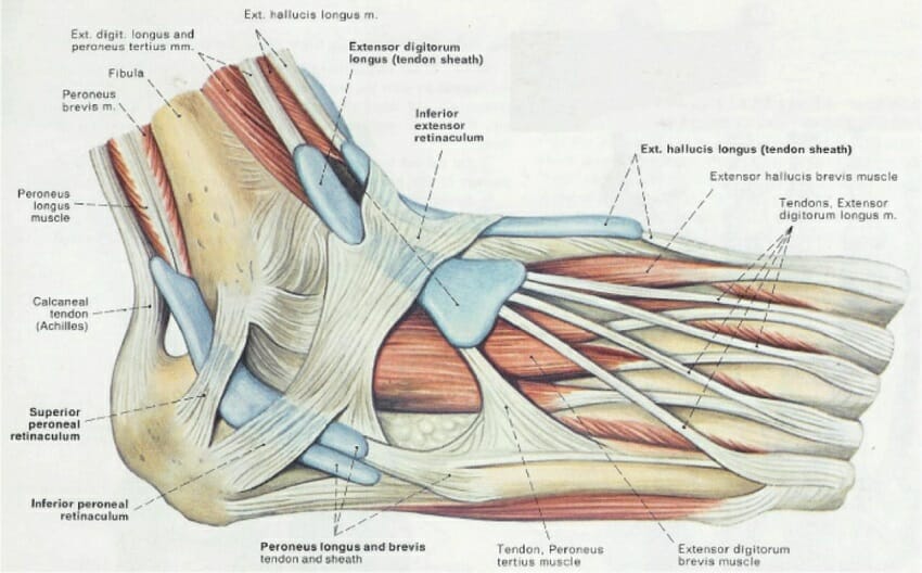
Here you can see the tendons that extend down the top of your foot toward your toes, allowing you to curl your toes upward if need be.
You can also see what is arguably the most important tendon in the foot – the calcaneal, or Achilles tendon, which allows the muscles of your calf to control the movement of your foot.
The Achilles tendon gets its name from the mythical Greek hero Achilles, who was invulnerable – except for his ankle. An injury to his ankle – possibly to the Achilles tendon – left him unable to stand and fight.
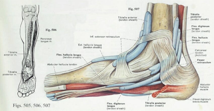
This image of the medial aspect of the foot shows tendons that run along the bottom of the foot. It is these tendons that allow you to curl your toes and grip surfaces with your feet, by permitting muscles on the bottom of the foot to pull tight.
Injuries may happen to any tendon in the foot, and these may cause pain or impair balance. Achilles tendon injuries are one of the most common tendon injuries that can occur, as the body relies on the Achilles tendon to support its weight.
Lesser injuries to tendons can be treated with rest, ice, compression, elevation, and over-the-counter anti-inflammatory medications. Doctors may recommend prolonged periods of rest, and prescribe a supportive wrap or cast for substantial tendon injuries.
Severe injuries to the Achilles tendon that may occur while playing sports can require surgery to repair.
In addition to acute injuries like strains and tears, tendons can become irritated due to chronic stress.
Tendonitis occurs when a tendon – a touch cord of tissue which attaches a muscle to a bone – becomes irritated over time. This can occur from overuse or misuse if a person is moving in a way that causes stress to the tendon.
Tendonitis often appears slowly, manifesting as a sharp pain when a person performs a certain movement. People with tendonitis in the foot may find that it is painful to put weight on the foot, despite the absence of a clear injury like trauma or strain.
Tendonitis can be treated with RICE and over-the-counter anti-inflammatory drugs. Physical therapy can also be extremely beneficial, as this can gently exercise and stretch the tendon, and correct any movement habits that may have caused the irritation.
Foot Arches
Normally, tendons in the foot pull the bones of the foot in toward each other, resulting in distinctive arches between the heel and toes, and between the inner and outer toes. This arch is important for ensuring that weight is properly distributed among the strongest muscles of the leg and foot, and to ensure we can shift our weight as needed to keep our balance or move quickly.

Fallen arches, or “flat feet,” can occur when the tendons of the foot do not pull the foot’s bones together with a normal amount of strength. This results in the foot becoming “flat,” which can lead to pain, balance problems, and tiredness in the leg or foot.
Flat feet can occur as a result of injury, or some people’s tendons simply never pull together properly. Rarely, other health problems such as arthritis or problems with the nerves going to the feet can cause flat feet.
Flat feet may ache and tire easily. Back pain and leg pain may also result as muscles in the back and legs may work to overcompensate for the normal balancing functions of the arch.
Treatment for flat feet may depend on the cause. If you believe you have flat feet, see a doctor to find out what is the best treatment for you!
Skin and Toenails
The internal parts of the foot are not the only important parts! The skin on the bottom of our feet protects our muscles, bones, tendons and ligaments from injury. It also prevents infection.
Toenails protect the top of our toes, which, as we all know, can sometimes be vulnerable to being stubbed, stepped on, or having things dropped on them.
However, there are things that can go wrong with each of these and lead to problems.
Plantar warts – Plantar warts are growths that appear on the bottom of the foot, and may become painful. They are caused by a strain of human papillomavirus that infects skin of the feet and causes unusual growth of skin and blood vessels.
The strain of human papillomavirus that causes plantar warts and other warts is very common in the environment. It is not known why some people develop warts and others don’t. Avoiding sharing shoes and socks with people who have plantar warts may help protect against them, but many people develop plantar warts with no known instances of person-to-person transmission.
If plantar warts remain small, they might not cause pain, and no treatment may be needed. If they become painful, however, they may need to be removed. Several options exist for doing this, including over-the-counter applications, and procedures to freeze the wart tissue which can be performed by a doctor.
Corns and calluses are hard areas of skin which build up as a result of frequent friction against the skin. The body creates corns and calluses to “toughen” the skin against repeated stress.
People who work with their hands such as carpenters, gardeners, and musicians often develop calluses on their hands in areas where they frequently rub against their instruments. People who walk often or whose feet rub against the insides of their shoes may develop corns and calluses on their feet.
People with conditions that cause fragile skin or impaired blood flow to the feet, such as diabetes, should talk with their doctor as soon as corns or calluses develop. This may be a sign of an underlying problem, and treatments that are appropriate for healthy people may cause harm to people with these conditions.
For people who do not have such health conditions, over-the-counter corn-removal and exfoliation treatments can help relieve discomfort caused by corns and calluses. Changing one’s shoes or walking habits may also prevent them from forming in the future.
Once, human toenails served a similar function to those of fingernails or animals’ claws. However, the foot has undergone some important changes in evolutionary history. Toenails have not always kept up.
Ingrown toenails occur when a toenail inappropriately curves, causing it to stab into the flesh of the toe. This is a painful condition, and may become serious if injury and infection occur.
Ingrown toenails can sometimes be managed at home through frequent clipping. But in serious cases, medical attention may be necessary to avoid dangerous infections.
See your doctor immediately if an ingrown toenail causes severe pain, or if a toe with an ingrown toenail becomes red and swollen.
Quiz
1. Why is it important to see a doctor if a foot injury is suspected?
A. Because different types of injuries such as broken bones, sprains, and strains may have similar symptoms but require different treatments
B. Because prompt treatment can make for a slower, easier healing process than if an injury is ignored or treated improperly at first
C. Because doctors can prescribe helpful devices like supportive wraps, casts, and crutches that decrease pain and help with healing
D. All of the above
2. Which of the following is NOT a bone found in the foot?
A. The calcaneus
B. The cuboid bone
C. The metacarpals
D. The phalanges
3. Which of the following is not a foot-related health problem?
A. A broken toe
B. An Achilles tendon injury
C. A sprained ankle
D. Carpal tunnel syndrome
References
- Hoffman, M. (n.d.). Picture of the Feet. Retrieved July 05, 2017, from http://www.webmd.com/pain-management/picture-of-the-feet#1
- Gray, H. (2012). Anatomy of the human body. London, England: Bounty.
- Foot Injuries | Foot Disorders | MedlinePlus. (n.d.). Retrieved July 05, 2017, from https://medlineplus.gov/footinjuriesanddisorders.html
- Topographic anatomy of the lower extremity, part II: knee, leg, ankle, and foot.[Video file]. (n.d.).
- Muscles of the foot. (n.d.). Retrieved July 05, 2017, from https://www.kenhub.com/en/videos/muscles-foot
- Calluses and Corns – Topic Overview. (n.d.). Retrieved July 05, 2017, from http://www.webmd.com/skin-problems-and-treatments/tc/calluses-and-corns-topic-overview#1