What is Meiosis?
Meiosis is the process in eukaryotic, sexually-reproducing animals that reduces the number of chromosomes in a cell before reproduction. Many organisms package these cells into gametes, such as egg and sperm. The gametes can then meet, during reproduction, and fuse to create a new zygote. Because the number of alleles was reduced during meiosis, the combination of two gametes will yield a zygote with the same number of alleles as the parents. In diploid organisms, this is two copies of each gene.
Function of Meiosis
Meiosis is necessary for many sexually-reproducing animals to ensure the same number of chromosomes in the offspring as in the parents. The act of fertilization includes two cells fusing together to become a new zygote. If the number of alleles of each gene is not reduced to 1 in the gametes that produce the zygote, there will be 4 copies of each gene in the offspring. In many animals, this would lead to many developmental defects.
In other organisms, polyploidy is common and they can exist with many copies of the same gene. However, if the organism cannot survive if they are polyploidy, meiosis must occur before reproduction. Meiosis occurs in two distinct divisions, with different phases in each.
Phases of Meiosis
Before meiosis, the DNA is replicated, as in mitosis. Meiosis then consists of two cell divisions, known as meiosis I and meiosis II. In the first division, which consists of different phases, the duplicated DNA is separated into daughter cells. In the next division, which immediately follows the first, the two alleles of each gene are separated into individual cells.
The following are descriptions of the two divisions, and the various phases, or stages of each meiosis. Remember, before meiosis starts the normally diploid DNA has been duplicated. This means there are 4 copies of each gene, present in 2 full sets of DNA, each set having 2 alleles. In the diagram below, the red chromosomes are the ones inherited from the mother, the blue from the father.
At the start of the following diagram, the DNA has already been replicated, which is why the red and blue chromosomes look like the letter “X”. Each one of these “X” chromosomes consists of two sister chromatids – cloned DNA from replication. They are connected at the centromere for storage but can separate into individual chromosomes.
Phases of Meiosis I
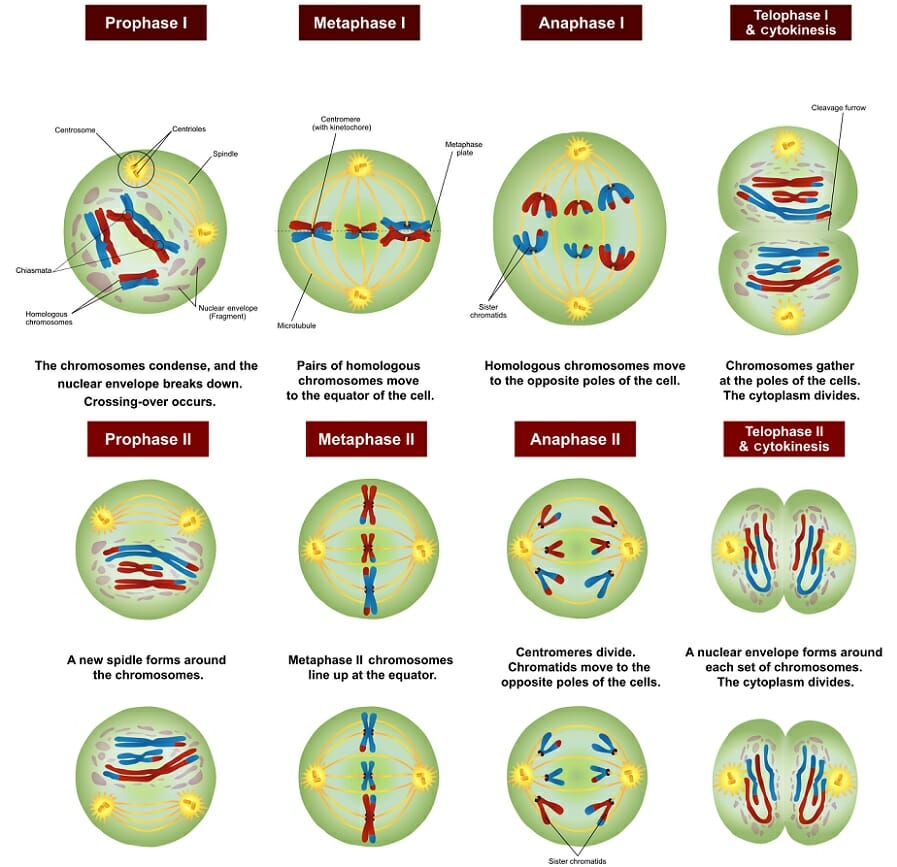
Prophase I
Prophase I, the first step in meiosis I, is similar to prophase in mitosis in that the chromosomes condense and move towards the middle of the cell. The nuclear envelope degrades, which allows the microtubules originating from the centrioles on either side of the cell to attach to the kinetochores in the centromeres of each chromosome. Unlike in mitosis, the chromosomes pair with their homologous partner. This can be seen in the red and blue chromosomes that pair together in the diagram. This step does not take place in mitosis. At the end of prophase I and the beginning of metaphase I, homologous chromosomes are primed for crossing-over.
Between prophase I and metaphase I, homologous chromosomes can swap parts of themselves that house the same genes. This is called crossing-over and is responsible for the other law of genetics, the law of independent assortment. This law states that traits are inherited independently of each other. For traits on different chromosomes, this is certainly true all of the time. For traits on the same chromosome, crossing-over makes it possible for the maternal and paternal DNA to recombine, allowing traits to be inherited in an almost infinite number of ways.
Metaphase I
In metaphase I of meiosis I, the homologous pairs of chromosomes line up on the metaphase plate, near the center of the cell. This step is referred to as a reductional division. The homologous chromosomes that contain the two different alleles for each gene are lined up to be separated. As seen in the diagram above, while the chromosomes line up on the metaphase plate with their homologous pair, there is no order upon which side the maternal or paternal chromosomes line up. This process is the molecular reason behind the law of segregation.
The law of segregation tells us that each allele has the same chance of being passed on to offspring. In metaphase I of meiosis, the alleles are separated, allowing for this phenomenon to happen. In meiosis II, they will be separated into individual gametes. In mitosis, all the chromosomes line up on their centromeres, and the sister chromatids of each chromosome separate into new cells. The homologous pairs do not pair up in mitosis, and each is split in half to leave the new cells with 2 different alleles for each gene. Even if these alleles are the same allele, they came from a maternal and paternal source. In meiosis, the lining up of homologous chromosomes leaves 2 alleles in the final cells, but they are on sister chromatids and are clones of the same source of DNA.
Anaphase I
Much like anaphase of mitosis, the chromosomes are now pulled towards the centrioles at each side of the cell. However, the centrosomes holding the sister chromatids together do not dissolve in anaphase I of meiosis, meaning that only homologous chromosomes are separated, not sister chromatids.
Telophase I
In telophase I, the chromosomes are pulled completely apart and new nuclear envelopes form. The plasm membrane is separated by cytokinesis and two new cells are effectively formed.
Results of Meiosis I
Two new cells, each haploid in their DNA, but with 2 copies, are the result of meiosis I. Again, although there are 2 alleles for each gene, they are on sister chromatid copies of each other. These are therefore considered haploid cells. These cells take a short rest before entering the second division of meiosis, meiosis II.
Phases of Meiosis II
Prophase II
Prophase II resembles prophase I. The nuclear envelopes disappear and centrioles are formed. Microtubules extend across the cell to connect to the kinetochores of individual chromatids, connected by centromeres. The chromosomes begin to get pulled toward the metaphase plate.
Metaphase II
Now resembling mitosis, the chromosomes line up with their centromeres on the metaphase plate. One sister chromatid is on each side of the metaphase plate. At this stage, the centromeres are still attached by the protein cohesin.
Anaphase II
The sister chromatids separate. They are now called sister chromosomes and are pulled toward the centrioles. This separation marks the final division of the DNA. Unlike the first division, this division is known as an equational division, because each cell ends up with the same quantity of chromosomes as when the division started, but with no copies.
Telophase II
As in the previous telophase I, the cell is now divided into two and the chromosomes are on opposite ends of the cell. Cytokinesis or plasma division occurs, and new nuclear envelopes are formed around the chromosomes.
Results of Meiosis II
At the end of meiosis II, there are 4 cells, each haploid, and each with only 1 copy of the genome. These cells can now be developed into gametes, eggs in females and sperm in males.
Examples of Meiosis
Human Meiosis
Human meiosis occurs in the sex organs. Male testis produce sperm and female ovaries produce eggs. Before these gametes are made, however, the DNA must be reduced. Humans have 23 distinct chromosomes, existing in homologous pairs between maternal and paternal DNA, meaning 46 chromosomes. Before meiosis, the DNA in the cell is replicated, producing 46 chromosomes in 92 sister chromatids. Each pair of sister chromatids has a corresponding (either maternal or paternal) set of sister chromosomes. These pairs are known as homologous chromosomes. During meiosis I, these homologous chromosomes line up and divide. This leaves 23 chromosomes in each cell, each chromosome consisting of sister chromatids. These chromatids may no longer be identical, as crossing-over may have occurred during metaphase I of meiosis I. Finally, meiosis II takes place, and the sister chromatids are separated into individual cells. This leaves 4 cells, each with 23 chromosomes, or 4 haploid cells.
Fruit Flies
Fruit flies have 4 pairs of chromosomes or 8 chromosomes in regular cells. Before meiosis takes place, each chromosome is replicated, leaving 8 chromosomes and 16 sister chromatids. Meiosis I takes place, and there are 2 cells, each with only 4 chromosomes. Each chromosome is still made of sister chromatids, and some crossing-over may have occurred during metaphase I. Meiosis II now takes place on those two cells. In total, 4 cells are created, again. However, these cells have 4 chromosomes. When two gametes meet to create a new fruit fly, the resulting zygote will have 8 chromosomes of 4 pairs of sister chromosomes, 4 coming from each parent.
Related Biology Terms
- Haploid – Organism with only one copy of each gene in each cell, or gametes with such.
- Diploid – Two copies of each gene, per cell.
- PolyploidDominance – Multiple (more than two) copies of each gene per cell.
- Sister Chromatids – The replicated DNA that exist as a single chromosome until separated in anaphase.
Quiz
1. A cell is going through meiosis. The sister chromatids are lined up on the metaphase plate. What phase of meiosis is this?
A. Metaphase I
B. Prophase II
C. Metaphase II
2. An adult organism has 60 chromosomes or 30 homologous chromosomes. 30 are maternally derived, 30 are paternally derived. How many chromosomes are in each cell after mitosis?
A. 60 chromosomes, 30 homologs.
B. 120 chromosomes, 60 homologs.
C. 30 chromosomes, no homologs.
3. An adult organism has 60 chromosomes or 30 homologous pairs of chromosomes. 30 are maternally derived, 30 are paternally derived. How many chromosomes are in each cell after meiosis?
A. 30 chromosomes, no homologous chromosomes.
B. 60 chromosomes, 30 homologous chromosomes.
C. 120 chromosomes, 60 homologous chromosomes.