Heart Definition
The heart is a muscular organ that pumps blood throughout the body. It is located in the middle cavity of the chest, between the lungs. In most people, the heart is located on the left side of the chest, beneath the breastbone.
The heart is composed of smooth muscle. It has four chambers which contract in a specific order, allowing the human heart to pump blood from the body to the lungs and back again with high efficiency. The heart also contains “pacemaker” cells which fire nerve impulses at regular intervals, prompting the heart muscle to contract.
This animation shows the functioning of this extraordinarily complex pump in action. As you read this article, try scrolling back up and seeing if you can spot the chambers, valves, and blood vessels we’re discussing in action:
The heart is one of the most vital and delicate organs in the body. If it does not function properly, all other organs – including the brain – begin to die from lack of oxygen within just a few minutes. As of 2009, the most common cause of death in the world was heart disease.
Most heart disease occurs as a result of age or lifestyle. Cholesterol can build up in the arteries as a person gets older, and this is more likely for people who have diets high in saturated fat and cholesterol. Rarely, however, heart disease can also occur due to a virus or bacterium that infects the heart or its protective tissues.
Scientists have had some success replicating the heart’s pumping action with artificial pumps, but these pumps can be rejected by the body, and they break down over time.
The four-chambered heart found in mammals and birds is more efficient than the one, two, or three-chambered hearts found in some other animals. It is thought that warm-blooded animals need highly efficient circulation to satisfy their cells’ high demand for oxygen. This is especially true of humans, whose huge brains require a near-constant supply of oxygen to function!
Function of the Heart
The heart pumps blood through our immense and complicated circulatory systems at high pressure. It is a truly impressive feat of engineering, as it must circulate about five liters of blood through a full 1,000 miles worth of blood vessels each minute! We will talk more about how the heart accomplishes this remarkable task under the “Heart Structure” section below.
The pumping action of the heart allows the movement of many substances between organs in the body, including nutrients, waste products, and hormones and other chemical messengers. However, arguably the most important substance it circulates is oxygen.
Oxygen is required for animal cells to perform cellular respiration. Without oxygen, cells cannot break down food to produce ATP, the cellular currency of energy. Soon, none of their energy-dependent processes can function. Without its energy-dependent processes, a cell dies.
Neural tissues, including the brain, are particularly sensitive to oxygen deprivation. Neural tissues maintain a special cellular chemistry which must be constantly maintained through the consumption of lots and lots of energy. If ATP production stops, neural cells can begin to die within minutes.
For this reason, the body has taken many special measures to protect the heart. It is located below the strongest part of the ribcage and cushioned between the lungs. It is also surrounded by a protective membrane called the pericardium, which is filled with additional cushioning fluid.
Heart Structure
The heart’s unique design allows it to accomplish the incredible task of circulating blood through the human body. Here we will review its essential components, and how and why blood passes through them.
Layers of the Heart Wall
The heart has three layers of tissue, each of which serve a slightly different purpose. These are:
- The Epicardium. The epicardium is also sometimes considered a part of the protective pericardial membrane around the heart. It helps to keep the heart lubricated and protected.
- The Myocardium. The myocardium is the muscle of the heart. You can remember this because the root word “myo” comes from “muscle,” while “cardium” comes from “heart.”
The myocardium is an incredibly strong muscle that makes up most of the heart. It is responsible for pumping blood throughout the body. - The Endocardium. The endocardium is a thin, protective layer on the inside of the heart. It is made of smooth, slippery endothelial cells, which prevent blood from sticking to the inside of the heart and forming deadly blood clots.
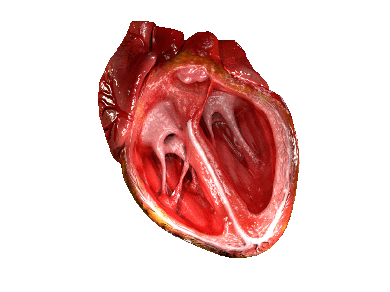
Chambers of the Heart
The heart has four chambers, which are designed to pump blood from the body to the lungs and back again with extremely high efficiency. Here we’ll see what the four chambers are, and how they do their jobs:
- The Right Atrium. The right and left atria are the smaller chambers of the heart, and they have thinner, less muscular walls. This is because they only receive blood from the veins – they don’t have to pump it back out through the whole circulatory system!
The right atrium only has to receive blood from the body’s veins and pump it into the left ventricle, where the real pumping action starts. - The Right Ventricle. The ventricles are larger chambers with stronger, thicker walls. They are responsible for pumping blood to the organs at high pressures.
There are two ventricles because there are two circuits blood needs to be pumped through – the pulmonary circuit, where blood receives oxygen from the lungs, and the body circuit, where oxygen-filled blood travels to the rest of the body.
Maintaining these two separate circuits with two separate ventricles is much more efficient than simply pumping blood to the lungs and allowing it to flow to the rest of the body from there. With two ventricles, the heart can generate twice the force, and deliver oxygen to our cells much faster.
The right ventricle is the one attacked to the pulmonary circuit. It pumps blood through the pulmonary artery and to the lungs, where the blood fills with oxygen, at high pressure. The blood then returns to… - The left atrium receives oxygenated blood from the pulmonary veins. It pumps this blood into the left ventricle, which…
- The left ventricle pumps blood throughout the rest of the body.
After the blood has circulated through the body and the oxygen has been exchange for carbon dioxide waste from the body’s cells, the blood re-enters the right atrium and the process begins again.
In most people, this whole circulatory path only takes about a minute to complete!
Valves of the Heart
You may be wondering how the heart ensures that blood flows in the right direction between these chambers and blood vessels. You may also have heard of “heart valves” referred to in a medical context.
Heart valves are just that – biological valves that only allow blood to flow through the heart in one direction, ensuring that all the blood gets to where it needs to be.
Here is a list of the most important valves in the heart, and an explanation of why they are important:
- The Tricuspid Valve. The tricuspid valve is what is called an “atrioventricular” valve. As you might guess by the name, it ensures that blood only flows from the atrium to the ventricle – not the other way around.
These atrioventricular valves have to stand up to very high pressures to ensure that no blood gets through, as the ventricle contracts quite powerfully to squeeze blood out.
The tricuspid valve is the valve that ensures that blood in the right ventricle goes into the pulmonary artery and reaches the lungs, instead of being pushed back into the right atrium. - The Pulmonary Valve. The pulmonary valve is what is called a semilunar valve. Semilunar valves are found in arteries leaving the heart. Their role is to prevent blood from flowing backwards from the arteries into the heart’s chambers.
This is important because the ventricles “suck” blood in from the atria by expanding after they have expelled blood into the arteries. Without properly functioning semilunar valves, blood can flow back into the ventricle instead of going to the rest of the body. This drastically decreases the efficiency with which the heart can move oxygenated blood through the body.
The pulmonary valve lies in between the pulmonary artery and the left ventricle, where it ensures that blood pumped into the pulmonary artery continues to the lungs instead of returning to the heart. - The Mitral Valve. The mitral valve is the other atrioventricular valve. This one lies between the left atrium and the left ventricle. It prevents blood from flowing back from the ventricle into the atrium, ensuring that that blood is pumped to the rest of the body instead!
The mitral valve lies at the opening of the aorta, which is the largest blood vessel in the body. The aorta is the central artery from which all other arteries fill. It is thicker than a garden hose, extends all the way from our hearts down to our pelvis, where it splits in two to become the femoral artery of each leg. - The Aortic Valve. As you might have guessed, the aorta needs a semilunar valve just like the pulmonary artery does. The aortic valve prevents blood from flowing backwards from the aorta into the left ventricle as the left ventricle “sucks” in oxygenated blood from the left atrium.
Many people have minor irregularities with these valves, such as mitral valve prolapse, which make their hearts less efficient or more prone to experiencing problems. People with minor valve issues can often lead a normal, healthy life.
However, total failure of any of these valves can be catastrophic for the heart and for blood flow. That’s why people with conditions like mitral valve prolapse are often turned down by the military and other programs that involve conditions which can be very taxing for the heart.
The Sinoatrial Node
The sinoatrial node is another very important part of the heart. It is a group of cells in the wall of the right atrium of the heart – and it is what keeps the heart pumping!
The cells in the sinoatrial node produce small electrical impulses in a regular rhythm. These impulses are what drive the contractions of the four chambers of the heart.
Artificial pacemakers replicate the action of the sinoatrial node by making similar electrical impulses for people whose sinoatrial node isn’t functioning properly. However, healthy people have a natural pacemaker built right into the heart!
Quiz
1. Which of the following is NOT true of the human heart?
A. It has four chambers.
B. It has a built-in “pacemaker” called the sinoatrial node.
C. It is responsible for pumping oxygen-filled blood around the body.
D. It is the only organ in the body you cannot live without.
2. Which of the following is NOT a layer of the heart?
A. The myocardium
B. The endocardium
C. The myometrium
D. The epicardium
3. Why does the heart need valves?
A. To keep out pathogens and toxins that could damage the heart
B. To ensure that blood goes not flow backward into the wrong chamber of the heart
C. To ensure that the heart does not pump too much blood at once
D. None of the above
References
- Moore, K. L., Agur, A. M., & Dalley, A. F. (2018). Clinically oriented anatomy. Philadelphia: Wolters Kluwer.
- Heart. (n.d.). Retrieved July 08, 2017, from http://www.innerbody.com/image/card01.html
- (n.d.). Retrieved July 08, 2017, from https://training.seer.cancer.gov/anatomy/cardiovascular/heart/structure.html
- Blood Vessels. (2017, May 19). Retrieved July 08, 2017, from https://www.fi.edu/heart/blood-vessels