Brain Definition
The brain is an organ that coordinates nervous system function in vertebrate and most invertebrate animals. The brain is typically located inside the head, within a protective covering such as an exoskeleton or skull.
In humans, the brain weighs about three pounds and consumes a stunning 20-25% of all the body’s energy!
The brain is primarily made up of neurons, which send nerve impulses and store information, and various support cells which nurture, insulate, and protect neurons so they can do their jobs reliably.
Neurons are an extremely high-maintenance cell-type, requiring large amounts of oxygen and fuel to keep them alive. In order to produce action potentials quickly, allowing for thought, movement, and other survival functions, neurons create an ion gradient that must be actively maintained at all times.
If the brain is deprived of oxygen or cellular fuel such as glucose, the ion pumps which maintain this ion gradient will shut down. This will lead to an influx of ions and fluid into the cells, which will actually cause the neurons to burst open.
This is why the brain is so vulnerable to oxygen deprivation. For a human, brains cells can begin to burst and die within minutes of oxygen deprivation.
Because it is so vital to the body’s functioning, brain tissue is separated from the blood stream by a “blood-brain barrier” through which only certain substances can pass. The blood-brain barrier filters out bacteria, some viruses, and some chemicals while allowing nutrients and oxygen to reach the brain tissues.
Brain Functions
The brain is involved with virtually every aspect of our experience. It is the fascinating arena where human experience meets biology.
Our experiences and feelings are processed, stored, and sometimes created by physical and chemical processes in the brain. Our thoughts and feelings can be measured as action potentials; our memories and personalities have physical form as synapses, which are branches that connect nerve cells to each other and determine how they interact.
Our brain perceives colors, sounds, and sensations. It perceives and creates emotional states. It contains our motor skills and our language center. It also releases hormones that regulate unconscious functions of our bodies. A part of the brain called the brain stem even sends nerve impulses which control and maintain our breathing!
Some general categories of functions of the brain include:
- Receiving and processing sensory information
- Directing movement through nerve impulses
- Directs breathing through the brain stem
- Helping to maintain the body’s homeostasis
- Helping to direct the body’s reproductive cycle
- Forming and storing memories
- Storing skills and conceptual information
- Creating, processing, and regulating emotional states
- It is thought that one particular part of the brain may create consciousness by unifying all of these functions into a single matrix. This is a relatively new finding, and needs more study.
Because the brain’s functions are many, it’s easiest to discuss them in more detail by looking at the function of each of the many structures in the brain.
Structure of Brain
The brain is a complex and intricate organ, with numerous functions. The easiest way to list those functions and discuss how they are accomplished is by studying the structure of the brain, which is the “machine” that accomplishes all of these functions.
Neurons
To understand the function of the brain, it helps to understand the structure of neurons. Just like other cells, neurons have a nucleus, cytoplasm, and a cell membrane. But unlike other cell types, neurons also have long, long arms called “axons,” and they constantly create and destroy tiny intercellular connections called “dendrites.”
The structure of a neuron can be seen here:
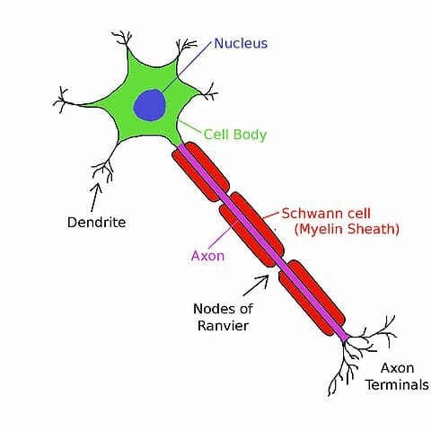
“Dendrites” can be thought of as the neuron’s receivers. They receive input from other nerve cells, to be processed in the cell body. This input can be received either in the form of a nerve impulse – that is, a direct electrochemical impulse – or in the form of neurotransmitters, chemical messengers which interact with receptors on the receiving cell. Neurons can “decide” whether or not to fire an action potential in response to an action potential or neurotransmitters received from another cell.
The means by which dendritic inputs are processed by the cell are not well understood. Some nerve cells appear to use simple summation – that is, the amount of stimulating and inhibiting input is added up by the cell when deciding not to fire. But other cells may fire different patterns of action potentials depending on which dendrites are stimulated, suggesting that there may be additional internal processing.
The “axon” of the neuron is the part of the neuron that fires an action potential of its own, if the cell “decides” to do so. Axons can be long, spanning the whole length of the brain or even the length of an arm or leg! They are insulated by a special layer of lipids called a “myelin sheath,” which prevent the ions that carry the nerve’s signal from leaking out as the nerve impulse travels the length of the axon.
What all of this means is that neurons in the brain are able to receive input from many other neurons and make “decisions” about what sort of action to take. This complex web of input, processing, and firing is what allows our brain to turn simple colors and lines into images of faces that we recognize, among other wonders!
Within the brain, there are numerous distinct structures that perform unique tasks. For the sake of brevity, we won’t talk about every single one of them here. Instead, we’ll focus on the major regions of the brain and the functions that these regions perform:
Frontal Lobe
The frontal lobe is part of the cerebral cortex. This cerebral cortex, or “cerebrum” is the largest part of the human brain, and is thought to be the most recently evolved.
Most other animals have a much smaller cerebral cortex than humans. In humans, the lobes of the cerebrum are responsible for “higher” tasks such as thought, language, action, and impulse control.
The frontal lobe is the region of cerebrum found at the front of the head, immediately behind the eyes and forehead. It contains the regions of the brain that can perform math and speech, as well as those responsible for planning, problem-solving, regulating the emotions, and making conscious decisions.
People with damage to the frontal lobe from injuries can show traits like volatile emotions, lack of self-control, and lack of socially appropriate behavior. They can also have trouble solving problems and making and sticking to plans.
Some neurologists have gone so far as to assert that, due to its connection to language, math, problem-solving, emotional regulation, and conscious decision-making, it is the frontal lobe of the brain that makes us uniquely human. Other neurologists, however, say it’s far more complicated than that!
Temporal Lobe
The temporal lobe is where our brain processes sounds, including the sound of speech. It is found on either side of the brain below and behind the cerebral cortex. A good reference point for the location of the temporal lobe is the place where the hinge of your jaw meets the braincase of your skull.
The temporal lobe contains complex circuitry for analyzing the sounds we hear for pitch, tone, and meaning. It even sends auditory data to the limbic system to determine a sound’s emotional content, and to the language center to determine its verbal content. The temporal lobe can even determine roughly where a sound is coming from through triangulation, by comparing when the sound reached one ear vs. the other.
The temporal lobe can temporarily store auditory memory, and may play a role in the formation of long-term memories through its connection to the hippocampus.
Parietal Lobe
The parietal lobe is located on top of the brain toward the back. It spans roughly from the top of your head to about halfway down the back of your skull. This lobe processes sensory input from the body, and also contains the circuitry for movement.
Once, it was thought that there was really only one sense contained in most parts of the body: that of touch. Now, however, we know that there are at least two distinct senses: touch, and proprioception. Proprioception uses motion and position sensors in the body to tell us where different parts of our body are in space. This is essential to allowing us to execute complicated movements, and to move at all without losing our balance!
The parietal lobe also contains circuitry which can process visual input from the occipital cortex to help us recognize faces and objects.
Occipital Lobe
The occipital lobe is the smallest in the cerebral cortex. It is located at the very back of the head, near the base of the skull.
The occipital lobe processes visual information. The optic nerves from the eyes pass deep into the brain, through a processing center, and finally deliver their information to the occipital lobe, which decodes visual information into colors, shapes, and objects.
Because we have two eyes facing the same direction – a trait called “binocular vision” – our occipital lobe can produce a three-dimensional image of the world by comparing the slightly different views from our two eyes.
The occipital lobe sends visual information through many steps of processing, ultimately linking up with memory circuits to allow us to recognize objects, people, and places in our environment.
Cerebellum
We are now leaving the cerebrum – the most recently evolved part of the brain – and moving into older structures. The cerebellum is a small structure at the base of the brain, directly below the parietal and occipital lobes. It is responsible for regulation of movement, posture, and balance – very important functions for any organism to have!
People with damage to the cerebellum can have difficulty walking, executing complex movements, and even standing. We often take this little part of our brain for granted, but walking on two legs is no easy task!
Limbic System
The limbic system is sometimes referred to as “the emotional brain. It lies at the center of the brain, with the cerebral cortex wrapped around it and the cerebellum tucked behind it. It is an evolutionarily old structure; it is also extremely vital. The limbic system includes:
- The hippocampus, which creates and stores memories. This structure nestled in the center of the brain has connections which can stimulate most other brain regions, allowing us to recall the sights, sounds, emotions, and other aspects of events in our past.
It is not known exactly how the hippocampus creates memories, or whether it is truly the storage site of memories. Some studies have suggested that people with hippocampal damage cannot form new memories, but may still be able to access memories from before the damage.
It is thought that the amygdala can affect how the hippocampus stores memories, resulting in stronger and more vivid encoding of memories that take involve fear, trauma, or other strong emotions. This may have given our ancestors an evolutionary advantage by ensuring that they avoided dangerous or harmful situations in the future. - The amygdala monitors and helps to create emotional states.
Emotional states are now thought to be a team effort between the brain and the body. The activity of the amygdala is influenced by cues from the body such as heart rate, posture, and adrenaline. But the amygdala also influences the body in return by triggering fear responses when a threatening sight, sound, or other stimulus associated with a dangerous or painful memory is detected.
The amygdala can also send signals to the hippocampus which cause a memory to be encoded more vividly if it is made under circumstances of acute fear or pain. This is thought to be a survival adaptation to allow us to avoid fear and pain more effectively in the future. It is also thought to be the reason why traumatic memories tend to be very vivid, and can sometimes be “triggered” by similar sensory stimuli in conditions such as PTSD.
The amygdala is most often said to be related to fear and pain, as these are some of the easiest emotional states to identify and are commonly studied by psychiatrist and neurologists seeking to help people heal from trauma. However, it is possible that the amygdala may also play a role in positive emotions that is not yet well-understood. - The thalamus acts as the “switchboard” for the cerebral cortex. All sensory information except for smell passes through the thalamus before it goes on to the cerebral cortex processing centers. This might be why you cease noticing some parts of your environment, such as the way your clothes feel against your skin, while stimuli that are new or important to what you are doing attract your attention.
It is thought that the thalamus might help the brain to “decide” which sensory stimuli to pay attention to. This might assist us with survival by ensuring that we prioritize relevant things in our environment, while ignoring parts of our environment that are not affecting us at the moment. - The hypothalamus is a tiny structure located under the thalamus. It plays a vital role in releasing chemical messages from the brain to the body, which regulate many of our body’s involuntary functions.
Chemical messages released by the thalamus include messages which make us hungry, thirsty, and sleepy; messages tell our kidneys when to conserve water; and messages that can effect our emotional states.
Problems with the hypothalamus can result in a wide array of illnesses where organs do not function as they should, even though the organ itself is healthy and undamaged.
Organs which need signals from the hypothalamus to function properly include the adrenal glands, the thyroid glands, the kidneys, and the reproductive organs.
Brain Stem
The brainstem is responsible for the most basic of life functions. Its duties include causing the diaphragm to expand and contract so that we can breathe; regulating the heartbeat; and regulating blood pressure. The parts of the brain stem include:
- The midbrain. This intriguing structure assists with many purposes, and may have been relied on more heavily by our very ancient ancestors early in our evolutionary history. It plays a role in vision, hearing, eye movement, and body movement. Its most crucial functions include:
The midbrain contains the substantia nigra, which produces all the dopamine used by the motor system to allow movement. Parkinson’s disease is most typically caused by deterioration of the substantia nigra, resulting in a lack of dopamine in the motor cortex.
The midbrain also contains the superior colliculus, which has a remarkable ability. Some people who cannot see because of damage to the occipital lobe can perform basic visual tasks using the superior colliculus. Although the superior colliculus does not consciously register visual data, the body seems to be able to use it to move appropriately! - The pons is involved in auditory processing, motor control, and sensory analysis. Perhaps its most unique function is its role in sleep.
During sleep, the pons sends signals to the rest of the brain which activate the processes of REM sleep – making dreams, as well as the consolidation of learning and memory possible! - The medulla oblongata is responsible for maintaining our breathing and regulating our heart rate. It also contains cells which can detect poisons in the bloodstream and trigger vomiting in response.
When death occurs from brain injuries, it is most often because the brain swelled to the point of crushing the brainstem, which lies underneath the other brain structures. Disruptions to medulla activity can cause breathing to stop, resulting in death.
This is why doctors recommend that patients with certain serious head injuries be awakened every few hours when they sleep. In cases of brain swelling and internal bleeding, unconsciousness typically occurs before breathing stops. Discovering that a person with a brain injury is unconscious and cannot be woken up can sometimes allow doctors to take action to prevent death from medulla compression.
Clostrum
The “clostrum” is a part of the brain which was only recently discovered, and about which little is known at present. It is intriguing because some scientists believe it might be the part of the brain where input from all of the above functions are combined into the experience of consciousness.
For many years, the answer to the question “how does consciousness occur?” as “we have no idea.” It still is – scientists don’t know how exactly the clostrum might produce consciousness – but with the clostrum’s discovery, at least one small piece of the puzzle has been solved.
Previously, a puzzle for doctors was that there seemed to be no single area of the brain that interfered with consciousness when damaged. Damage to different areas of the brain could cause many different symptoms, but people would continue to seem awake and aware unless most or all of the brain stopped functioning. What part of the brain, then, was responsible for consciousness?
The very existence of the clostrum was missed for many years because it is tiny. The clostrum is a thin sheet of tissue lining each hemisphere of the brain, which receives inputs – and sends outputs – to virtually every part of the brain.
While attempting to treat a patient with epilepsy, it was quite accidentally discovered that disrupting the activity of her clostrum resulted in a cessation of consciousness. She did not react to, experience, or remember anything from periods of time where her clostrum was being disrupted.
All of these discoveries have been made only in the last few years, so much more research is needed. But that makes it a very exciting area of research to follow!
Two Hemispheres
One of the most remarkable and underappreciated things about our brain is that it has two hemispheres. Our cerebral cortex is essentially separated into two halves, each of which have very similar wiring. The two halves of our cerebral cortex can only communicate with each other directly through the corpus callosum – a band of fibers which sends information back and forth between the two sides.
They can communicate very basic information, such as emotion and survival, indirectly through the limbic system and brainstem which receive input from both hemispheres.
For many years this was thought to simply be a biological oddity, but recently scientists are beginning to think that it’s very important to who we are. Our brain hemispheres often have slightly different wiring, which gives each one slightly different abilities. In most people, for example, a speech center is only found in the left hemisphere; the right brain may have little or no language capability, but is more sensitive to the emotional content of sensory stimuli.
It’s not quite as simple as the pop culture myth that math and science live in the left brain, while art and music live in the right. But it is true that the different brain hemispheres have some different abilities – and may even have different desires, and come to different solutions when solving problems.
One patient whose corpus callosum had been cut to control severe seizures was interviewed by scientists. He was found to have some language function in both his left and right brain hemispheres, which allowed each side to be verbally interviewed separately. This was done by only letting one hemisphere “see” or “hear” the questions, since each eye and each ear send their sensory input to only one hemisphere of the brain.
The results were rather astounding! This patient’s right brain hemisphere gave different answers from is left brain when asked about his ambitions, political feelings, and religious beliefs.
Later experiments showed that other patients with their corpus callosum cut would display similar “differences of opinion” between their brain hemispheres, such as having each of their hands try to solve a puzzle in a different way. Sometimes the two hands would even appear to fight over the best solution, undoing each other’s work!
For most people, the two hemispheres are united by a corpus callosum which allows us to take both of their perspectives into account. But the implications of the split brain for healthy people are still being studied!
Quiz
1. Which of the following is NOT a function of the brain?
A. To process and integrate sensory input
B. To permit movement
C. To coordinate essential life functions
D. None of the above
2. Which of the following is the part of a neuron responsible for firing an action potential and sending a message to other neurons?
A. The dendrites
B. The nucleus
C. The axon
D. The myelin sheath
3. Which of the following symptoms might be expected of someone with frontal lobe damage?
A. Blindness
B. Deafness
C. Lack of impulse control
D. Inability to regulate bodily functions
References
- Shepherd, G. M. (1994). Neurobiology. New York: Oxford University Press.
- Brain Structures and their Functions. (n.d.). Retrieved July 14, 2017, from http://serendip.brynmawr.edu/bb/kinser/Structure1.html
- Sporns, O. (2010). Networks of the Brain. MIT Press.
- (n.d.). Retrieved July 14, 2017, from http://www.nytimes.com/health/guides/disease/hypothalamic-dysfunction/overview.html
- Kandel, E. R., Schwartz, J. H., & Jessell, T. M. (1995). Essentials of neural science and behavior. Norwalk, CT: Appleton & Lange.
- Rogers-Ramachandran, V. S. (n.d.). When Blindness Is in the Mind, Not the Eyes. Retrieved July 14, 2017, from https://www.scientificamerican.com/article/when-blindness-is-in-the-mind/