Definition
The parietal lobes of the left and right hemispheres of the cerebral cortex are essential for sensory perception and language processing. Damage or degeneration of the parietal lobe produces typical symptoms that include aphasia (impaired speech), tactile agnosia (inability to recognize an object by touch), and agraphesthesia (difficulty recognizing a shape, number or letter traced on the skin).
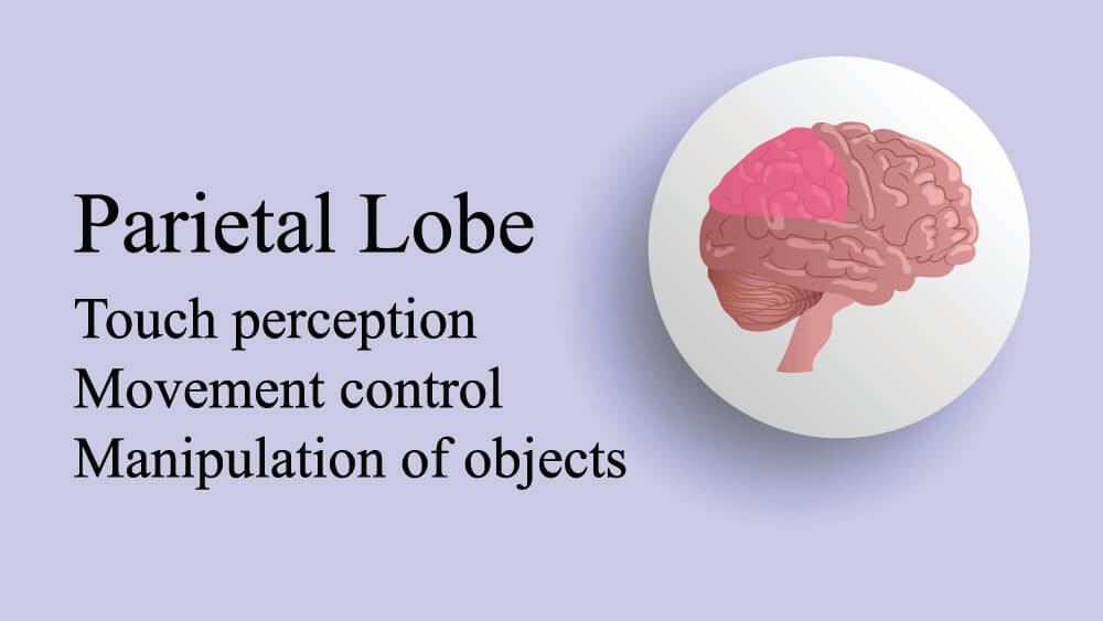
Parietal Lobe Function
Parietal lobe function depends on two factors – the dominant side of the brain and the subarea of the parietal lobe involved. Dominance allows the brain to have asymmetrical functions and, in the parietal lobe, this enables better motor, language, perception, and emotion processing. What side of your brain is dominant depends on genetic factors, although right brain hemispheres are dominant in human infants. Limited asymmetry, where there is not as much difference in the function of each side of the brain as normal, is associated with neurological disorders such as schizophrenia and bipolar disorder. So when we talk about parietal lobe function and which part of each lobe does what, there are no guarantees that this applies to everyone.
The parietal lobes are extremely complex and best categorized according to specific locations. The main functions of the parietal lobes are to receive sensory stimuli, to associate these stimuli with certain actions, and to synchronize them with other sensory impulses that happen at the same time.
The parietal lobe is split into five functional sections. These are the:
- Primary receiving area
- Somesthetic association area
- Polymodal receiving area
- Granular insular area
- Polymodal assimilation area
All of these regions are heavily interconnected with each other and with other structures of the central nervous system. After all, processing information in the brain is much more than a one-step process. The groups of cells and tissues that connect to the parietal lobes and the lobes themselves work together so that we can respond to all kinds of sensory stimuli. Exactly how much we respond also seems to depend on the parietal lobes. For example, studies on violent men show various brain abnormalities like larger areas of parietal white matter and smaller areas of gray matter.
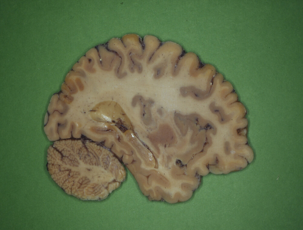
In very simple language, gray matter houses the control areas of the brain, and white matter provides millions of connected pathways. So, in the example of violent tendencies, there are too many connections and too few control mechanisms. Exactly how this works we don’t quite know. The image below is a sliced-through section of a human brain that shows the distinctive gray and white layers.
Primary Receiving Area
The primary receiving area of the parietal lobe receives input from our muscles, joints, and skin. It responds to touch, texture, shape, movement, and the direction of movement. Each area of input differs histologically – that is, different types of sensory neurons are found in each input area. These specific groups of neurons respond to temperature differences felt on the skin, to pain, to touch, and so on.
It is up to the parietal lobe to prioritize these inputs according to how much they affect (arouse) us. If we hardly react to a stimulus, it will fade into the background. That is why smells disappear and why we don’t always hear background music. And can you feel your shoes or socks as you are reading this? You probably can now, as you have just been reminded.
The reasons why you stopped feeling them is because you stopped taking notice of them; your parietal lobe understood that you were not reacting to this touch stimulus as much as you were to your screen and your reading skills, and the touch sensation was pushed to the back of the queue.
It is a good thing the parietal lobes organize stimuli according to our arousal to them – if they didn’t, we would have so many sensations going on at any one time that we would find it practically impossible to function. People suffering from severe chronic pain are unable to concentrate on a book, music, a good meal, social contact, or any range of enjoyable and not so enjoyable distractions. The brain is overwhelmed by pain stimuli that the person cannot ignore because the parietal lobes make pain processing a priority. Sometimes deep breathing exercises, massage, and certain scents seem to reduce pain. This is partially because they take the focus away from the pain stimuli and the parietal lobes may reduce their priority…at least for a short time.
The primary receiving areas of the two halves of the parietal lobes are split into four Brodmann’s areas – 1, 2, 3a, and 3b. Brodmann’s areas are regions of the brain that differ according to specific cell types. The order of Brodmann area (BA) numbers has nothing to do with rank or organization but simply lists the order in which each cell group was first identified.
You will rarely be expected to learn every Brodmann area as these are becoming less significant, especially as the results of new neurological research surfaces. However, considering the role of Brodmann’s areas helps us to form a clearer overview of brain function. This also applies to the parietal lobe.
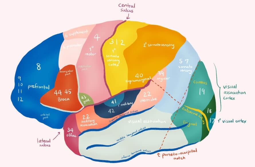
The primary type of tissue in the primary receiving area of the parietal lobe is BA 3; it receives most of its sensory input from the thalamus. The electrical messages that arrive from the nerve endings on the skin and inside the body are then processed in area 3b. In area 3a, electrical messages (evoked potentials) arrive from nerves that measure the body’s position in space (proprioception). BA 3 receives this information and passes it on.
Evoked potentials from BA 3 are sent to areas 1 and 2. Brodmann area 1 is important for processing texture, and area 2 processes information about shape, size, and position. For example, your cat suddenly bumps its head on your elbow. Different senses send information to BA 3 – these might be the texture of the fur, the form of the skin and bones of the skull, the pressure with which the cat bumps its head, the area of skin that is affected, and the position of your elbow. All of these messages are split into appropriate groups and forwarded to BA 1 and BA 2.
The primary receiving areas also process groups of stimuli. The more we use certain muscles and senses in combination, the more likely the primary receiving area will group these together. So it is also an important part of the brain in terms of memory. This explains why you didn’t jump a mile when the cat suddenly bumped your elbow – it has probably happened many times before and your primary receiving area recognizes the group of stimuli and files it as at cat-related incident.
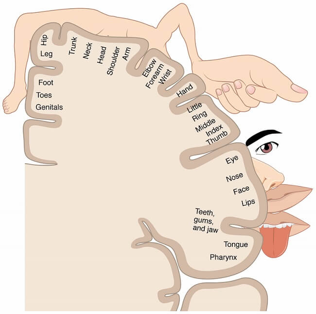
As eating is necessary for survival, the areas of sensory input and motor coordination for hands and mouth are extremely close to one another. And the more sensitive a body part, the more space it is allocated in the primary receiving area. You have probably come across strange-looking Picasso-like illustrations of the brain that show the association between different brain areas and parts of the body. These are called sensory and motor homunculi. These pictures visually describe the primary receiving areas of the parietal lobe that correspond with sensory input from various sensitive and not so sensitive areas of the body.
Another interesting point to make is that neurons in this area of the parietal lobe process information that comes from the contralateral side of the body. For example, the right-hand parietal lobe receives information from the left hand and vice versa.
Somesthetic Association Area
The next functional area of the parietal lobe is the somesthetic association area. This part of the parietal lobe is the next step in processing the information from the primary receiving area. But it also has access to another source of information – motor stimuli from the frontal lobe. These messages can also pass between the hemispheres via the corpus callosum – a central band of nerve fibers that connect both hemispheres.
The somesthetic association area is nearly all composed of Brodmann area 5 tissue. This tissue type plays a major role in limb movement and our sense of proprioception. Damage to this part of the brain usually provides signs of disturbed limb movement and an inability to reach for or grasp objects.
Polymodal Receiving Area
The information gathered from the primary receiving and somesthetic association areas, together with motor, visual, and auditory stimuli from the receiving areas of other lobes such as the frontal lobe, are sent to the polymodal receiving area. Polymodal means multiple functions, and this part of the brain certainly has a lot of them. The polymodal receiving area is mostly composed of Brodmann area 7 tissue and is connected to a huge range of specialized neurons.
The nerve cells in BA 7 are also polymodal – each neuron can respond to multiple sensory stimuli. A single neuron might transmit information from the skin, the nose, and the ears. This is why damage to the polymodal receiving area often leads to multiple symptoms that can include partial loss of vision, pain, absence of pain, impaired speech, unusual eye movement, reduced proprioception, and/or abnormal limb movements.
Granular Insular Area
The second-to-last link in the parietal lobe chain is the granular insular area. Insular regions are deep-lying and not always attributed to a specific Brodmann group.
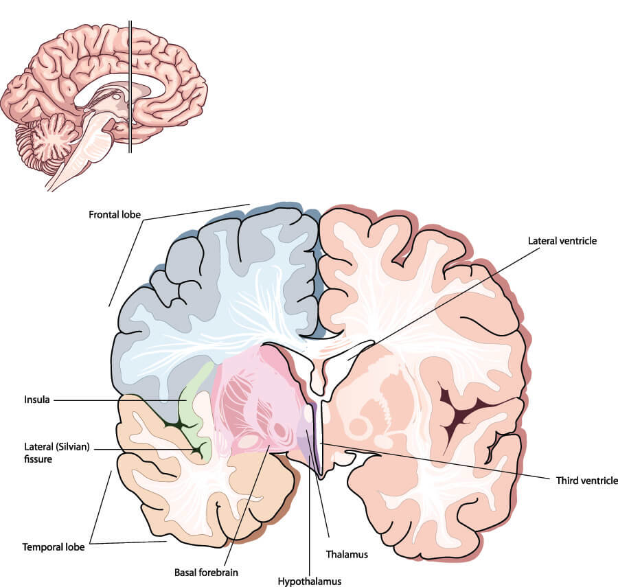
The insular cortex of the brain is shared between the parietal, temporal, and frontal lobes. It is the left-hand pale green structure in the below image, deep within the brain. It organizes various stimuli and creates complex reactions that combine emotion, cognition, motivation, and memory with action and reaction. Stimuli enter the insular cortex from all over the central nervous system.
Post-traumatic stress disorder (PTSD) is linked to smaller amounts of insular cortex tissue, especially in young girls. PTSD is an anxiety disorder – the sufferer re-experiences previous traumatic events. At the other end of the scale, people who regularly practice yoga or meditation have greater quantities of insular cortex tissue. People with PTSD can benefit from relaxation techniques. This is a very simple example, as both PTSD and relaxation techniques involve multiple areas of the brain; however, it is great to know that we can improve its function.
Polymodal Assimilation Area
The final functional unit of the left and right parietal lobe consists of Brodmann area 39 and Brodmann area 40 tissue.
Damage to Brodmann area 40 in the left hemisphere causes lexical agraphia or the inability to write words, and anomia – if you cannot recall the names of everyday objects you are suffering from anomia. If Brodmann area 39 is damaged on the same (left) side, phonological dyslexia (the inability to sound out words when reading), and phonological agraphia where you cannot sound out the words you are writing are common. Damage to all of the polymodal assimilation area of the left hemisphere can result in Gerstmann’s syndrome.
Gerstmann’s syndrome has four main symptoms and these describe both shared and unique functions of the polymodal assimilation area. These are the inability to write, calculate, know right from left, and – very strangely – determine whether observed fingers are the person’s own or belong to someone else.
Parietal Lobe Damage
Parietal lobe damage can lead to both sensory and motor symptoms that include all of the functions mentioned in this article and probably many more. Damage to the parietal lobe might be caused by increased intracranial pressure (ICP) due to infection, burst or blocked cerebral blood vessels (stroke), edema, or a tumor. Any of these will cause a portion of nervous tissue to die and affect brain function, depending on where the damage occurs.
Damage to the dominant side of the parietal lobe at Brodmann areas 1, 2, and 3 (the primary receiving area) produces a loss of proprioception and fine-touch skills. Ever had someone trace a number or a letter on your back? If the parietal lobe is damaged in the primary receiving area you won’t know what they are writing. This is called agraphesthesia.
The owner of the feet in the photograph below should at least know in what direction the complicated drawing is being done and will probably recognize a number of the shapes – perhaps a square or circle on the big toe. Chances are the person will figure out what the drawing is of without needing to see it (or notice the skeleton foot clue).

If you find it difficult to identify a familiar object you are holding in your hand through your sense of touch (not looking at or smelling it), you are suffering from astereognosia. Astereognosia is also associated with parietal primary receiving area damage.
Damage to the non-dominant side of the parietal lobe leads to hemineglect or not being aware of objects or even body parts on one side of the body; the damaged primary receiving area side will be opposite to the side of neglect.
Left hemisphere damage in the somesthetic association cortex means you will be unable to move with purpose. Signals are lost that give us the motivation to move. Right-hand side damage can lead to visuospatial disorders that affect our ability to perceive movement, depth, and distance.
In the polymodal receiving area, many different functions overlap. Tactile localization (knowing that your cat bumped your elbow), language and comprehension, reading, calculation, verbal creativity, processing of emotions, and decision making can all be affected. Emotion processing in combination with action and reaction responses is also the main function of the insular tissue. If damaged, you may have problems recognizing which position your limbs are in and so be unbalanced and uncoordinated. Moving objects might not be properly processed, and you won’t be able to dodge a low-flying Frisbee. Your sense of creativity will be negatively affected and you might also present with the symptoms of Gerstmann’s syndrome.

Quiz