Definition
The parietal bone or os parietale is a paired, flat cranial bone that covers the mid portion of the skull. Both bones cover the left and right parietal lobes of the brain respectively. As part of the neurocranium, the parietal bone helps to form the shape of the head and protect the brain. More specifically, both bones form part of the calvaria (skull cap) and skull base (basicranium).
Parietal Bone Location
So, where is the parietal bone located?
Each bone has sutures with the:
- Greater wing of the sphenoid bone
- Temporal bone
- Frontal bone
- Occipital bone
This means it is very easy to picture the exact location of the parietal bone.
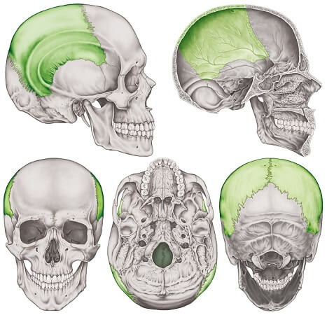
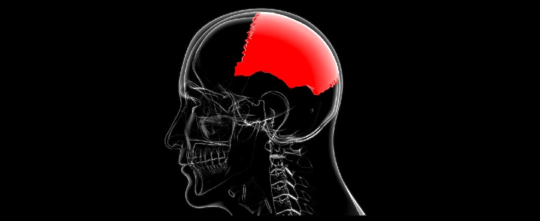
The parietal bones make up the majority of the top of the head.
Parietal Bone Anatomy
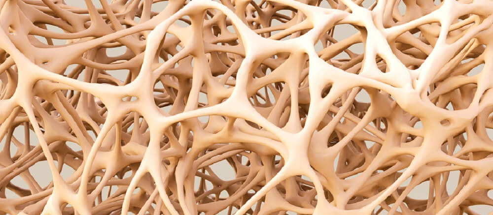
Parietal bone anatomy involves its borders, angles, and surfaces. It is a flat bone composed of two layers of compact bone with spongy bone (cancellous bone) between.
Cancellous bone is honeycomb-like with a network of spaces that contain red bone marrow. Red bone marrow produces blood cells; much smaller quantities are produced in the skull than in the long bones.
Cancellous bone also acts as a shock absorber – extremely important for the protection of the soft and delicate tissue of the brain.
Parietal Bone Borders (Sutures)
Parietal bone articulations with the sphenoid, temporal, frontal, and occipital bones are called sutures.
Immovable joints with a slight amount of give, sutures slowly fuse during childhood.
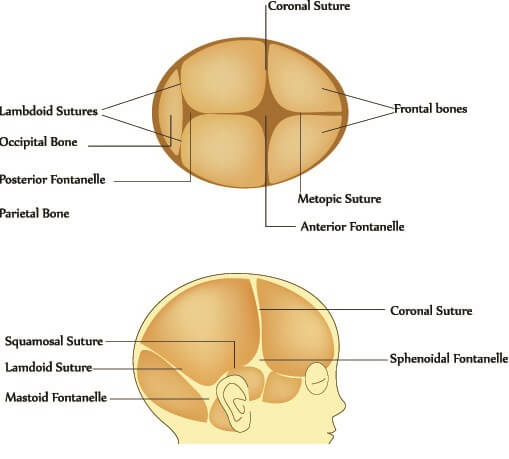
We all know how delicate a baby’s skull is – this is because many sutures have not yet fused. Areas of the skull that have not properly joined are called soft spots or fontanelles.
The largest fontanelle is the anterior fontanelle where the frontal bone, left parietal bone, and right parietal bone have not yet fused. Other soft spots are the sphenoid, mastoid, and posterior fontanelles.
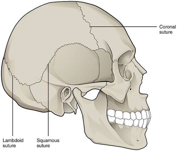
Once fused, the bones of the skull are said to be sutured. The parietal bones have five sutures:
- Sagittal: joins the left parietal bone to the right parietal bone
- Lambdoidal: joins the posterior parietal with the top of the occipital bone
- Sphenoparietal: smallest suture where parietal bone meets the greater wing of the sphenoid bone
- Coronal: front of parietal and back of frontal bone
- Squamosal: bottom of parietal and top of temporal bone
Parietal Bone Surfaces
The external surface of any skull bone is the surface we can feel.
Together, the convex external surfaces of the parietal bones form the rounded casing of the last two-thirds of the top of the skull.
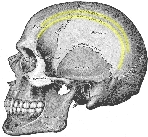
The external surface is not only a protective casing. It also provides attachment points for muscles. The superior temporal line – the upper curve that borders the temporal fossa – provides a place of attachment for the temporal fascia. The temporal fascia covers the temporalis muscle – a mastication muscle.
The inferior temporal line that lies under the posterior line is where the temporalis muscle originates.
The internal surface is a little more complicated.
An internal view shows three grooves:
- Sulcus arteriae meningeae mediae (grooves for the middle meningeal artery)
- Sulcus sinus sagittalis superioris (midline groove for the superior sagittal sinus)
- Sulcus sinus sigmoidei (part of the groove for the sigmoid sinus)
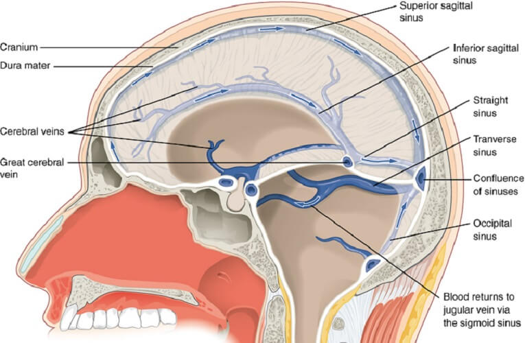
A cranial sinus or dural venous sinus is a space that absorbs blood and cerebrospinal fluid. Sinuses empty into larger veins that return these fluids to the central venous system.
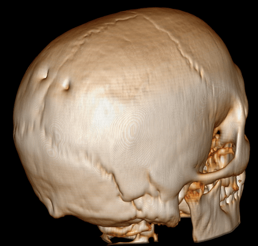
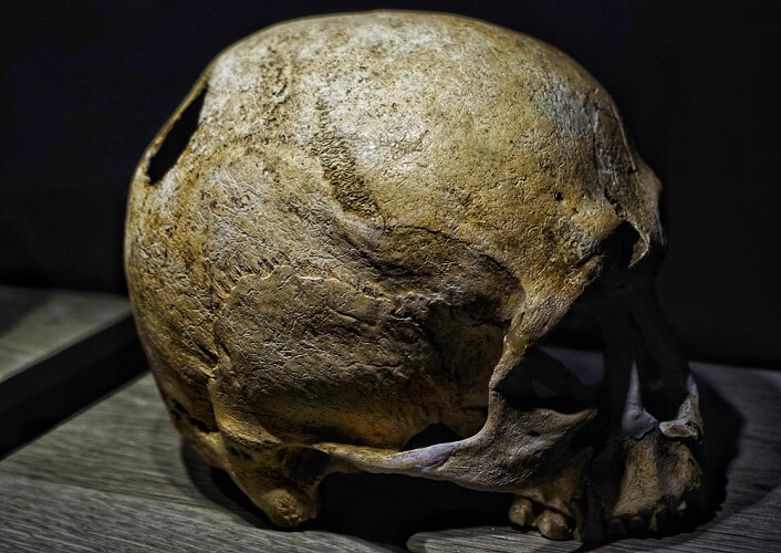
Another feature of this paired bone is a set of two holes. These holes are parietal foramina. They allow blood vessels to pass through the bony plates.
The parietal emissary vein (and sometimes the occipital artery) pass through each parietal foramen. The parietal foramen is found near the lambda – a midline skull landmark where the lambdoid and sagittal sutures meet.
Not everybody has these holes – they are “inconsistent” foramina. Alternatively, they can become smaller over time or be overly large, meeting to form one large hole.
Some syndromes and genetic disorders present with enlarged parietal foramina. In these cases, something stops the ossification process.
Archeologists and paleontologists have often mistaken large parietal foramina for signs of trephination (trepanation) – an early surgical procedure.
Other marks on the inside surface (of all cranial bones) are granular foveolae – small recesses where the arachnoid mater membrane protrudes into the bone. Smaller versions of parietal grooves called sulci arteriosi provide space and protection for smaller arteries.
Parietal Bone Function
The primary parietal bone function is structural. This pair of bones helps to form a strong, rounded casing over the brain. The skull naturally protects the soft tissue of the brain from trauma – at the same time, it stops expansion when infection, increased production of cerebrospinal fluid, and bleeding occurs. These situations can lead to increased pressure inside the cranium.
Without surgical treatment, cases can quickly become fatal.
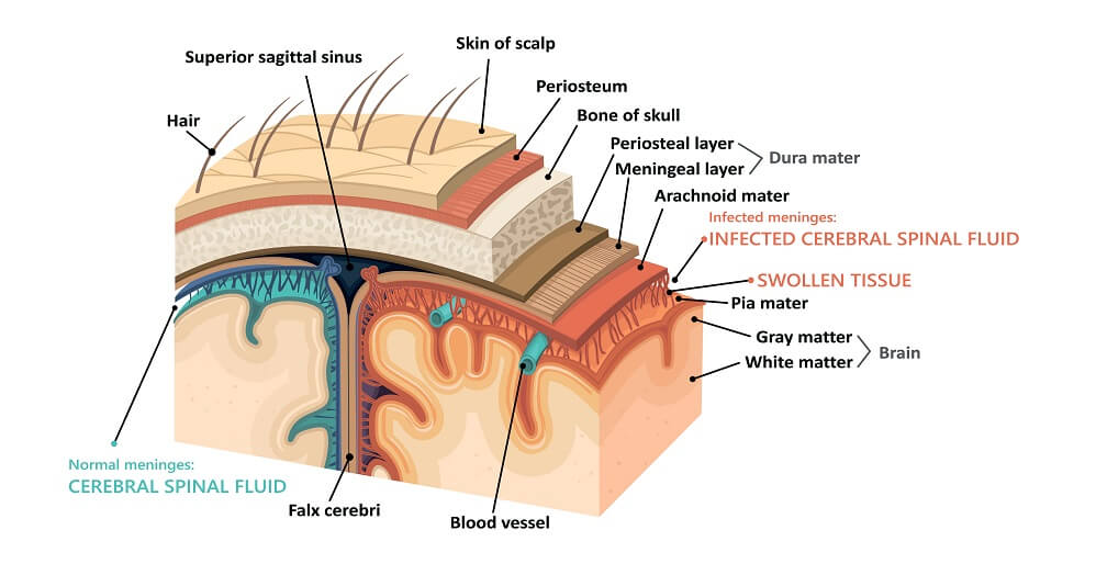
The skull’s internal surface also provides space for the meningeal blood supply – providing oxygen and nutrients to the three membranes that cover the brain.
Parietal Bone Lumps and Bumps
A parietal bone lump is not a normal sign. It may indicate a benign skull osteoma – an extremely slow-growing condition where the two compact bony plates (but not the central cancellous bone) increase in size. Most cranial osteomas form in the parietal plates and rarely occur alone.
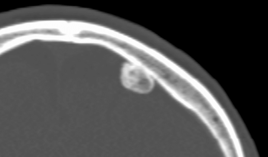
As an external parietal bone bump or osteoma slowly increases in size, it can affect the shape of the head. These thickened areas of bone can be surgically removed. Bone thickening rarely occurs on the internal surface, where pressure to the brain can occur; most skull lesions are discovered incidentally.
Parietal Bone Fractures
A parietal bone fracture can cause serious consequences. The bones cover the parietal lobes of the brain but edge over parts of the frontal lobe, occipital lobe, and temporal lobes.
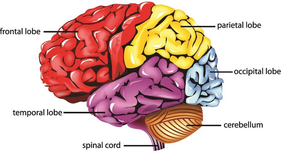
If a parietal bone fracture damages the parietal lobe, several senses may be affected, as well as cognition.
Damage to the left parietal lobe can cause Gerstmann’s syndrome with symptoms like agraphia (difficulty writing), acalculia (trouble doing math), aphasia (speech defects), and agnosia (recognizing objects).
If both parietal lobes are damaged, Balint syndrome may be the result.
Damage to the right parietal lobe can cause memory and personality disorders.
Parietal fractures aren’t always visible. Symptoms can take time to develop. If a skull fracture causes bleeding inside the brain, it may take a few days for intracranial pressure to cause neurological symptoms.
These symptoms depend on the area of the brain that is affected. A stroke – bleeding inside the brain caused by blocked or broken blood vessels – is not the result of a fracture but can lead to similar symptoms.
If a parietal fracture is not displaced – a hairline fracture or mild fracture – no treatment is necessary. A depressed fracture will push into the brain tissue below and an open fracture can allow bacteria to cause infection of the brain membranes (meningitis) and spread to the brain (encephalitis).
As the parietal plates are very strong, this type of fracture is usually the result of extreme assault (kicks to the head and blunt force trauma) and side-on vehicle collisions.
A darker side of parietal bone fractures is abusive injury in very young children. Before the fusion of the sutures, the cranium is more susceptible to injury.
Children are also more likely to suffer skull fractures after falling down the stairs, off playground equipment, or out of a stroller.
Quiz