Eyes Definition
Eyes are animal organs that are specialized for sight. These may be as simple as proteins or cells which can tell light from darkness – like the “eyes” found in many microorganisms – or they may be complex assemblies of lenses, filters, light-sensitive tissues, nerves, and support structures.
Most animals, including humans, have complex and highly specialized visual systems. Different animals have found several different ways to gather light and use it to accomplish complex visual processing.
Mammals, for example, have a single lens and retina which gather light and turn that light into information that the brain can read; insects, on the other hand, have “compound eyes” which use many separate lenses to gather light and put together a mosaic-like view of the world.
Here, we will focus on the details of the human eye.
Eye Parts and Functions
The eye has many parts which work together to accomplish vision, and to keep the structures required for vision safe from infection and injury. These parts include:
The Conjunctiva
The surface of the eye and of the inner eyelids is covered by a clear, protective membrane called the “conjunctiva.”
This is where the word “conjunctivitis” – the scientific name for “pink eye” – comes from. Conjunctivitis means simply “inflammation of the conjunctiva.”
The conjunctiva is lubricated by several substances produced by the body to keep the eye in good working order. These substances, which include mucuous, oils, and a watery solution, prevent the eye from drying out and protect it from surface irritants.
The Sclera
The sclera is also known as the “white of the eye.” It is – as you may have guessed – the white part of the eye that surrounds the iris and pupil.
The sclera does not collect visual data itself. Instead it acts as a tough, protective membrane for the eyeball. Only the outer part of the sclera is white; the interior of the membrane is brown, and wraps around the clear inner chambers of the eye which allow light to pass through.
The Cornea
Light starts its journey into the eye by passing through the cornea. This layer of transparent tissue sits on top of the iris and pupil. It helps to focus light to produce a clear image on the retina, and acts as an additional protective layer for the eye.
Although the cornea looks curved, it is usually actually a flat sheet of uniform thickness. The rounded bulge is the anterior chamber, which will be discussed next.
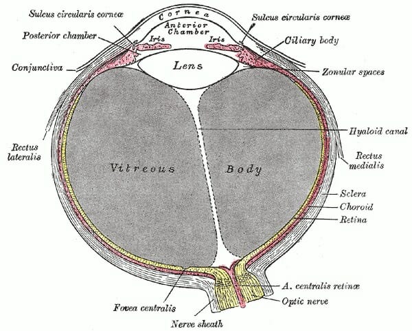
The cornea can be seen in this diagram:
When you use contact lenses, these lenses essentially augment or re-shape your cornea to focus light properly. Some people also undergo laser surgery to change the shape of the cornea so that it will focus light better.
Because it is such a valuable part of the eye, the body wants to know when the cornea has been injured! For that reason it has many nerve fibers running through it, and may hurt a lot of it is scratched, irritated, dried out, or infected.
Because the cornea has almost no blood vessels in it – these would get in the way of light passing through – it can take a long time to heal from injury, and can have a difficult time fighting infection.
For that reason, people who suspect they may have an eye injury or infection should see a doctor right away. Eye injuries and infections can permanently damage vision if they are not treated properly.
Anterior Chamber
The anterior chamber of the eye refers to a small pocket of fluid that lies between the cornea and the iris. This fluid is “aqueous humor” a watery solution that helps the cornea and pupil to focus light.
Just like focusing light through a glass of water or a solid transparent lens, the aqueous humor helps the eye to form an image by refracting light at a constant rate.
The aqueous humor is made from blood plasma, using a special filtering process that removes proteins and other impurities that may cloud vision.
Posterior Chamber
The posterior chamber refers to the aqueous fluid-filled chamber behind the iris and pupil. The posterior chamber sits between the iris and the lens, which completes the job of focusing light.
It can be seen here:
Glaucoma – a condition which leads to gradually impaired eyesight, and eventually blindness if untreated – occurs when aqueous fluid cannot drain properly from the anterior and posterior chambers.
When aqueous humor is unable to drain, the fluid’s pressure builds until permanent damage is caused to parts of the eye essential for vision.
Iris
The iris is the colored ring around the pupil. Different people have different amounts of pigment in their iris, resulting in eye colors ranging from blacks to very pale blues and greens.
Interestingly, there is actually no blue or green pigment produced by the human eye. All human eyes have brown pigment melanin, the same pigment that is found in our skin. But those with very small amounts of melanin reflect a lot of light, which is scattered as it reaches the surface of the eye.
Light that is scattered through a transparent substance tends to appear blue, because more red and green wavelengths are absorbed by the apparently transparent medium, while blue light tends to scatter and reflect. This scattering of blue light is the same reason that the sky is blue, that water in swimming pools looks blue, and the same reason that your veins look blue under your skin even though they are actually dark red.
Green eyes occur when someone with a very small amount of pigment in their iris – producing a blue color through scattering – also produces a yellow pigment that mixes with the blue color.
The iris has a sphincter muscle, which allows it to expand or contract, making the pupil larger or smaller. This is important for controlling the amount of light our eyes receive. If you ever have your pupils artificially dilated by an eye doctor, you’ll notice that having an overly-dilated pupil causes blurred vision, and can make bright lights painful.
Pupil
The pupil is the opening to the inner chamber of the eye. Pupils appear black because light passes through them and does not return. The pupil, then, is our actual “window to the world.”
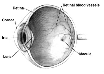
Once it has passed through the pupil, light is focused by the lens. It then travels through the rest of the eyeball to the retina, which lies at the back of the eye. The retina turns the light into signals our brain can understand.
Lens
The lens of the eye lies immediately behind the pupil. Some people think that the lens of the eye is found on the outside, where the cornea is – perhaps because of the use of the word “contact lenses.” But the lens that performs the final focusing of light is found inside the eye, behind the pupil.
The lens is a complex structure. It is made of an elastic capsule containing proteins and water, which refract light at a constant rate just like the lenses used in glasses. It has layers of soft tissue surrounding a firm “nucleus.”
The softness of its outer layers allow the lens to change shape when pushed or pulled by the surrounding ciliary muscles, making it an “adjustable” lens that can change the way it focuses light depending on how close or far away an object is.
Many people’s lenses lose the ability to change shape around the age of 50. This is why many older people need reading glasses in order to focus light to read small print.
Vitreous Humor
The vitreous humor is a thick, gelatinous fluid that fills most of the eyeball. Like the aqueous humor, it refracts light at a constant rate – but unlike the aqueous humor, it is thick and jelly-like.
The jelly-like thickness of the vitreous humor helps the eye to retain its round shape. The precise maintenance of this shape is essential for vision, because light is focused by the cornea and lens with the intent of hitting the retina a set distance away. If the retina moves closer or further to the lens due to changes in eye shape, the light will not be properly focused when it reaches the retina.
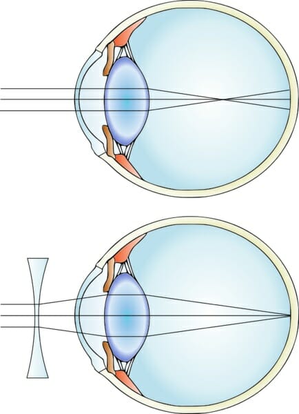
Eyes that have “elongated” or “squashed” shapes are the causes of nearsightedness and farsightedness. Nearsighted eyes are elongated, causing light to focus on a point in front of the retina instead of on the retina itself. Likewise, farsighted eyes are too short, causing light to focus on a point behind the retina.
Glasses correct for nearsightedness or far sightedness by adjusting the focus of light before it enters the eye, so that light is properly focused when it hits the retina.
The focus point of light in a nearsighted eye, along with a nearsighted eye with a corrective lens, can be seen below:
Retina
The retina is a light-sensitive layer of tissue that covers the back of the inner eyeball. It contains light-sensitive cells which can determine light, dark, and color to assemble images of the world. The retina then converts that color information into neural information and sends it to the brain for processing.
The retina contains two major types of light receptors: cone cells, and rod cells.
Cone cells allow us to see color. There are three types of cone cells (or more, in some people with rare mutations). Each type of cone cell responds to a certain wavelength – or color – of light.
S-type cone cells respond to short wavelengths of light, and allow us to see the colors blue and violet. M-type cone cells respond to medium wavelengths, and allow us to see the color green. L-type cone cells respond to long wavelengths of visible light – the red and orange wavelengths.
The color yellow is produced by the activation of both green M-type cone cells and red L-type cone cells. The color pink is produced by the activation of both blue S-type cone cells, and red L-type cone cells. The color white occurs when all cone cells are activated equally, indicating an object that is reflects all wavelengths in the visual spectrum.
Colorblindness occurs when a mutation prevents one or more types of cone cells from working properly. Often, these cone cells do respond to light – but not at the normal wavelength. This can lead to gaps in color perception.
Colorblindness can sometimes be treated using special glasses that filter out wavelengths of color which may confuse mutant cone cells, causing different colors to look the same. While wearing these glasses, many people with colorblindness report seeing all colors clearly and vibrantly.
Because cone cells only respond to a portion of the visual spectrum, they do not work well in low-light conditions. We see in the dark using rod cells, which cannot distinguish color, but which are more sensitive to overall light levels.
Rod cells respond to all wavelengths of visible light. They can tell us how much light is coming at us – but not what wavelength it is. That’s why we don’t see color in the dark; we are getting all of our information from cone cells, which cannot distinguish between different colors.
The retina can only extract information from the light that hits it. This means that in order for the retina to see a clear image of the world, the light that hits it must have been properly focused by the other parts of the eye. As discussed above, failure to focus light properly can lead to blurry vision and other impairments.
Optic Nerve
The optic nerve is a bundle of neural fibers that travel from the retina to the brain. Each optic nerve encodes the image data recorded by the retina in the form of neural signals that can be read by the brain.
The brain then reads the data and performs complex processing, including looking for associations with known objects. This is how we’re able to identify faces and other objects in our environment.
Interestingly, although the eyes are at the front of the head, the brain’s processing of visual signals occurs at the “occipital lobe” in the back of the head.
This means that the optic nerve must travel back into the brain, and then through special channels all the way through it. In the process, the optic nerves “cross over” – meaning that the left side of the occipital lobe interprets visual data from the right eye, and vice versa.
The “crossing over” point can be seen in this image from a brain scan, which has been artificially colored to show the path taken by the optic nerve. Note the red “X” that is formed where the optic nerves cross over each other behind the eyes:

Damage to the optic nerve or visual processing areas of the brain can result in permanent blindness, even if the eye itself is fine. Conversely, people with intact optic nerves can sometime be allowed to “see” by visual prostheses that stimulate the optic nerve, even if the rest of the eye is missing or nonfunctional.
It is hoped that as technology advances, our ability to artificially stimulate the optic nerve will continue to advance so that people with damaged eyes can have near-normal vision.
The Irreducible Complexity Argument
The complexity of the human eye has often been singled out as evidence for “irreducible complexity.”
The idea of irreducible complexity holds that some structures found in nature could not have evolved through mutation and natural selection, because they would not have functioned at all if even one piece were missing. Proponents of irreducible complexity have asked how the eye could have evolved through random mutation, since slight changes to its structure or the removal of any of its parts would render the eye useless.
However, in recent years new insight into the evolution of eyes has been discovered, which suggests a natural way in which all the structures of the eye could have come about through random mutation.
The historical tendency for things “science can’t explain” to later be explained by new discoveries has led many scientists who are also religious to warn against use of the “irreducible complexity” argument.
In his book “The Language of God,” geneticist Francis Collins warns readers that citing “things science can’t explain” as evidence of God creates a “God of the gaps” idea, in which the existence of God is seen to rely on the existence of things science can’t explain. As these “gaps” in scientific understanding continue to shrink, Francis argues, so to will the importance of a “God of the gaps.”
As a result, Collins argues that it’s better for science and religion alike to see the two fields as compatible, than to have a religious view that requires science to fail in order to work. Himself a devout believer in God, Collins has urged his fellow religious people to embrace scientific theories and see these as a means to learn more about God rather than as challenges to dogmatic ideas about God.
Quiz
1. Which of the following is NOT true of eyes?
A. All eyes must have, at minimum, a lens, a retina, and an optic nerve.
B. All eyes must contain cells or proteins that are responsive to light.
C. Almost all animals have some sort of eyes.
D. None of the above.
2. Which of the following is a gel-like substance that helps the eye keep its shape?
A. Aqueous humor
B. The conjunctiva
C. Vitreous humor
D. None of the above
3. Which of the following is NOT true of the cornea?
A. It has almost no direct blood supply, so it is vulnerable to injury and infection.
B. It has almost no nerves, so it is hard to tell when it is injured or infected.
C. It is a clear, flat membrane of uniform width.
D. None of the above.
References
- Land, M. (1992). The Evolution Of Eyes. Annual Review of Neuroscience, 15(1), 1-29. doi:10.1146/annurev.neuro.15.1.1
- Montag, E. D. (n.d.). Parts of the Eye. Retrieved August 10, 2017, from https://www.cis.rit.edu/people/faculty/montag/vandplite/pages/chap_8/ch8p3.html
- Liz Segre; eye illustration by Stephen Bagi. (n.d.). Human Eye Anatomy – Parts of the Eye Explained. Retrieved August 10, 2017, from http://www.allaboutvision.com/resources/anatomy.htm
- The New Encyclopaedia Britannica. (1987). Chicago: Encyclopaedia Britannica.