Endoskeleton Definition
An endoskeleton is a skeleton found within the interior of the body; it provides structural support and protection for the internal organs and tissues of an organism.
Endoskeletons can take several forms, and can differ in complexity, shape and function, depending on the requirements of the animal. Most vertebrates have an endoskeleton, which is comprised of mineralized tissue in the form of bone and cartilage. This ‘true skeleton’ is formed from the mesoderm during embryogenesis.
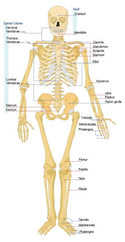
The image shows a diagram of a human endoskeleton with the major bones labeled.
Fish within the class chondrichthyes (sharks, rays and chimaeras) have an endoskeleton; although, rather than bone, their skeletons are made up of cartilage, muscle and connective tissues.
While the majority of invertebrates have a non-cartilaginous exoskeleton, a select few invertebrates have endoskeletons, including squid and octopus, as well as echinoderms such as starfish and sea urchins.
Porifera (sponges) and cnidarians (jellyfish) are invertebrates that have a form of endoskeleton called a hydrostatic skeleton. Instead of bone or cartilage, it consists of a cavity called the coelom, which is filled with a gelatinous substance called mesohyl, and is supported by fluid pressure.
An advantage that endoskeletons have over exoskeletons is that—as living tissue— the endoskeleton grows in tandem with the rest of the body. In order to grow from infancy to adulthood, organisms with exoskeletons must shed or ‘molt’ their outer skeletons and then grow a new one. This is not necessary with an endoskeleton. During the molting process, an animal is without an exoskeleton and is therefore particularly vulnerable. Additionally, it can be very costly in terms of resources to grow or acquire a new exoskeleton,
While still being lightweight, endoskeletons are also able to support greater body weights than exoskeletons. This enables vertebrate organisms to grow to much larger sizes than those with external skeletons, such as insects.
Bones
There are two types of bone tissue within the endoskeleton of humans:
The Cortical Bone
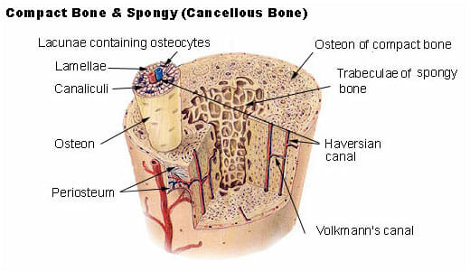
The cortical bone—also called the ‘compact bone’— is the dense bone tissue that forms the hard exterior and gives long bones their strength.
Compact bone is formed of a calcified matrix containing very few spaces, although it does contain many small cylindrical columns of only a few millimeters wide called lamellae. These lamellae form the osteon or the haversian system.
Within the osteon is the haversian canal, the central canal which surrounds blood cells and nerves.
Surrounding the haversian canal are the osteocytes, which store the mineral tissue of bones such as calcium. These osteocytes are connected to each other in a network of tiny canals called canaliculi, which allows them to transport minerals, fatty acids and waste and between each other.
The Cancellous Bone
The cancellous bone, also known as trabecular bone or ‘spongy bone’, makes up the interior of the bone structure. Cancellous bone is typically found at the ends of the long bones as well as the rubs, skull, pelvic bones and the vertebrae of the spinal column.
It is a lightweight and porous bone with the tissue arranged into a honeycomb-like matrix with large spaces; these spaces are often filled with blood vessels and bone marrow. The main structure of the cancellous bone is formed of thin rod-like bones called trabeculae.
Functions of the Endoskeleton
Protection and Support
The endoskeleton provides the structural support for the body, enabling its owner to stand up; without it, the body would have no shape.
Although the skeleton does not necessarily prevent damage to outer organs such as the skin, it provides a great deal of protection for the inner organs.
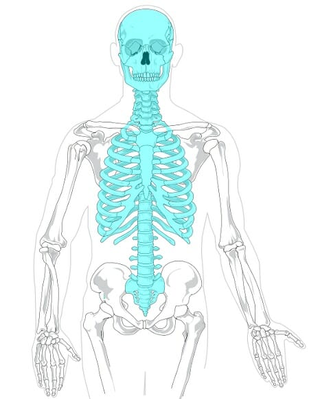
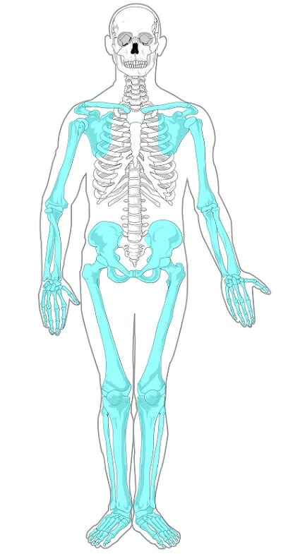
The vertebrate skeleton is formed of two different parts:
The axial skeleton is the ‘inner skeleton’. This is comprised of the skull, the ribcage and the vertebral column. Its main protective function is for the central nervous system and the vital organs such as the lungs, heart, kidneys and liver.
The appendicular skeleton consists of the pelvic girdle, the shoulder blades and arm bones and the legs and feet. This part of the endoskeleton protects and supports the limbs.
Movement
Bones, when supported by the function of muscles, deliver the capacity of locomotion (movement). The muscles are attached to the bone via tendons or ligaments.
Since the structure of bones is mostly rigid, movement of the skeleton is made possible by connecting bones called joints. There are several different types of joint, allowing different ranges of movement.
The hip and shoulder have ‘ball and socket’ joints. The ‘ball’ part of the joint is a spherical bone, which fits within the ‘socket’, and can move in almost all directions.
The wrist has a ‘condyloid’ joint. This is similar in structure to the ball and socket, and although it has a wide range of movements, it does not allow the wrist to rotate 360-degrees.
A ‘saddle’ joint is the joint that allows movement in the thumb. This provides the same range of movements as the condyloid joints although cannot bend backwards.
The ‘hinge’ joint is found within the fingers and toes. This allows movement like the hinge of a door—bending in and straightening, although not backwards or sideways. The knee and ankle joints, although hinges, allow a degree of movement when the limb is held in a certain position.
A ‘pivot’ joint allows rotational movement. This joint can be found at the elbow, and at the vertebrae directly under the skull allowing the head to move in a rotation.
Storage
The osteocyte cells—star shaped cells that form a network surrounding the haversian canals—are the cells that are responsible for the maintenance of mature bone.
The bone is made up of calcium, phosphorus and other fatty acids, all of which are stored within the osteocytes in the compact bone. When the body is in need of these nutrients, they can be taken from these stores and utilized.
Manufacturing
Within the cancellous bone is the flexible tissue called bone marrow. There are two types of bone marrow: yellow marrow and red marrow.
Within the bone marrow, there are special cells called ‘stem cells’. These are unique in that they have the ability to become any other type of cell.
Yellow bone marrow consists primarily of fat, which gives it the yellow color. This fat contains a source of energy that can be used in times of starvation. The yellow marrow contains stem cells called stroma, which can produce fat, cartilage and bone tissue).
Red bone marrow—also called myeloid tissue—contains hemopoietic stem cells, which produce an assortment of different blood cells through haematopoiesis. This system typically produces around 500 billion blood cells per day.
Some of these blood cells are the red blood cells associated with carrying oxygen around the body, while others, such as lymphocytes, are essential for support of the immune system.
Homeostasis
The bones of the endoskeleton hold around 99% of the body’s calcium, so they play a key part in the regulation of calcium levels within the body through the process of homeostasis.
When blood calcium levels become too high, the hormone calcitonin is released from the thyroid gland. Calcitonin inhibits the osteoclast cells (those responsible for the break down of bone tissue) within the osteon, and stimulates the osteoblast cells (responsible for the building of bone tissue), thus absorbing calcium to the bone and decreasing the calcium levels in the blood.
When calcium levels are too high, the thyroid gland releases parathyroid hormone, which acts to inhibit osteoblasts and stimulate osteoclasts, as well as reducing the output of calcium from the kidneys and increasing the amount of calcium absorbed by the small intestine, thereby increasing the blood calcium levels.
Related Biology Terms
- Exoskeleton – A rigid external skeleton, which acts to protect the soft interior of an animal.
- Cartilage – A smooth malleable tissue with elastic qualities that forms part of the endoskeleton, or the whole endoskeleton in the case of chondricthyes.
- Hydrostatic Skeleton – The endoskeleton of certain soft-bodied animals, which consists of fluid-filled cavities rather than bone or cartilage.
- Notochord – The skeletal rod structure made of cartilage, which supports the body in chordate embryos and some adults.
Quiz
1. The calcium-storing osteocytes are found within the:
A. Bone marrow
B. Cancellous bone
C. Cortical Bone
D. Joints
2. Which of the following properties least describes a potential advantage of having an endoskeleton, rather than an exoskeleton?
A. It is harder, providing more protection for tissues
B. Can support more body weight
C. Is relatively lightweight
D. Grows with the other tissues