Definition
The cerebrum of the central nervous system is the uppermost part of the brain. It is composed of the basal ganglia, cerebral cortex, and olfactory cortex. The cerebrum is divided into a left and right hemisphere on either side of a central fissure. As the largest part of the brain, the cerebrum sits in front and on top of the brainstem. It receives sensory information, processes it, regulates motor and conscious activity, and is responsible for a broad range of cognitive processes.
What Is The Cerebrum?
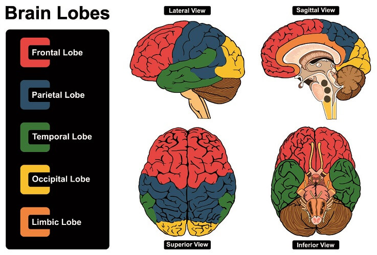
The cerebrum is the largest part of the brain. Its outer surface is covered by the cerebral cortex spread over five paired lobes: frontal lobe, parietal lobe, temporal lobe, occipital lobe, and (to a lesser degree) limbic lobe.
The brain in its entirety is, together with the spinal cord, part of the central nervous system. It can be divided into eight anatomical structures, a few of which overlap. These are the:
- Prosencephalon: forebrain.
- White matter: colored by the myelin sheaths of neurons.
- Gray matter: colored by neuronal cell bodies (soma).
- Limbic system: emotions and memory, behavior, and some autonomic functions.
- Glymphatic system: waste removal network of blood and lymph vessels.
- Cerebral ventricles: production of cerebrospinal fluid.
- Brain stem: controls vital autonomic functions, includes the cerebellum.
- Blood-brain barrier: protective barrier between the brain and the rest of the body.

In the above list, the cerebrum is part of the prosencephalon (forebrain). The prosencephalon is divided into two main structures – the diencephalon and the telencephalon. The cerebrum is a subcategory of the telencephalon. These develop from the fetal brain (neural tube), as seen in the above image.
Telencephalon
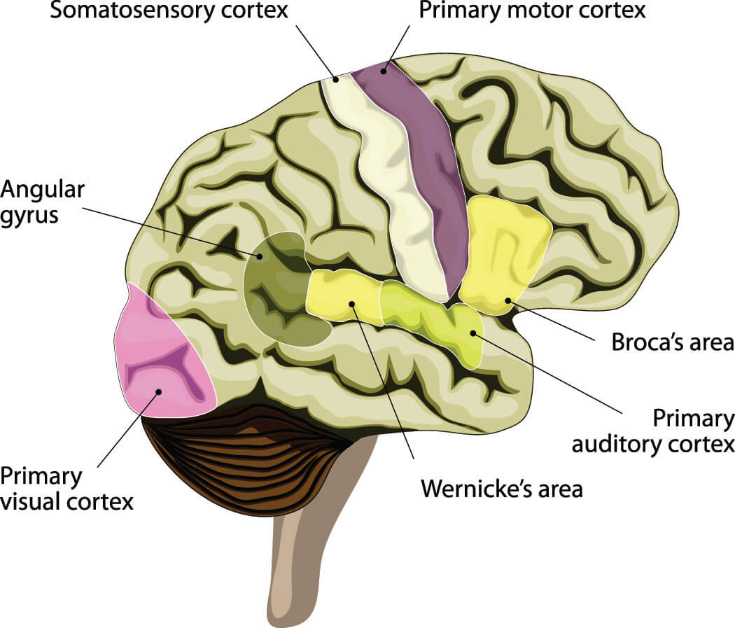
The telencephalon is the most developed and largest part of the forebrain. Each hemisphere is divided by the longitudinal fissure and lateral ventricle but is well-connected to its opposite partner. Specific sides can be dominant for certain functions. For example, a specialized region of brain tissue called Broca’s area (see below) is most commonly located at the side of the left frontal lobe when the left-hand side of the brain is dominant for communication.
The telencephalon is divided into seven main structures:
- Cerebrum
- Diagonal band of Broca: contains Broca’s area.
- External capsule: pathway for neurons traveling between the lower forebrain and cortex.
- Internal capsule: pathway for neurons traveling between the thalamus, cortex, brainstem, and spinal cord.
- Olfactory bulb: sensory stimuli collection area involved in our sense of smell.
- Brain septum: contains septal nuclei (specialized neurons in large quantities) and the septum pellucidum running through the center of the cerebrum.
- Telencephalic commissures: connection points between the two hemispheres.
The cerebral cortex is covered with gray matter which gives the brain its distinguishable color. The neocortex of high primates and humans covers white matter made of concentrated neuron soma (bodies). In the average human cerebral cortex, there are approximately ten billion neurons.
Cerebrum
The primary three parts of the cerebrum are the basal ganglia, cerebral cortex, and olfactory cortex (not the olfactory bulb). In terms of cerebrum vs cerebral cortex, the latter is part of the former; they are not the same structures.
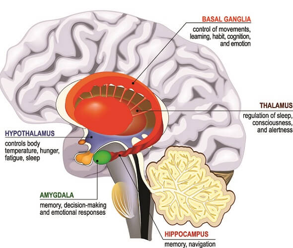
Basal ganglia refer to a group of specialized neurons that form part of the extrapyramidal system of the central nervous system. This tract of nerve fibers starts in the brainstem and brings motor stimuli to the spinal cord, controlling muscle tone, balance, and movement. The basal ganglia are composed of the:
- Amygdala: part of the limbic system that lies within the cerebrum.
- Claustrum: coordinates the connections that regulate consciousness.
- Corpus striatum: primary input area for motor and action planning, reward, and decision-making.
- Substantia innominata: integral to associative learning and memory.
The cerebral cortex is the second main area of the cerebrum and is present in four paired lobes of the brain: frontal, temporal, parietal, and occipital. It is, therefore, involved in all of the sensory and motor actions associated with each lobe. Other parts of the cerebral cortex are:
- Hippocampus: part of the limbic system that regulates motivation, emotions, learning, and memory.
- Limbic lobe: contains the amygdala, hippocampus, and brain septum and forms the part of the limbic system that lies within the cerebrum.
- Neocortex: a large, highly-cognitive area of the primate brain connected to the limbic system and responsible for sensory perception, emotion, reasoning, conscious thought, language, and information processing.
- Sensorimotor cortex: processes sensations of touch, the position of the body (proprioception), pain (nociception), and temperature and connects these to motor areas.

The third and final area of the cerebrum is the olfactory cortex responsible for processing our sense of smell. As this is close to the areas of the brain that regulate emotion and memory, it is understandable why a certain scent can affect our emotions. The olfactory cortex is composed of four main structures:
- Basal forebrain: produces the neurotransmitter (acetylcholine) used by the majority of cerebral neurons. This area is often damaged in neurodegenerative disease.
- Entorhinal cortex: connects the hippocampus and neocortex to regulate memory and also plays a role in conditioning and eye and ear nerve impulses.
- Olfactory tubercle: receives information from the piriform cortex and olfactory bulb and connects this to sensory, cognitive, endocrine (hormone), and reward areas.
- Piriform cortex: involved in our senses of smell and taste and also seems to activate seizures in epilepsy.

It is, therefore, apparent that the cerebrum controls in at least some aspect every nerve pathway in the central nervous system. If you are asked, “What does the cerebrum do?”, it is important to include the collection, processing, and regulation of sensory and motor stimuli from the four paired lobes, involvement in cognitive function in terms of memory, reward, and learning, and its role in consciousness, balance, and our senses of smell and taste in your answer.
Cerebrum Location
Cerebrum location should always include the cerebral cortex, basal ganglia, and olfactory cortex. Drawing a line around these structures is not as clear-cut as it may seem, especially as the limbic and olfactory systems cross between different regions of the telencephalon.
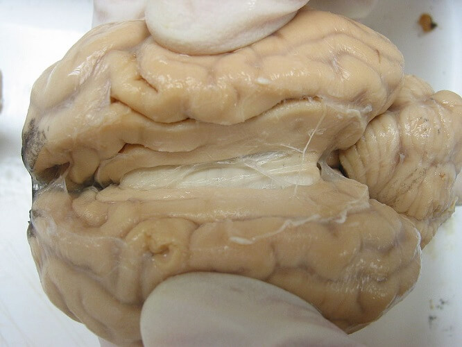
The cerebrum is located in the superior part of the cranial cavity inside the skull. It is divided by the longitudinal fissure (easily recognized when looking at the top surface of the brain) with each hemisphere linked by the corpus callosum (the white structure in the below image).
Within the brain, this location is less apparent. The basal ganglia lie deep inside the cerebral cortex close to the midline and around the corpus callosum. The olfactory cortex sits under the basal ganglia.
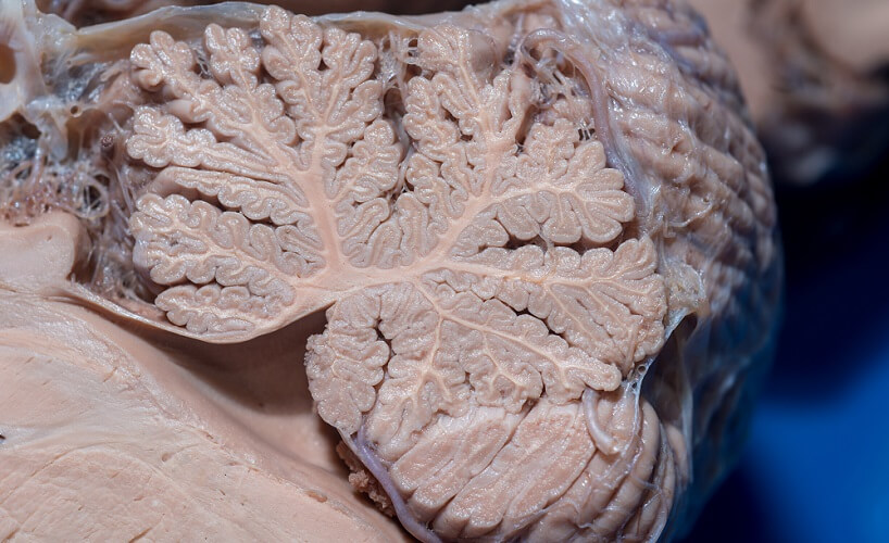
Cerebrum vs Cerebellum
In cerebrum versus cerebellum, the cerebrum is a subcategory of the brain telencephalon that is part of the forebrain. The cerebellum is a subcategory of the rhombencephalon or hindbrain. These are fetal brain forms that eventually develop into multiple complex structures.
Cerebrum functions have been discussed in basic detail above. We can compare these functions with that of the cerebellum and note the differences between both structures.
The first obvious difference is size; the cerebrum is much larger than the cerebellum. This, however, disguises the fact that the cerebellum contains more neurons than the cerebrum. It only makes up 10% of total brain mass but contains over 50% of the total neurons of the brain.
If we look at the reptilian brain, it is easier to have a better indication of cerebrum vs cerebellum. This is because the cerebellum is most developed in mammals – especially humans – and reptiles have brains composed of brainstem and cerebellum but not cerebrum. Most theories agree that early forms of the brain evolved from reptilian-like structures to include first a limbic system and, finally, a neocortex. The more intelligent the animal the greater the volume of the neocortex.
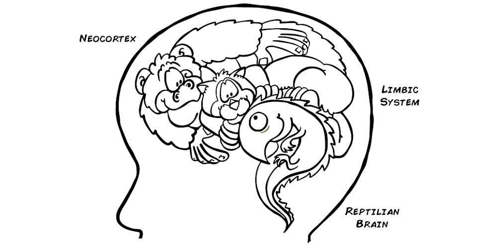
The reptilian brain controls the vital functions of the body and is, therefore, part of the autonomic (involuntary) nervous system. Our heart rate, respiration, body temperature, movement, and balance are controlled here. Reptiles act in the same way and learning is the result of repetition and conditioning.
Over time, mammals involved: The earliest of these had brains consisting of the brainstem, cerebellum, and limbic system – or emotions and memory. Only when the neocortex of the cerebellum evolved in mammals did functions such as decision making, communication, imagination, and thought become possible.

Even so, all of these evolving structures work closely together. For example, the human cerebellum is also involved in decision-making and working memory.
Together, the cerebrum, brainstem, and cerebellum make up the mammalian brain. The brain and spinal cord form the central nervous system. All parts are interconnected, making mammals the most intelligent creatures on earth. The human brain has the largest cerebrum, separating us from animals through our ability to carry out conscious thought and communicate at an extremely high level.