Definition
The temporal lobes of the brain run from the temples to the backs of the ears and are involved in a broad range of cognitive and sensory functions. The temporal lobe is divided into the superior, medial, and inferior temporal lobes, with wide-ranging functions that include visual recognition, memory, written and spoken language, and auditory, cognitive, and emotional processing.
Temporal Lobe Function
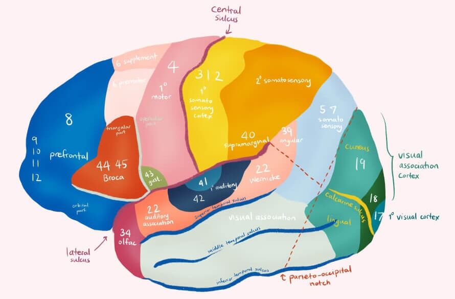
Temporal lobe function includes many different processes. These processes are very loosely represented by specific regions known as Brodmann areas. Brodmann areas are groups of functional nervous tissue. The Brodmann areas (BAs) of the temporal lobe are represented by BAs 20, 21, 22, 37, 38, 41, 42, and 52. These areas are found at random positions throughout the cerebral cortex or ordered according to function but were named according to the order in which they were first discovered by scientists. This can make their positions difficult to memorize but never mind, you don’t have to know them all!
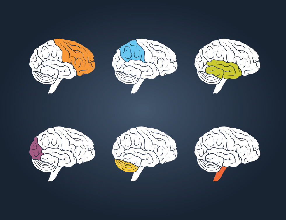
Different regions of the temporal lobe are responsible for different functions. As Brodmann areas often overlap and seem to be spread much more randomly than previously believed, looking at more obvious sections is not only simpler but more likely to be accurate. These are the superior, medial, and inferior temporal lobe regions. Each region’s function is described in more detail below.
Superior Temporal Lobe Function
Superior temporal lobe functions involve hearing and speech. This upper area of the temporal lobe is located slightly above the external ear and contains Brodmann areas 22, 41, 42, and 52.
The largest area of the superior temporal lobe contains mainly BA22 cells, a portion of which make up Wernicke’s speech area. Wernicke’s area – most commonly found in the left hemisphere of the brain – enables us to put meaning to the spoken word. Without this extensive region we would hear words but not be able to understand them or reproduce them in the correct context. Damage to this area of the brain due to stroke, infection, increased intracranial pressure, or trauma can cause the individual to speak ‘gobbledygook’ without realizing that they are not using the correct words. The vocabulary used when affected by this disorder – also known as fluent or receptive aphasia – is either made-up or incorrect. As the main cause of fluent aphasia is damage to Wernicke’s area, this speech problem is also called Wernicke’s aphasia.

Other functions of BA 22 which spread outside of Wernicke’s area include the processing of non-verbal sounds, melodies, and words. This is an auditory association region with countless connections to regions of the brain that deal with how we experience noises. How selective we are to speech and noise – for example paying attention to a lecture when people behind you are talking – and a degree of deductive reasoning are also made possible through the cells of Brodmann area 22. Saccades or the rapid movement of both eyes between remembered fixation points – specifically without any visual stimuli – is another function. To test yourself, close your eyes and think of a room in your home or your garden and move your eyes to where you know a table, chair, picture, tree, or plant is located. Another example of memory-guided saccade could be a monkey who spots a food source in a tree, is temporarily distracted, and then looks up to the same location expecting to see that food source.
Brodmann areas 41 and 42 are both associated with hearing and make up what is known as the auditory cortex. The auditory core (primary) and belt (secondary) are represented by BA 41 and BA 42 respectively. The core receives high, medium, and low-frequency stimuli via the cochlea and sends them to the belt for further processing. The belt forwards auditory electrical signals to BA 22 and directly to the frontal cortex of the brain. Finally – or perhaps not so finally, as research shows us how little we currently know about the brain – Brodmann area 52 is made up of auditory neurons. Very little research on this part of the brain exists and no one has agreed to its exact function; you might not even find it on every Brodmann area diagram. Tests on primates show that BA 52 cells might respond to different tones and pitches.
As the left hemisphere is nearly always the dominant auditory processing area, when different words of the same volume are spoken into each ear at the same time, those spoken into the right ear are most likely to be heard. This is because the right ear connects to the dominant left hemisphere (where Wernicke’s area makes sense of the word). You can try this experiment at home, too.

Medial Temporal Lobe Function
Medial temporal lobe function is represented by Brodmann areas 21 and 38. These areas have extremely complex features. We will try to keep it simple!
BA 21 has multiple functions that include language, visual, auditory, and deductive reasoning processing. Thanks to area 21, our language is rich; we can categorize spoken words and written texts into meaningful phrases and sentences – a process known as semantics. While some people think language is unique to humans, many young animals slowly learn to call or sing, just as human young learn to speak. An interesting report from Columbia University lists many reasons why speech semantics and rich language can also be attributed to non-human species.

BA 21 helps us to recognize speech attributes such as word emphasis, intonation, and rhythm (prosodic speech). Prosody is best described with the classic human skill of sarcasm. “Wow, you’re really clever” is positive when the voice that tells you is upbeat, slightly singsong, and high in pitch. If the sentence is said more slowly and with a lower pitch than normal, that person is probably being sarcastic. It is BA 21 that helps us to distinguish whether someone is being sarcastic or not. Of course, this also has a lot to do with our memories. If we have never encountered sarcasm, we’ll take it as a well-deserved compliment!
The cell types found in BA 21 also play important roles in how we observe moving objects and process complex sounds. They enable us to have opinions that relate to how we expect others to react based upon their behavior (attribution of intention). For example, when the person you are talking to frowns, you might come to the conclusion that they disagree. In other words, you attribute the other person with disagreement when you see them frown. Again, this skill is closely linked to memory and learning. And this is where BA 38 comes in.
Brodmann area 38 of the medial temporal lobe has a similarly broad range of language and auditory functions but also helps in the processing of visual stimuli, emotion, cognition, memory, our levels of enjoyment when listening to certain sounds, and our personal responses to humor and irony. As with all of the temporal lobe, the dominant hemisphere is most commonly the left side.
Thanks to BA 38, you know who is speaking as you can identify familiar voices. Selective hearing is improved; background noise does not actually fade away – our brains simply stop responding to auditory stimuli when we cease paying attention to them. BA 38 is also essential for memory retrieval – not just sensory memory but also emotional and cognitive memory. These memory functions overlap with auditory processing (familiar voices), visual processing (familiar objects), and word comprehension (familiarity of language). It is the next step in the chain as far as the processes of the superior temporal lobe are concerned.
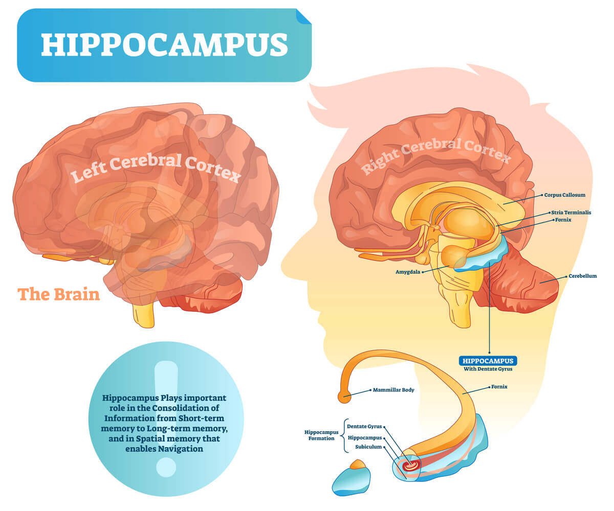
The deeper levels of the medial temporal lobe are where the hippocampus and amygdala are found. The hippocampus and amygdala are not part of the neocortex (like the temporal lobe) but of the archicortex – the most primitive area of the brain. They are not dealt with in this article but, in short, the hippocampus is very much involved in memory and our capacity for learning; the amygdala is a center for our emotions and motivation. Both are part of the limbic system and often the first areas to be affected during the early progression of Alzheimer’s disease.
Inferior Temporal Lobe Function
Inferior temporal lobe function is mostly due to the cell types of Brodmann areas 20 and 37. BA 20 possesses similar functional elements as Wernicke’s area and extends this region from the top of the temporal lobe to the bottom. Specific functions in the inferior temporal lobe include language (comprehension, selective processing, semantic processing, and the understanding of metaphors), vision (fixed observation, using separate visual elements to imagine the whole), working memory, and attribution of intention.
Where the dominant (usually left) side of Brodmann area 37 of the temporal lobe is damaged, problems finding the right word – word retrieval – occur. Damage to the right side has produced strange drawings of unrecognizable objects and people. Students who prefer to make visually-attractive study cards that incorporate more than simple words may have a well-developed left temporal lobe. If these students can actually remember what’s on those study cards on examination day, the right temporal lobe is well-developed, too!
More specific functions of BA 37 mean we can use sign language, understand gestures, categorize separate words into even more specific groups, recognize faces and objects, put names to faces, and predict motion. In terms of memory, the chronology of past, personal experiences, and true and false memories begin here. Don’t lose sight of the fact that no complete function is the result of a single Brodmann area – every temporal lobe function is a complex, coordinated collaboration between multiple locations across the central nervous system.
Temporal Lobe Epilepsy
Temporal lobe epilepsy (TLE) is a recurrent, one to two-minute episode of impaired awareness that is caused by repeated bursts of electrical activity in this part of the brain. The Epilepsy Foundation provides a detailed look at the various forms of epilepsy that may or may not involve the temporal lobe. There are two temporal lobe epilepsy subtypes that are associated with different symptoms.

Neocortical Temporal Lobe Epilepsy (nTLE)
Epilepsy that originates in the lateral (surface) areas of the temporal lobe usually causes auditory symptoms such as buzzing or ringing in the ears. This type of epilepsy rarely lasts for more than a few seconds. Another symptom is the presence of focal impaired awareness seizures (FIAS) that cause confusion as the person is unable to process what they hear. Neocortical TLE might lead to aggressive behavior towards caregivers and family members; this is due to wrongly-made associations in the attribution of intentions to others. A smile might be translated as a threat, for example. If they can’t process gentle, calming words, a situation might become dangerous through no fault of their own.
All of the above are complex partial seizures and may often look as if the person is simply daydreaming. It is possible that this seizure type can develop into the tonic-clonic form on both sides of the brain (see the temporal lobe seizures paragraph below).
Medial Temporal Lobe Epilepsy (mTLE)
Medial temporal lobe epilepsy occurs in the deeper structures of the temporal lobe and has more diverse symptoms that develop slightly more slowly and last a little longer than neocortical (surface) seizures. These include autonomic nervous system reactions such as abdominal pain, cognitive effects such as déjà vu or quite the opposite – a sudden unfamiliarity with a common situation – and sensory distortions (particularly the senses of taste and smell).
Other symptoms are behavioral arrest and automatisms. Automatisms can involve all kinds of actions such as repetitive or unusual hand movements, swallowing, lip-smacking, and chewing. Caregivers sometimes observe one dilated pupil which indicates that the seizure is occurring on the same side of the brain as the dilated pupil (ipsilateral mydriasis). One-sided limb movements, on the other hand, point to the contralateral side of the brain, as does a sideways gaze – “Look Left” means repeated bursts of abnormal, heightened electrical activity in the right hemisphere.

Temporal Lobe Seizures
Temporal lobe seizures are not always epilepsy, although the symptoms are often very similar. A single seizure is called just that; however, when these seizures recur at least once due to a neurological condition, they are renamed epilepsy. A single temporal lobe seizure is most commonly caused by non-neurological conditions such as atherosclerosis, trauma, brain tumor, infection, diabetic ketoacidosis, drug abuse, electric shocks, and congenital birth defects. What all of these causes have in common is that they limit oxygen and energy (glucose) transport to the brain.
Any degenerative process (the aging process) in, or trauma (head injury) to, the central nervous system can cause neurons to become oversensitive. These disrupted neurological pathways cause seizures that range from symptomless, brief episodes to fatality. Temporal lobe seizures that result in death are known as Sudden Unexpected Death in Epilepsy or SUDEP; the cause of death is most likely to be apnea, heart arrhythmia, or a combination of both.
Seizures are classified according to their range of symptoms – generalized and focal. Generalized seizures affect both hemispheres of the brain and are split into two groups – absence (petit mal) seizures that are often confused with daydreaming, and tonic-clonic (grand mal) seizures. Tonic-clonic (status epilepticus) seizures are most likely to cause SUDEP as they cause loss of consciousness and violent muscle contractions that may affect breathing and heart function.
Focal seizures affect a single area of the brain and are grouped into three types. Simple focal seizures are the least intrusive, causing a person to smell an odor that is not there or twitching, for example. Complex focal seizures affect cognition to cause confusion and an inability to communicate. Secondary generalized seizures start as focal seizures but go on to spread through the brain and even into both hemispheres.
Quiz