Peripheral Nervous System Definition
The peripheral nervous system consists of all neurons that exist outside the brain and spinal cord. This includes long nerve fibers as well as ganglia made of neural cell bodies. The peripheral nervous system connects the central nervous system (CNS) to various parts of the body.
Peripheral Nervous System Overview
Functionally, the peripheral nervous system (PNS) is divided into sensory (afferent) and motor (efferent) nerves, depending on whether they bring information to the CNS from sensory receptors or carry instructions towards muscles, organs or other effectors. Motor nerves can be further classified as somatic or autonomic nerves, depending on whether the motor activity is under voluntary conscious control.
Anatomically, the PNS can be divided into spinal and cranial nerves, depending on whether they emerge from the spinal cord or the brain and brainstem. Both cranial and spinal nerves can have sensory, motor, or mixed functions. The enteric nervous system, which surrounds the gastrointestinal tract, is another important part of the peripheral nervous system. While it receives signals from the autonomic nervous system, it can function independently and contains nearly five times as many neurons as the spinal cord.
Peripheral Nervous System Function
The primary function of the peripheral nervous system is to connect the brain and spinal cord to the rest of the body and the external environment. This is accomplished through nerves that carry information from sensory receptors in the eyes, ears, skin, nose, and tongue, as well as stretch receptors and nociceptors in muscles, glands and other internal organs. When the CNS integrates these varied signals and formulates a response, motor nerves of the PNS innervate effector organs and mediate the contraction or relaxation of skeletal, smooth or cardiac muscle.
Thus, the PNS regulates internal homeostasis through the autonomic nervous system, modulating respiration, heart rate, blood pressure, digestion reproduction, and immune responses. It can increase or decrease the strength of muscle contractility across the body, whether it is sphincters in the digestive and excretory systems, cardiac muscles in the heart, or skeletal muscles for movement. It is necessary for all voluntary action, balance, and maintenance of posture.
The peripheral nervous system also controls the release of secretions from most exocrine glands. The PNS innervates the muscles surrounding sense organs, so it is involved in chewing, swallowing, biting and speaking. At the same time, it mediates the response of the body to noxious stimuli, quickly removing the body from the injurious stimulus, whether it is extremes in temperature, pH, or pressure.
Sensory Nervous System
The functional classification of the PNS divides it into three categories. The first is the sensory nervous system, carrying signals from the viscera, sense organs, muscles, bones and joints towards the CNS. Nerve fibers that carry this information are part of the afferent division. Sensory receptors can transduce a physical stimulus such as pressure, sound waves, electromagnetic radiation, or chemical composition, into an electrochemical signal.
This signal, when it reaches a certain threshold, is transmitted as an action potential along an afferent neuron, and relayed to the CNS, where the signal is perceived and interpreted. Thus the sensory nervous system consisting of the receptor and neural pathway deliver information about the intensity, location, type, and duration of a stimulus to the CNS.
Somatic Nervous System
The second functional division of the PNS is the somatic nervous system. It controls the voluntary muscular movement of skeletal muscles in the limbs, back, shoulders, neck, and face. It also mediates reflex actions, where an afferent nerve fiber is nearly directly connected to a motor nerve fiber, to quickly generate a response to a stimulus. These include protective responses, like the movement of the body away from acute injurious stimuli like extremes in temperature, as well as those like the patellar ‘knee-jerk’ response when the patellar ligament is struck.
Autonomic Nervous System
The autonomic nervous system is related to all the involuntary visceral activity of the body. It consists of the sympathetic and parasympathetic nervous systems, and their effector organs include cardiac muscle, smooth muscle, and various glands. The anatomy of the autonomic nervous system is distinct because the effector arm involves two neurons that synapse with each other at specific ganglia.
The neurons of the sympathetic nervous system have short preganglionic neurons that can excite multiple postganglionic nerve fibers. The sympathetic nervous system is said to have a thoracic and lumbar outflow. The parasympathetic nervous system, on the other hand, uses cranial and sacral nerves and their ganglia are situated close to the target organ.
Peripheral Nervous System Parts
General Nerve Structure
The peripheral nervous system is made of nerves, ganglia, and plexuses. A nerve contains the axons of multiple neurons bound together by connective tissue. The axon itself is often myelinated, containing a phospholipid secreted by a glial cell called the Schwann cell. The thin covering of Schwann cell cytoplasm forms the innermost layer protecting an axon and is called the neurilemma or neurolemma.
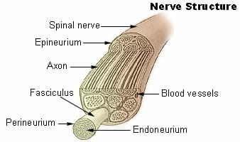
The image above depicts the structure of a nerve. Blood capillaries and other connective tissue around the neurilemma form the endoneurium. When multiple axons are bundled together to form structures called fascicles, fibrous tissue called the perineurium holds them together. Finally, the whole nerve containing numerous axon bundles is encased in fibrous epineurium.
The cell bodies or soma of these neurons also cluster together and are covered by the epineurium to form ganglia that look like swellings on the nerve fiber. In the autonomic nervous system, these ganglia become the sites for synaptic transmission between two neurons. Branching networks of intersecting spinal and autonomic nerves form structures called plexuses that have both sensory and motor functions and serve a particular region of the body.
Cranial Nerves
The PNS can be said to consist of 12 pairs of cranial nerves and 31 pairs of spinal nerves. Cranial nerves emerge in pairs on either side of the base of the skull, through small openings called foramina. Cranial nerves are numbered using Roman numerals I-XII, depending on their position while exiting the cranium. A potentially vestigial nerve called cranial nerve zero emerges anterior to the first cranial nerve.
Cranial nerves also have a Latin or Greek name, based on their structure or their effector organ. They primarily innervate the head and neck, with the significant exception of the tenth cranial nerve, also known as the vagus nerve. Some cranial nerves have only sensory functions, such as the olfactory and optic nerves. The structure of these nerves also occasionally leads to their classification under the central nervous system. Cranial nerve VIII is another sensory nerve relating to hearing and balance. Motor nerves contain nerve fibers that carry signals to muscles of the pupil of the eye or external eye muscles. The rest are mixed nerves containing both sensory and motor nerve fibers. Among these, the XI and XII cranial nerves mostly serve a motor function. They innervate the neck, back, and tongue.
The Vagus nerve is another mixed nerve that carries signals from internal organs to the brain and conducts impulses to the organs of the thorax, abdomen and respiratory muscles of the pharynx and larynx. It plays an important role in the parasympathetic innervation of the body.
Spinal Nerves
There are 31 pairs of spinal nerves, arising from different regions of the spinal cord. There are 8 that emerge from the cervical region, 12 from the thoracic region, 5 each from the lumbar and sacral regions and 1 pair of spinal nerves from the coccygeal region. Each spinal nerve is a mixed nerve formed by a combination of afferent and efferent neurons.
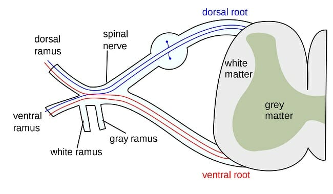
The image shows the region near the spinal cord, where every spinal nerve has a posterior and anterior root. The anterior or ventral root contains motor neurons while the dorsal or posterior root has ganglia containing the cell bodies of afferent sensory neurons. Distal to the spine, the nerve again splits into an anterior and posterior ramus in addition to forming a small meningeal branch. The posterior ramus leads to the muscles, joints, and skin on the back. The anterior ramus is involved in innervating the skin and muscles of the trunk and leads towards the limbs. Often, the anterior ramus forms a network of intersecting nerve fibers to create plexuses.
Examples of the Peripheral Nervous System Response
The pupils of the eyes enlarge in a dimly lit room. This allows maximum light to fall on the retina. When a bright light is suddenly turned on, sensory receptors in the eye communicate this to the CNS. The PNS mediates the response to this stimulus. The pupils contract and the external eye muscles squint. Your body may even move the skeletal muscles of the arm to shield the eye.
Similarly, when a sharp or pointed object is stepped on, pain and stretch receptors in the skin send signals to the CNS, which immediately brings about a change in posture, and balance, protecting the foot from potential injury.
Quiz