Peptidoglycan Definition
Peptidoglycan, also called murein, is a polymer that makes up the cell wall of most bacteria. It is made up of sugars and amino acids, and when many molecules of peptidoglycan joined together, they form an orderly crystal lattice structure. Bacteria are classified as being either Gram-positive or Gram-negative based in differences in the structure of their peptidoglycan cell wall. Archaea do not have a cell wall consisting of peptidoglycan, but some do have a layer of pseudopeptidoglycan (pseudomurein), which a is similar polymer.
Function of Peptidoglycan
Peptidoglycan is the main component of the cell wall in most bacteria. Cross-linking between amino acids in the layer of peptidoglycan forms a strong mesh-like structure that provides structure to the cell. Peptidoglycan provides a very important role in bacteria because bacteria are unicellular; it gives strength to the outer structure of the organism.
It is also involved in binary fission, which is how bacteria reproduce. Bacteria undergo asexual reproduction and divide themselves into two cells. For this to occur, the peptidoglycan in the cell wall must grow as the bacterium elongates before dividing. Then, when the bacterium has split into two, the cell wall must reform so that the two new bacterial cells can become enclosed.
Structure of Peptidoglycan
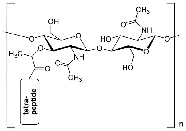
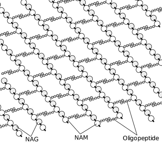
Two alternating amino sugars make up the crystal lattice structure of peptidoglycan; they are N-acetylglucosamine (shortened to NAG) and N-acetylmuramic acid (shortened to NAM). Amino sugars are sugar molecules that have an amine group (-NH2) replacing one of their hydroxyl groups. Each NAM molecule has an attached chain of four or five amino acids. Crosslinking between these amino acids gives peptidoglycan its strong structure.
These diagrams show the structure of one molecule of peptidoglycan and the structure of peptidoglycan when many molecules join together.
Gram Staining
Gram staining is a method of staining bacteria, and it is used to classify them into two groups: gram-positive, which show the stain, and gram-negative, which do not. The technique was developed in the 19th Century by Hans Christian Gram, whom it is named for. Gram originally developed the method while searching for a way to make sectioned cells more visible, but it was later discovered that bacteria could be classified into two groups based on whether or not they retain the stain’s color.
Peptidoglycan cell wall differences are responsible for whether a bacterium is gram-positive or gram-negative. The gram staining method basically involves staining the slides that the cells are on with crystal violet dye, rinsing with water, and adding iodine. Then, safranin, which imparts a red color, is used to counter-stain. Crystal violet dye forms a complex with iodine. Gram-positive bacteria have thick peptidoglycan layers which trap the crystal violet-iodine complex. This makes the cell walls of these bacteria appear purple; the purple masks the color of the lighter red safranin counter-stain. Gram-negative bacteria, however, have thin cell walls that the crystal violet-iodine complex cannot adhere to. In these bacteria, the purple stain washes off but the red counter-stain remains, making gram-negative bacteria appear red under Gram staining.
Gram staining cannot be used to identify bacteria specifically, such as at a species level. Instead, it is used for general identification. Some gram-positive bacteria include the genera Listeria, Streptococcus, and Bacillus. Most types of bacteria are gram-negative, such as Proteobacteria, green sulfur bacteria, and cyanobacteria. Some species of bacteria are gram-variable or gram-indeterminate, meaning that they either show both violet and red colors or do not respond in a predictable way to the stain. Mycobacterium tuberculosis, which causes tuberculosis, is one example of a gram-indeterminate bacterium.
Difference Between Peptidoglycan and Proteoglycan
Peptidoglycan should not be confused with the similar-sounding word proteoglycan. While peptidoglycan refers to the bacterial cell wall, a proteoglycan is a protein that has been glycosylated, which means that it has had carbohydrates attach to it. Proteoglycans are found in connective tissue, a category which includes cartilage, bone, blood, fibrous tissues, and adipose tissue (fat).
Related Biology Terms
- Polymer – A large molecule made up of many repeated units.
- Binary fission – The method by which bacteria reproduce asexually through dividing.
- Amino sugar – A sugar molecule with an amine group attached instead of one of its hydroxyl groups.
- Proteoglycan – A protein that has been glycosylated.
Quiz
1. Which of the following is a component of peptidoglycan?
A. N-acetylglucosamine
B. N-acetylmuramic acid
C. Peptide chain of amino acids
D. All of the above
2. Which substance is NOT a component of Gram staining?
A. Crystal violet dye
B. Safranin
C. Iodine
D. These are all components of Gram staining
3. What color will a gram-negative bacterium appear on a slide that has undergone Gram staining?
A. Red
B. Violet
C. Blue
D. Green