Definition
A motor neuron is a cell of the central nervous system. Motor neurons transmit signals to muscle cells or glands to control their functional output. When these cells are damaged in some way, motor neuron disease can arise. This is characterized by muscle wasting (atrophy) and loss of motor function.
Overview
Neurons are specialized cells of the nervous system that transmit signals from the brain and spinal cord to the body. There are over 80 billion neurons in the human brain! Generally, there are three types of neurons: sensory neurons, interneurons, and motor neurons.
- Sensory neurons detect external stimuli and convert it into information that the rest of the nervous system can process. For example, if you put your hand on a hot stove, the sensory neurons detect this (ouch!) and pass along the signal to the rest of the nervous system.
- Motor neurons can use the information gathered by sensory neurons and translate it into action in your muscles and gland. It is your motor neurons that would actually initiate the muscles in your arm to lift it off of the hot stove, in response to the signals from the sensory neurons.
- Interneurons connect sensory and motor neurons, transmitting information between them in cases where the two neurons are not directly connected to one another.
Structure of a Motor Neuron
Neurons are single cells. Therefore, they contain the classic eukaryotic organelles such as the nucleus, cell membrane, ribosomes, mitochondria, and more. However, they have a far more interesting structure than the classic picture of a cell in textbooks!
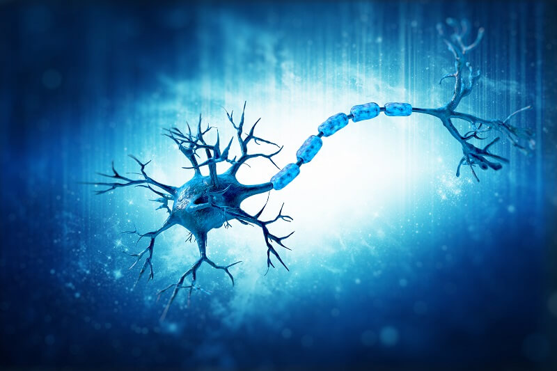
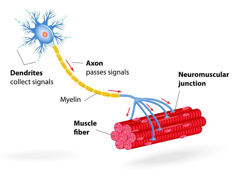
The structure of a motor neuron can be divided into three components: the dendrites, the cell body (soma), and the axon. They are multipolar in structure, which means they possess a single axon and multiple dendrites.
Dendrites
Dendrites are the branch-like extensions found at one end of a neuron. These are the structures that receive information from the other neurons, and pass it along to the cell body to transmit the signal and activate the cell. There are usually around five to seven dendrites per neuron, but some, such as the Purkinje neurons in the brain, have over a thousand!
Cell Body (Soma)
The cell body is where the organelles are contained. It controls all the functions of the cell, and this is where most of the protein synthesis occurs.
Axon
The axon is a (sometimes very long) projection from the cell body. It is tube- or cable-like in structure, and it transmits the information it receives from the dendrites via the cell body to the opposite end of the cell, called the axonal terminal. Here, the neuron passes the information to the next cell, which could be another neuron or an effector cell like a muscle cell.
There is usually only one axon per neuron, but it can contain many branches with many terminals, allowing it to communicate with several different cells. Axons can be very long, in fact, the longest axon in the human body belongs to the axons that make up the sciatic nerve. These run from the base of the lumbar spine to the big toe, and can be over 1 meter in length!
Synapses are where neurons connect to one another. They are the site between the axon terminal and dendrite where information transfer takes place.
Additionally, some neurons are coated with a myelin sheath, protecting the cell from external influences that might alter the transmission of signals.
Function of a Motor Neuron
The function of motor neurons is to transmit signals from the brain and spinal cord to muscle cells. Thus, they are responsible for voluntary and involuntary movements of all our muscle cells.
Motor neurons rapidly conduct electrical signals in order to cause these effects in our cells. Their specific function depends on the position of the cell body within the nervous system.
Locations of Motor Neurons
The cell bodies of motor neurons are found in the spinal cord, brain stem, and motor cortex of the brain, a region of the cerebral cortex. The motor cortex is involved in the planning and execution of voluntary actions.
Their projections then extend out to directly or indirectly communicate with effector organs, primarily muscles and glands, throughout the body.
Types of Motor Neuron
There are two types of motor neuron: upper motor neurons and lower motor neurons.
Upper Motor Neurons
Upper motor neurons originate in the motor cortex of the brain or the brain stem and transmit signals from the brain to interneurons and lower motor neurons. These are the main cells that initiate voluntary movement throughout the body by connecting the cerebral cortex to the brain stem or spinal cord.
Lower Motor Neurons
The lower motor neurons are found in the brain stem and spinal cord and are directly responsible for communicating with the effector organs, such as the muscle cells. They receive the signals from upper motor neurons (either directly or via interneurons) and stimulate their activity.
They can be classified as either alpha motor neurons, beta motor neurons, or gamma motor neurons.
- Alpha motor neurons are responsible for controlling muscle contractions involved in voluntary movement through contracting extrafusal muscle fibers, which make up most of the muscle tissue.
- Beta motor neurons are less common than alpha and gamma motor neurons and are less well-characterized. However, they are known to stimulate intrafusal muscle fibers (which are found deeper within the muscle).
- Gamma motor neurons control muscle contraction in response to external forces through the intrafusal fibers. They regulate the muscle response to stretch. For example, the knee-jerk reflex.
Motor Neurons in Action: The Knee-Jerk Reflex
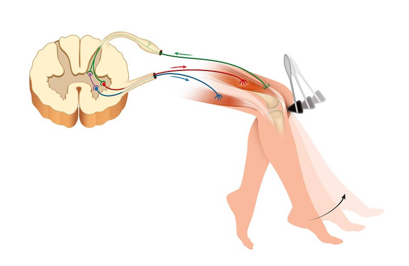
The knee-jerk reflex, sometimes known as the patellar reflex, is a medical test that doctors carry out to assess the function of your nervous system.
You can even try it yourself. Sit upright in a position in which your legs are not touching the floor. Then, firmly tap the tendon just below your kneecap. Your lower leg should make a sudden movement in an upwards kicking motion. This is the knee-jerk reflex!
This is an example of a reflex arc. The sharp tap causes your patellar tendon to slightly stretch your quadriceps muscles. This movement is detected by the muscle spindles, which go on to stimulate the sensory neurons. The sensory neurons transmit the information to the spinal cord, where a signal is sent to motor neurons.
The motor neurons then cause the quadriceps muscles to contract, causing the kicking motion. In this example, interneurons are not involved, as there is a direct connection between the sensory and motor neurons.
If this reflex does not happen properly (a feature called Westphal’s sign) or is over-exaggerated, it can indicate problems with the nervous system due to injury or disease.
Motor Neuron Disease
Motor neuron disease describes a collection of neurodegenerative diseases that specifically affect the motor neurons, causing the death of the cells. There are various different types of motor neuron diseases, including amyotrophic lateral sclerosis (ALS), primary lateral sclerosis (PLS), bulbar onset MND or progressive bulbar palsy (PBP), and progressive muscular atrophy (PMA).
Because the motor neurons are no longer effectively signaling the muscles, motor neuron disease causes muscle weakening, stiffening, and wasting. This causes a diverse range of symptoms that are dependent upon the specific disease and the individual.
The onset of motor neuron disease can be subtle, and the initial signs and symptoms include muscle weakness with potential cognitive and behavioral changes. The disease can impact a sufferers’ ability to eat and drink, talk, walk, and breathe. Eventually, most affected individuals will lose the ability to carry out these tasks entirely.
ALS, also called Lou Gehrig’s disease, is the most common type of motor neuron disease. Two notably individuals that died as a result of complications of the condition are Stephen Hawking and Christopher Reeve. In 2014, the viral video fad termed the “Ice Bucket Challenge” increased awareness of, and research funding for, the condition.
The causes of motor neuron disease are largely unknown. The disease is thought to be the result of both genetic and environmental factors, and sometimes there can be a familial link. Studies have identified mutations around the gene C21orf2 which are thought to be linked with some cases of ALS.

Upper Motor Neuron Lesion
An upper motor neuron lesion, also called pyramidal insufficiency, refers to damage to the motor neurons of the brain or brain stem that travel to the spinal cord. Such damage can occur as a result of a variety of disorders, including multiple sclerosis (MS), stroke, brain injury, or cerebral palsy.
The resulting effects are referred to as upper motor neuron disease (UMND). The symptoms include muscle weakness, poor motor control, poor posture, and exaggerated reflex responses.
Lower Motor Neuron Lesion
Lower motor neuron lesions are damage to the lower motor neurons that travel from the spinal cord to the effector muscles. The symptoms include muscle paralysis and weakness, and the lesions are usually caused by a systemic infection, such as Lyme disease, HIV, or the Herpes virus (which can cause Bell palsy).
Quiz