Definition
A foramen (plural foramina) is an opening or hole through tissue, usually bone. It allows nerves and blood vessels to travel from one side of the tissue layer to the other. Foramina are primarily found in the skull; others are located in the vertebrae, long bones, roots of the teeth, heart, and abdomen. A similarly-named aperture is also found is in the female reproductive organ of seed plants (the ovule); however, this is more correctly called a microphyle.
What is a Foramen?
The Latin word foramen means an aperture or opening. Most foramina are found in the facial bones and cranial bones.
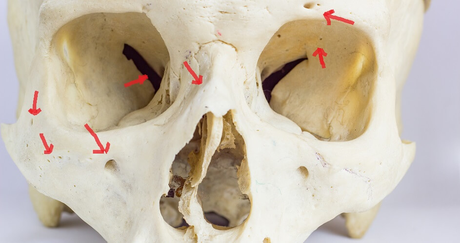
Not every skull foramen is present in everyone. These holes can also be different sizes and shapes according to individual anatomy. For example, the supraorbital foramen in the upper ridge of the eye socket can be a hole or, when positioned at the edge of the ridge, a notch.
Alternatively, parietal foramina can be so enlarged that, when found in ancient remains, archeologists think that the person has undergone an early surgical procedure called trepanning or trepanation.

Cranial Foramen
A cranial foramen allows important nervous and circulatory tissue to travel throughout the head and neck region.
The following headings list the singular or paired name, location, and the structures that pass through each aperture.
Some foramina are the result of mirrored notches in multiple bone articulations that, when put together, form a circular hole; in this case, the information is repeated.
Foramina of the Frontal Bone
- Paired supraorbital foramen (or supraorbital notch); inferior rim of the upper orbit; supraorbital nerve, artery, and vein.
- Single foramen cecum; shared between the frontal crest of the temporal bone and the ethmoid bone, between the cranium and nasal cavity; emissary veins.

Parietal Bone Foramina
- Paired parietal foramen; at the back of the parietal bone, close to where the parietal bones meet; parietal emissary vein and occasionally a branch of the occipital artery.
Foramina of the Temporal Bones
- Paired stylomastoid foramen; between the styloid and mastoid processes of the temporal bone; facial nerve (CN VII) and stylomastoid artery.
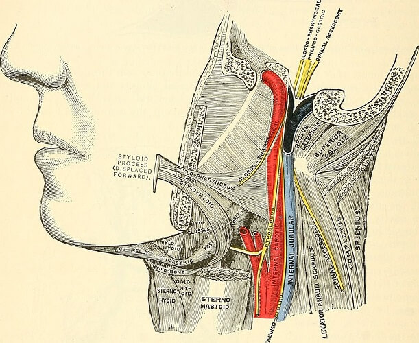
- Paired jugular foramen; shared between the temporal and occipital bones, close to the foramen magnum; inferior petrosal and sigmoid sinusus, glossopharyngeal nerve (CN IX), vagus nerve (CN X), accessory nerve (CN XI), and arterial branches of the meningial blood supply.
- Paired internal acoustic meatus; close to the ear; facial nerve (CN VII), acoustic nerve (CN VIII), and branch of the basilar artery.
- Paired carotid foramen; opens into a channel (carotid canal) through which the internal carotid artery and internal carotid plexus pass. The carotid canal ends at another aperture (f. lacerum) in the sphenoid bone.
Foramina of the Occipital Bone
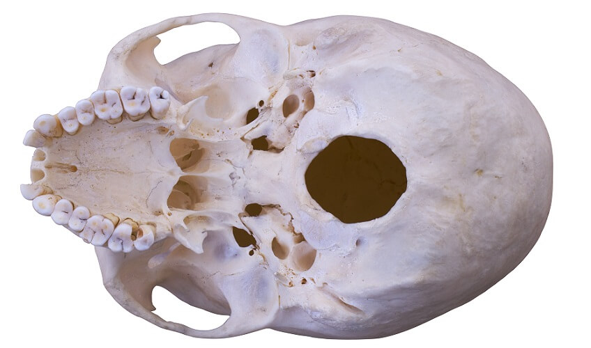
- Singular foramen magnum; large hole at the skull base that connects the brain stem to the spinal cord; spinal and vertebral arteries, meninges, and accessory nerve (CN XI).
- Paired jugular foramen; shared between the temporal and occipital bones, close to the f. magnum; glossopharyngeal nerve (CN IX), vagus nerve (CN X), accessory nerve (CN XI), inferior petrosal and sigmoid sinuses, and arterial branches of the meningial circulation.
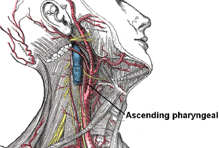
- Paired anterior condyloid foramen; above the f. magnum; hypoglossal nerve (CN XII) and branch of the ascending pharyngeal artery.
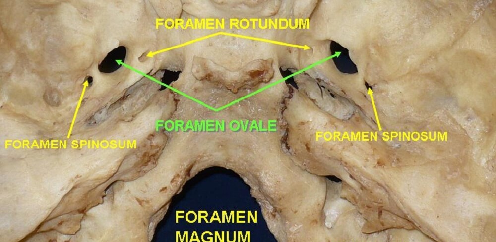
- Singular posterior condlyar foramen; close to the f. magnum; occipital emissary vein. This text includes photographs that show where several occipital foramina are located.
Foramina of the Sphenoid Bone
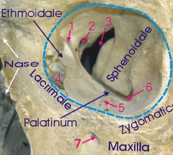
- Paired optic foramen; back of the eye socket; optic nerve (CN II) and ophthalmic artery. In the image, this aperture is labelled as number 2 (1 = ethmoid foramen; 3 = superior orbital fissure; 4 = sulcus for lacrimal sac; 5 = infraorbital sulcus; 6 = inferior orbital fissure; 7 = infraorbital foramen).
- Paired superior orbital fissure; between the lesser and greater wings of the sphenoid bone; oculomotor nerve (CN III), trochlear nerve (CN IV), branches of the opthalmic nerve, abducens nerve (CN VI), and ophthalmic veins.
- Paired foramen rotundum; greater wings of the sphenoid bone; maxillary nerve.
- Paired foramen sphenopalitinum; shared between the sphenoid and palatine bones; nasopalatine nerve, greater palatine nerve branches, and the sphenopalatina artery and vein.
- Paired inferior orbital fissure; shared with the maxilla bone (the opening into the maxilla is called the infraorbital f.); below the lower ridge of the eye socket on either side of the nose; infraorbital nerve, artery, and vein.
- Paired foramen ovale; greater wing of the sphenoid bone; mandibular nerve, accessory meningeal artery, and emissary vein.
- Paired foramen Vesalius; not always present; vein of Vesalius.
- Paired foramen spinosum; greater wing of the sphenoid bone; branch of the mandibular nerve and middle meningeal artery.
- Paired foramen lacerum; the end of the shared carotid canal that begins at the carotid f. (temporal bone); located next to the sella turcica; nerve and artery of the pyterygoid canal, internal carotid artery, and emissary vein.
Foramina of the Ethmoid Bone
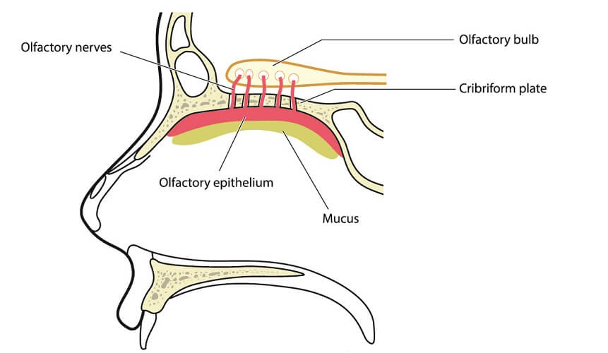
- Cribriform plate foramen; most of us have around 20 of these small holes that form the sieve-like portion of the ethmoid bone above the nasal cavity; multiple fibers of the olfactory nerve (CN I).
- Paired anterior ethmoidal foramen; back of the eye socket; anterior ethmoidal nerve, artery, and vein.
- Paired posterior ethmoidal foramen; back of the eye socket; posterior ethmoidal nerve, artery, and vein.
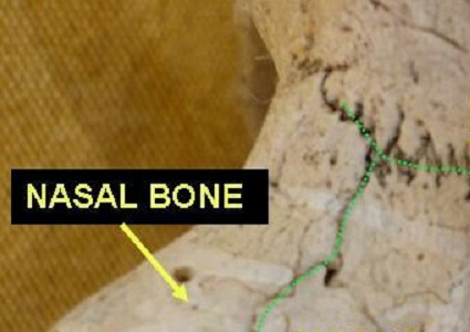
Nasal Bone Foramen
- Paired nasal foramen; tiny holes on either side of the bridge of the nose; small vein.
Foramina of the Palatine Bones
- Paired greater palatine foramen; palatine nerve, artery, and vein.
- Paired foramen sphenopalitinum; shared between the palatine and sphenoid bones; nasopalatine nerve, nasal branches of the greater palatine nerve, and the sphenopalatina artery and vein.
- Lesser palatine foramen; set of four foramina shared between the palatine and maxilla bones at the back of the hard palate; lesser and greater palatine nerves, and the lesser palatine artery and vein.
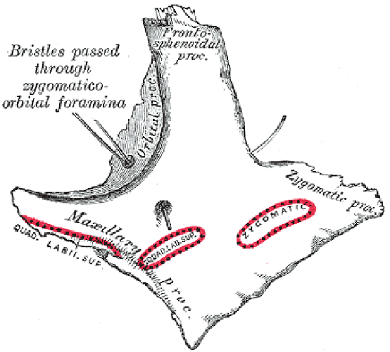
Foramina of the Zygomatic Bones
- Paired zygomatico-orbital foramen; orbital processes of the zygomatic bones – from here, a single canal splits to exit via two holes (see below image). These two holes are the paired zygomaticotemporal and zygomaticofacial foramina that convey similarly-named facial nerves and vessels.
Foramina of the Maxillae
- Paired infraorbital foramen; part of the inferior orbital fissure shared with the sphenoid bone that ends at the infraorbital f. of the maxilla; infraorbital nerve, artery, and vein.
- Lesser palatine foramen; set of four foramina shared between the maxilla and palatine bones at the back of the hard palate; lesser and greater palatine nerves, and the lesser palatine artery and vein.
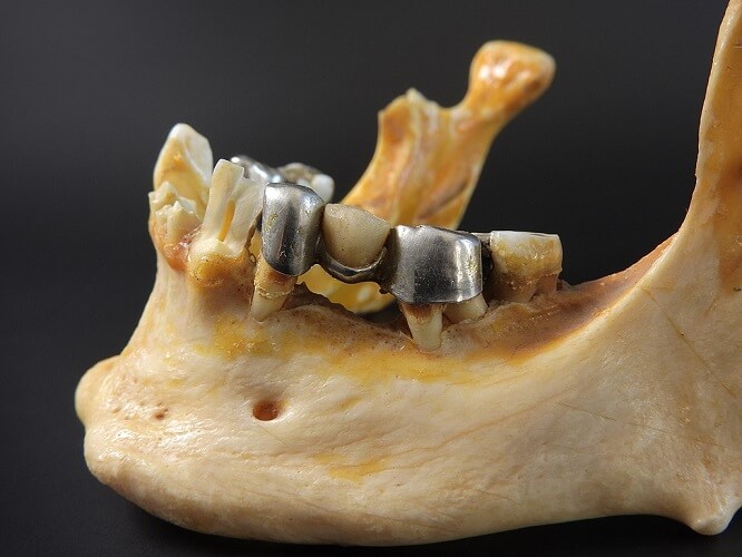
Foramen of the Mandible
- Paired mandibular foramen; ramus of the mandible (lower jaw); inferior alveolar nerve and vessels.
- Paired mental foramen; chin (body of mandible); mental nerve and vessels.
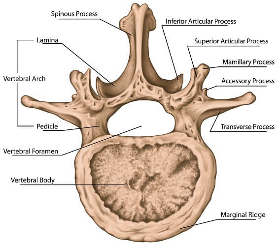
Spinal Foramen
There are three types of spinal foramina – vertebral, intervertebral, and transverse.
The vertebral foramen is the largest central gap in a vertebra. It lies between the body (anterior segment) and the arch (posterior part).
Vertebral foramina are only found in the section of the spine running from the first cervical vertebra (C1) to the fifth lumbar vertebra (L5). They allow the spinal artery, plexus veins, sinu-vertebral nerves, and ligaments to travel along the spine.
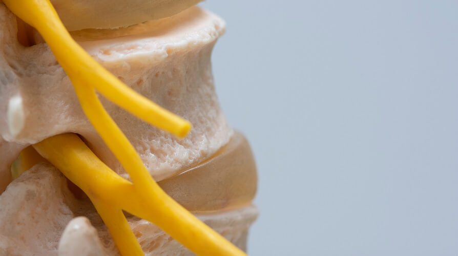
The intervertebral foramen or neural foramen refers to paired holes on either side of the spinal column. You can best see them when looking at a spine side view, as in the image below.
These provide space for the spinal root nerve, dorsal root ganglion, spinal artery, plexus veins, sinu-vertebral nerves, and transforaminal ligaments.
The transverse foramen lies in the transverse process of the upper six vertebrae and provides a channel for the vertebral artery, vertebral vein, and sympathetic nerves.
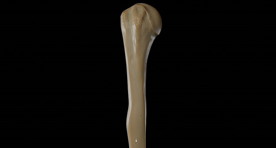
Long Bone Foramen
Long bone foramina or nutrient canals are found in many bones but are more common in long bones. They allow the medullary cavity to have access to a blood supply.
Other Foramina
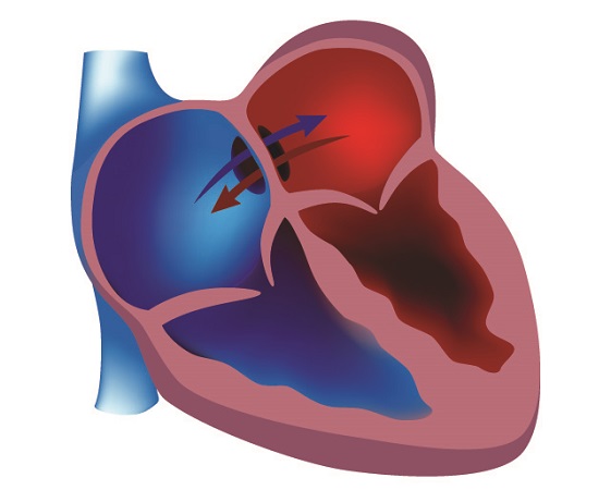
Some other tissue types also have foramina. The foramen ovale of the heart (not of the sphenoid bone) is located in the heart septum between the left and right atria. It allows oxygen-rich blood provided by the mother to flow throughout the fetal heart.
When a baby is born, it is separated from the placenta and this hole closes. Sometimes, it does not close at birth and is this becomes an anatomical abnormality called a patent foramen ovale. This can become an atrial septal defect.
Oxygenated and deoxygenated blood continue to mix across the atria. The f. ovale can close on its own at a later date but commonly requires surgery.
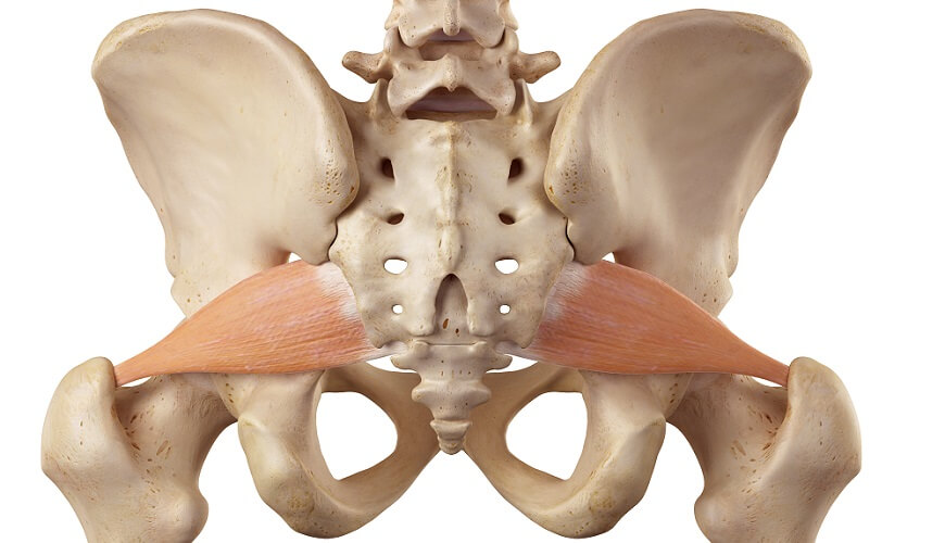
Another example is the sciatic foramen of which there are two: greater and lesser. The greater foramen, located in the pelvis, is filled with the piriformis muscle.
The lesser foramen is found between the back of the thigh and pelvis and allows tendons, nerves, and the internal pudendal artery to pass through.
The foramen obturator, also of the pelvis, is located between the ischium and pubis. It provides a channel for the obturator nerve and blood supply.
In the abdominal cavity, the foramen of Winslow lets the greater omentum under the stomach communicate with the lesser omentum located under the liver.
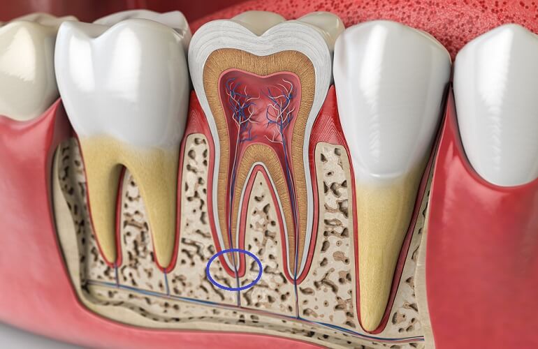
An apical foramen is a hole at the bottom of the roots of the teeth. Nerves and blood vessels passing through innervate, feed, and remove waste products from the dental pulp.
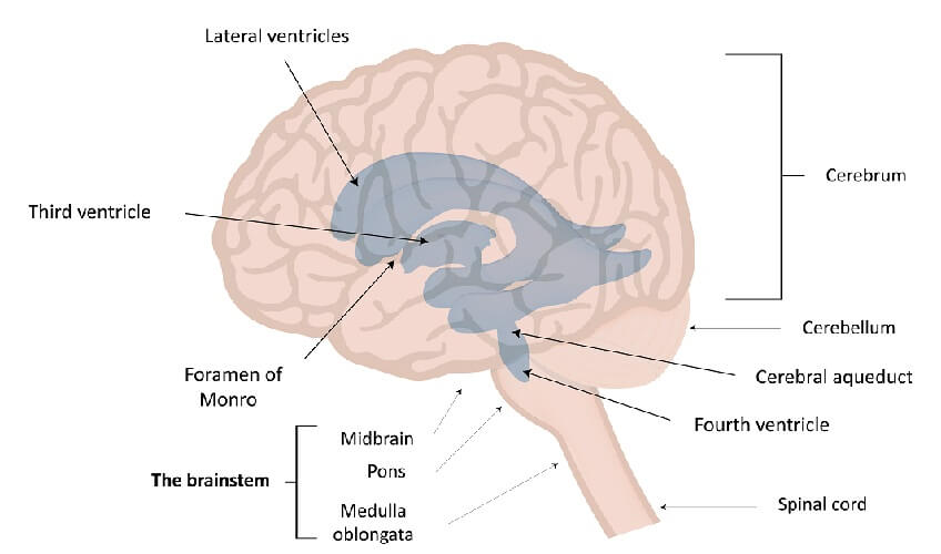
The last group is found in the ventricular system of the brain. An interventricular foramen allows cerebrospinal fluid to flow from one ventricle to the next.
The foramen of Monro is a paired aperture between the lateral ventricles and the midline third ventricle.
The paired foramen of Luschka links the fourth ventricle to the cerebellopontine cistern.
Finally, the singular foramen of Magendie links the fourth ventricle to the cisterna magna.