Definition
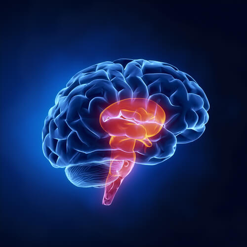
The brain stem (or brainstem) contains three structures- the midbrain, pons, and medulla oblongata- that connects the brain to the spinal cord. It has several roles in the autonomic nervous system, with ascending pathways to receive sensory information for brain processing and descending pathways to send motor information back to the body. It is also the location of ten of the twelve cranial nerves. Considering these roles, the brain stem is most notable for cardiovascular and respiratory control, pain and thermal regulation, sleep cycles, muscular movement, and sensory control in the cranial regions.
Background
Before looking into each of the sub-structures and their respective functions within the brain stem, let’s first look at the brain stem’s relation to the nervous system.
The Nervous System, Neurons, and Brain
The nervous system is a major system spanning the entire body that plays a key role in survival and regulation. It is responsible for relaying sensory information from the body to the brain, where the brain then sends appropriate responses back to the body. These responses can vary- from motor to physiological to storage.
The nervous system is made up of individual nerve cells (or neurons), which recognize signals from the body and its environment. The neurons pass along these signals to their respective destinations in the brain almost instantly via electrical signaling. When one nerve passes the signal to the next nerve, a synapse occurs. This is where electrical signals become chemical in the spaces between two neurons, before becoming electrical again at the next neuron.
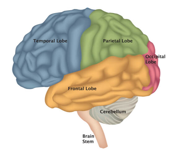
Neurons make up the entirety of the nervous system, which is broken into two physical sub-systems: the central nervous system and the peripheral nervous system. The central nervous system includes the brain and spinal cord, while the peripheral nervous system includes all other neurons throughout the body. The brain itself is made up of four regions: the cerebrum, cerebellum, diencephalon, and brain stem. While each region has distinct differences and roles in relation to the rest of the body, there are many interconnected pathways and neural connections that can pass through multiple structures.
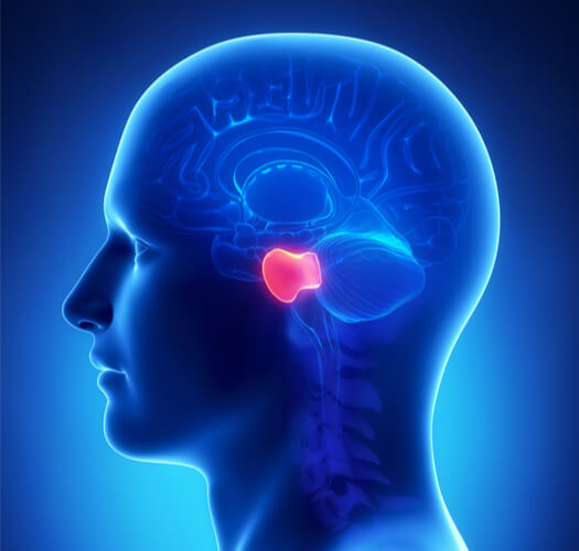
The Brain Stem
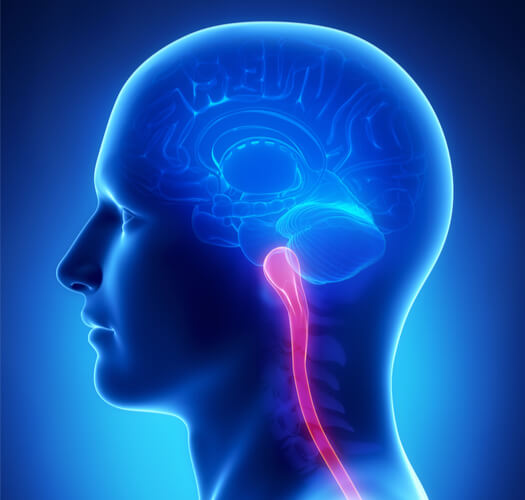
The brain stem is a central nervous system region that directly connects the brain to the spinal cord. Just like all other structures in the brain and nervous system, the brain stem is fully compromised of neurons. These neurons are sometimes referred to and broken down as fibers, axons, or nuclei, depending on the part of the neuron being highlighted.
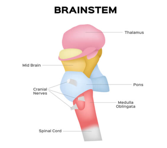
The brain stem as a whole is not a single structure. Instead, it contains three main structures- the midbrain, pons, and medulla oblongata. Each of these regions contain prominent sub-structures and roles that are centralized to each region, as well as overlapping between regions. These regions are also responsible for containing the origins of several cranial nerves.
The brain stem- with all its sub-structures- has many important functions in the autonomic nervous system (which will be described in detail in the next section). Specifically, the brain stem plays key roles in the cardiovascular, respiratory, and digestive systems, as well as in other involuntary functions throughout the body.
The brain stem is extremely vital for survival, where losing the neural connections is highly deadly. Medically, brain stem death is an “irreversible loss” in regaining consciousness and the ability to breath. During brain stem death, the brain stem fails to function, but there can still be projections present in the cortex. Once the cortical and brain stem projections are both lost however, the organism undergoes “biological death”. True death occurs when cardiopulmonary activity ceases as well. Ventilators can be used to extend the heart beating and oxygen circulation following brain stem death, but there is no true cure.
The Autonomic Nervous System
There are two major functional nervous systems in the body: the somatic nervous system and the autonomic nervous system. The somatic nervous system is responsible for regulating and carrying out voluntary responses throughout the body. Specifically, these are the responses that the conscious is aware of (such as lifting your arm to pick up a drink or kicking your legs to perform a dance routine). As a result, the somatic nervous system typically targets skeletal muscles.
However, voluntary movement is not the only action occurring in the body. The body also undergoes many involuntary movements, which are motions that are not conscious. Movements such as this include heartbeat- as controlled by cardiac muscle- and digestion- as controlled by smooth muscle. This category also includes glandular functions. All of these actions fall under the control of the autonomic nervous system.
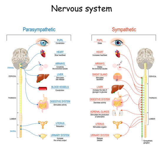
The autonomic nervous system is further broken down into two sub-categories: the sympathetic and parasympathetic nervous systems. As part of the autonomic nervous system, these two sub-systems also control parts of the body for involuntary movement. The sympathetic system (nicknamed the “fight or flight response”) allows the body to prepare itself for stressful situations. This can include increased heart rate, increased glucose release into the blood, and inhibited digestion. On the contrary, the parasympathetic system (nicknamed the “rest and digest response”) allows the body to increase and store energy. This can be accomplished by slowing down heart rate and increasing digestion.
The brain stem plays a large role in controlling the autonomic nervous system, including both the sympathetic and parasympathetic nervous systems.
Brain Stem Structures and Functions
The brain stem contains ascending pathways and descending pathways. The ascending pathways process sensory information while the descending pathways create motor responses to the sensory information received.
As mentioned previously, the three main structures that make up the brain stem are the midbrain, pons, and medulla oblongata. All three of these structures are separated into three regions: the tectum, tegmentum, and basis (from posterior to anterior). Generally, the tectum contains specialized functions to senses and movement. Meanwhile, the tegmentum contains the cranial nuclei, reticular formation, and pathways interconnecting the brain stem and additional structures throughout the brain. Lastly, the basis contains fibers from descending pathways from the cerebral cortex. The following sections of this article will go into detail of specific structures found in each of these three regions.
Midbrain Overview
The midbrain is the smallest structure and topmost portion of the brain stem. It is located between the cerebellum, cerebrum, diencephalon, and pons. As part of the brain stem, the overall midbrain function is to control sensory and motor pathways. Generally, it is important for relaying nerve impulses from the spinal cord to the rest of the brain, and vice versa. While there are many specific bodily functions that the midbrain either controls or assists with, its most noteworthy functions include visual and auditory processing, the internal reward system, and some muscular movements. Additionally, the midbrain contains the origins for cranial nerves III and VI.

At the center of the midbrain is the cerebral aqueduct, which forms a channel between the third and fourth ventricles where cerebrospinal fluid is released. The posterior and anterior portions of the midbrain are split into the tectum and tegmentum sectors, respectively, via the cerebral aqueduct.
Midbrain Regions and Functions
The midbrain’s posterior (the tectum) contains the corpora quadrigemina, which is a pair of protrusions on the midbrain. This pair includes the superior colliculi, which is the visual reflex center, and the inferior colliculi, which is the auditory relay center.
The midbrain’s anterior (namely the tegmentum) is responsible for many parts and roles, including the following:
The Reticular Formation: The most important role of the reticular formation is to filter sensory information relayed to the brain, allowing the conscious to pay attention to the most important senses present. As a result, this region is important for maintaining overall attention and alertness. In addition, the reticular formation is important for cardiovascular systemic control, respiratory regulation, and its relation to consciousness during the waking and sleeping cycles. Furthermore, it contains networks for mood and pain modulation. All of these networks start at the brain stem in the core and branch throughout the brain’s entirety, with ascending pathways into the thalamus and cortex and descending pathways into the spinal cord.
The Red Nucleus: The red nucleus is a sub-part to the reticular formation and is found in the midbrain. It is important for motor control, specifically in coordinating the autonomic movement between swinging arms and walking legs. This feature is important for ensuring balance.
The Periaqueductal Gray Areas: The periaqueductal gray areas (or PAG) is another structure in the midbrain that works to control pain. Neurotransmitters- such as dynorphin and serotonin- from neurons in the PAG moderate pain management. This region also plays an important role in the general autonomic system for survival by avoiding painful situations and inhibiting dangerous behaviors.
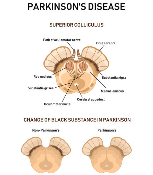
The Substantia Nigra: The substantia nigra contains dopamine-producing neurons that aid in motor control (alongside the basal ganglia- another brain structure). Sections of this area are responsible for inhibiting thalamic motor activity. This region degrades early on in patients with Parkinson’s Disease.
The Ventral Segmental Area: The ventral segmental area also contains dopamine- producing neurons. Unlike the substantia nigra however, this region is responsible for the reward system of the brain. It rewards motivational salience, associative learning, and positive emotions. This is also the region that is active in orgasms. Ventral segmental area neurons project to regions of the cortex associated with consciousness and sleep.
Pons Overview and Functions
The pons is the center structure of the brain stem, located between the midbrain and the medulla oblongata. It contains longitudinal fibers that connect with higher centers throughout the brain and spinal cord, as well as transversal and dorsal fibers that send information between the motor functioning regions of the cortex and cerebellum. Additionally, the pons is where cranial nerves V, VI, VII, and VIII originate.
The pons’ most noteworthy roles include functions related to the respective cranial nerves originating at this structure. This includes recognizing sensations to the head and facial areas as well as movement of the face, eyes, ears, and mouth. Furthermore, the pons is important for autonomic functions such as salvia production, in addition to maintaining equilibrium. Similar to the midbrain, the pons contains part of the reticular formation and thus accompanies functions in cardiovascular control and breathing rhythm.
Gliomas (or glial tumors) can occur anywhere in the brain stem and are therefore known as brain stem gliomas. However, gliomas occurring in the midbrain and medulla oblongata tend to be low grade. For unknown reasons, gliomas occurring in the pons experience rapid growth and are high grade tumors.
Medulla Oblongata Overview
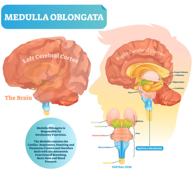
The medulla oblongata (also known as the medulla) is the bottommost region of the brain stem. The medulla directly connects the brain stem to the spinal cord, and there is no distinct separation between the two structures. Part of this structure forms the bottom of the fourth ventricle. Similar to the other brain stem regions, it is important for relaying descending motor control and ascending sensory information. Specifically however, the medulla is most notable for its control over the body’s vitals, including ensuring proper cardiovascular control and respiration. It is also noteworthy for stimulating many reflexes, such as vomiting, coughing, and sneezing. Many functions of the medulla overlap with hypothalamus, where the medulla is often the structure in which the hypothalamus relays instructions. Additionally, the medulla is the origin of cranial nerves IX, X, XI, and XII.
Just as with the other brain stem structures, the tectum, tegmentum, and basis separate the medulla into three sections. The tectum forms part of the fourth ventricle, while the tegmentum contains the inferior olivary nucleus and holds the medulla cranial nerves. The basis contains the pyramid decussation.
Medulla Oblongata Regions and Functions
Just as with the midbrain and pons, medulla contains part of the reticular formation as well. The cardiovascular and respiratory system connect as a single system within this structure of the midbrain. Afferent cardiorespiratory signals synapse at the medulla and work to regulate respiration. The ventral respiratory columnist, which controls respiratory rhythm and its oscillating pattern, is also present at the medulla. Furthermore, the medulla acts as the vasomotor center. This is because neurons present can stimulate blood vessel diameter adjustments while monitoring baseline arterial pressure.
The nucleus of the solitary tract is found in the medulla and is organized by information type being transmitted, as well as by activated pathways in response to the information. It largely coordinates afferent information. The most important functions of the medulla occur at this nucleus, including information from baroreceptors and chemoreceptors. Blood vessel baroreceptors send information to the nucleus of the solitary tract to adjust heart rate and blood flow. Blood vessel chemoreceptors sense oxygen and carbon dioxide levels, allowing the nucleus of the solitary tract to maintain proper respiration. Additionally, taste synapses at this nucleus first, before being sent to the thalamus and cortex for sensory processing.

The Area Postrema: The area postrema is located at the medulla’s dorsal surface. As the vomiting center, the cells located here lack a blood-brain barrier. Therefore, large and polar molecules are able to pass through. Innervation here can cause a person to feel nauseous. Research has found that this area has receptors for the human chorionic gonadotropic (hCG) hormone, which is a pregnancy hormone. This could indicate a possible explanation for increased sensitivity to morning sickness in pregnant women.
The Spinal Trigeminal Nucleus: The spinal trigeminal nucleus is important for sensing temperature pain and deep ipsilateral (or same side) face touches. It is the first place where the nerves for orofacial pain (the overall term for head and neck pain) synapse.
The Inferior Olivary Nuclei: The inferior olivary nuclei are important for receiving information about proprioception (or awareness of body position and movement), muscle and joint tension, and motor intention. As the cerebellum is notably responsible for skeletal movement and balance, the nuclei here directly synapse at the cerebellum. Additionally, these nuclei are responsible for swallowing, coughing, and sneezing.
The Pyramidal Decussation: The pyramidal decussation contains a majority of motor fibers from the motor cortex, forming the lateral corticospinal tract in the spinal cord. This is where the pyramidal tracts cross over each other to make connections with the opposite sides of the body. As a common trait in vertebrates, the advantage for this crossing over is still questioned by evolutionary biologists.
The Cuneate and Gracious Nuclei: The cuneate nucleus of the medulla receives information from the upper extremities, while the gracious nucleus receives information from the lower extremities. These fibers form the medial lemniscus, which is a pathway bringing proprioception, vibration, and fine touch information to the thalamus.
The Spinothalamic Tract: The spinothalamic tract is an ascending pathway where pain, temperature, and crude touch travels from the spinal cord throughout the brain. Eventually, the tract ends at the ventral posterior lateral nucleus in the thalamus. Pain and temperature travel on the anterior tract, while crude touch travels on the lateral tract.
Cranial Nerves of the Brain Stem
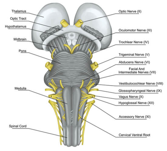
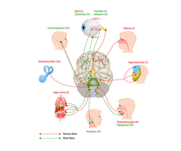
As mentioned over the course of this article, the brain stem contains the origins for ten of the twelve cranial nerves. Nerves III and IV originate at the midbrain, while nerves V, VI, VII, and VIII originate at the pons and nerves IX, X, XI, and XII originate at the medulla. These nerves can have sensory functions, motor functions, or both. The following sections will describe the general function of each brain stem cranial nerve with diagrams marking their locations.
Note: The two nerves that do not originate in the brain stem are the olfactory (I) and optic (II) nerves. These nerves instead originate in the cerebrum.
Nerves Originating in the Midbrain:
Oculomotor Nerve (III): The oculomotor motor nerve contains motor functions to control pupil and eye movements.
Trochlear Nerve (IV): The trochlear nerve has motor functions to control the superior oblique muscle. This muscle is located to the upper and medial regions of the obit (surrounding the eye), allowing for the eye to abduct, depress, and internally rotate.
Nerves Originating in the Pons:
Trigeminal Nerve (V): The trigeminal nerve contains both sensory and motor functions to the jaw and surrounding mastication muscles. Additionally, it contains sensory functions to many parts of the face, including the orbital structures, nasal cavity, forehead skin, eyebrows, eye lids, part of the nose, lips, gums, teeth, skeet, palate, pharynx.
Abducens Nerve (VI): The abducens nerve contains motor functions to the lateral rectus muscle, which is one of the six muscles responsible for eyeball movement.
Facial Nerve (VII): The facial nerve contains sensory functions involving taste on the first two thirds of the tongue. Additionally, it contains motor functions for muscles leading to facial expressions, the lacrimal glands (which contains fluids to lubricate the eye), and the submandibular and sublingual salivary glands (which are two of the three major glands that aid in saliva production within the mouth).
Vestibulocochlear Nerve (VIII): The vestibulocochlear nerve has special sensory functions in the cochlea for hearing and in the vestibule for motion and balance.
Nerves Originating in the Medulla Oblongata:
Glossopharyngeal Nerve (IX): The glossopharyngeal nerve contains sensory functions to the back third of the tongue, the pharynx and palate, blood pressure receipts, pH levels, oxygen levels, and carbon dioxide concentrations. Additionally, it contains motor functions for the pharyngeal muscles and parotid salivary gland (the final remaining major gland for saliva production in the mouth).
Vagus Nerve (X): The vagus nerve contains sensory functions to the auricle and external acoustic canal, the diaphragm, and visceral organs within the thoracic cavities. It also contains motor functions to the palatal and pharyngeal muscles and thoracic visceral organs.
Accessory Nerve (XI): The accessory nerve contains motor functions for the skeletal muscles of the palate, pharynx, and larynx [working with the vagus nerve (X)], as well as the sternocleidomastoid and trapezius muscles in the neck and spine.
Hypoglossal Nerve (XII): The hypoglossal nerve contains motor functions to control tongue musculature.
Conclusion
The brain stem is the connecting structure between the brain and the spinal cord of the central nervous system. Containing the midbrain, pons, and medulla oblongata, the brain stem has multiple functions in the autonomic nervous system, including in cardiovascular and respiratory control, as well as in sensory and motor functions. Additionally, the brain stem is responsible for holding the origins of ten of the twelve cranial nerves, including nerves III – XII.
Quiz