Definition
The skull is found in all vertebrates – a bony casing that protects the brain and brainstem and supports the face. Skull bones are joined by sutures or articulations. The entire construction is divided into two anatomical parts: the neurocranium (cranial bones) and the viscerocranium (facial bones). There are many differences in human and animal skulls – for example, only humans have a chin.

Skull Anatomy
Skull anatomy divides this patchwork of bones into two categories: the neurocranium (red in the below image) and the viscerocranium (blue).

Anatomy of the Neurocranium
The neurocranium or cranial bones are similarly split into two anatomical areas: the skull cap (calvarium) and skull base.
The calvarium consists of the occipital bone, parietal bones, and frontal bone. These are joined by sutures that will be described later on.
The skull base is composed of the frontal, occipital, and temporal bones, as well as the ethmoid and sphenoid. It is divided into three parts:
- Anterior cranial fossa
- Middle cranial fossa
- Posterior cranial fossa
The cranial fossae are best viewed when you flip the base over and look at its internal structure.
Close to the middle of the image below is the large gap of the foramen magnum where the occipital bone connects with the first cervical vertebra of the spine.
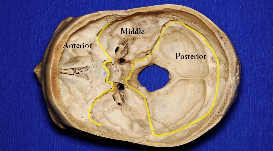
At the front is the indentation known as the anterior cranial fossa. In its center sits a crest of bone and an area of sieve-like bony material. This sieve allows the olfactory nerves to run through and connect the brain to our sense of smell. Other nerves such as the optic nerve also run through the anterior cranial fossa.
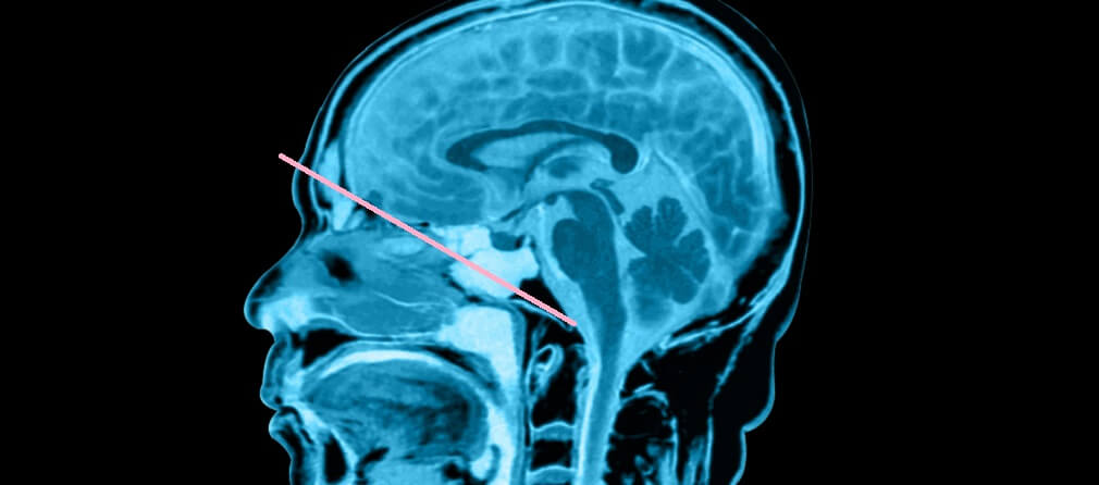
The anterior cranial fossa supports the frontal lobe of the brain. This part of the skull floor is shallow. When you look at a side view (see below), you can see that the base runs diagonally; it is easy to see how the temporal lobe fits into the anterior fossa at this shallower point.
In the middle of the skull base is the butterfly-shaped middle cranial fossa. This is a little deeper than the anterior part. The anterior fossa houses several cranial nerves and the sella turcica – the saddle-like protrusion located about halfway between the frontal crest and the foramen magnum. The sella turcica separates the pituitary gland of the brain from the nasal cavity.
The posterior cranial fossa houses the largest skull foramen (a foramen, in this case, is a hole through the bone tissue that allows nerves, arteries, and veins to pass through). This large foramen (foramen magnum) connects the skull with the spine. Again, cranial nerves pass through the foramina of the posterior cranial fossa.
Anatomy of the Viscerocranium
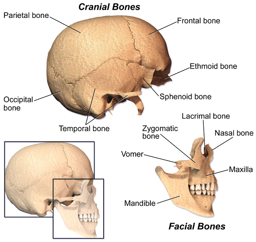
The viscerocranium or facial skeleton contains multiple small bones as well as the larger bones of the upper and lower jaw. The bones that construct the viscerocranium are the:
- Palatine bones
- Lacrimal bones
- Nasal conchae
- Nasal bones
- Vomer
- Zygomatic bone
- Maxilla
- Mandible
Some sources also include the sphenoid and ethmoid bones in the facial bones as these contribute to viscerocranial structures such as the orbits of the eyes and the nasal cavity. This is not incorrect but they must also be included in the bones of the neurocranium.
Sutures of the Skull
The sutures of the calvarium (cranial sutures) are threefold.
The coronal suture joins the back of the frontal bone to the front of the two parietal bones.
The sagittal suture joins the two parietal bones at the midline at the top of the head.
The lambdoidal suture (or lambdoid suture) runs diagonally at the back of the head to join the top of the occipital bone with the back of the parietal bones. The Greek letter lambda is an upside-down “V” that describes the shape of this suture.

A complex area of cranial sutures is the pterion. This is an H-shaped group of sutures that connect the frontal, parietal, temporal, and sphenoid bones at the side of the cranium. Because of these concentrated joins and thinner bone layers, this part is the weakest.
While there are sutures in the viscerocranium, such as the fused suture between the two mirrored parts of the lower jaw through the center of the chin, these are usually referred to as articulations.
Human Skull vs Animal Skull
Differences concerning human skull versus animal skull are obvious from a visual point of view. One of the most quoted facts about the differences in the human skull is that only humans have a chin.
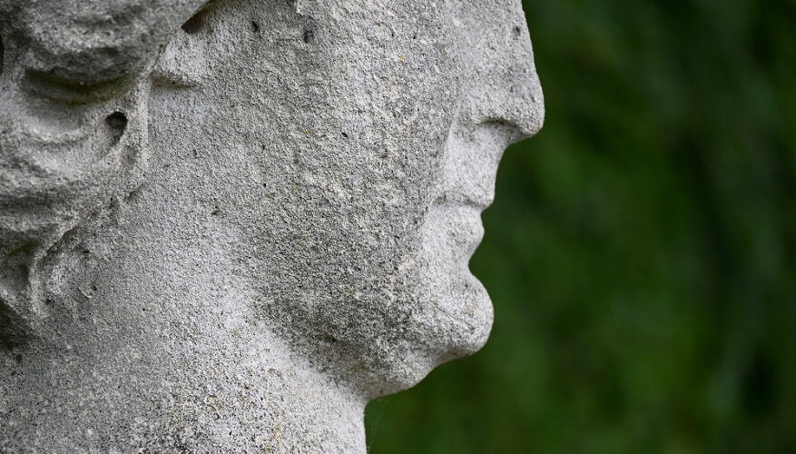
The human skull cap is bulbous because it provides extra space for our larger brain. In relation to the neurocranium, the face is small. Our U-shaped mandible also differs; the animal skull has a V-shaped lower jaw.
When you compare a Neanderthal human skull with a modern version, it is obvious that the main change is in the skull base.
Over millennia, this bone has changed position, bringing the face of modern man almost flush to the frontal lobe of the brain. This movement has also removed the heavier brow that early man is famous for.
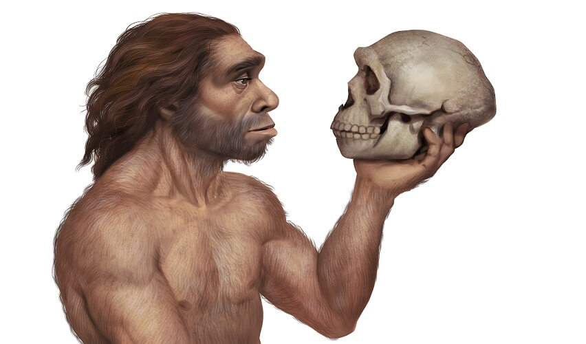
Theories say this is due to growth in the temporal lobes that play, among many others, important memory roles. The size of our frontal lobe is similar to the size of the average chimpanzee and, even more interestingly, we have smaller brains than Neanderthals.
Today, the average human brain weighs about three pounds or 1400 grams – just 2% of the average human’s body weight. You can find a list of different average brain weights of various species here, together with other brain trivia. As the neurocranium houses the brain, brain size affects skull shape.
Dog Skull
The first thing we notice when we look at a dog skull is the long canine teeth, although teeth are not considered to be part of the viscerocranium as they are not bones.
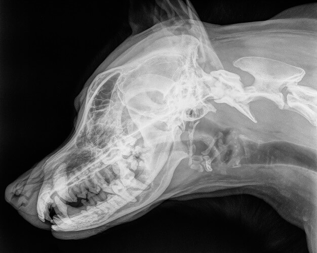
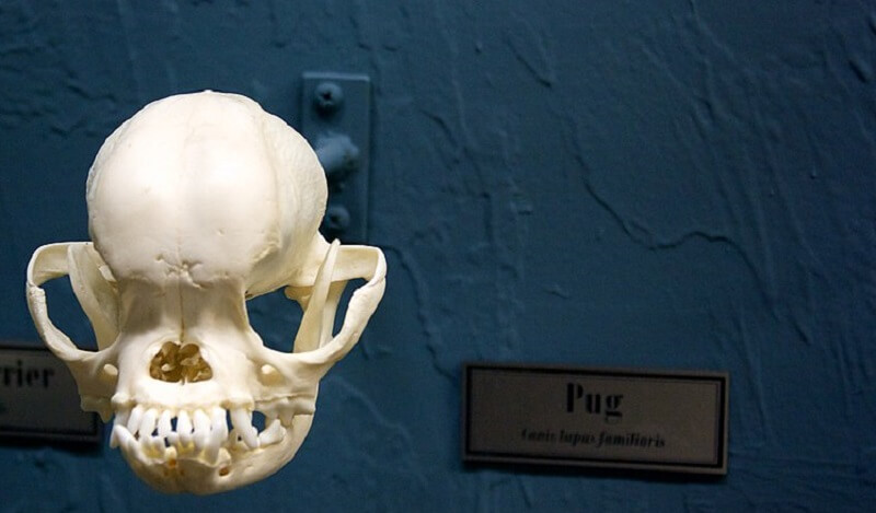
Dogs come in all shapes and sizes so the cranium of a greyhound cannot be likened to a pug skull.
A dog skull (if it is not a pug or bulldog) is elongated; the frontal lobe of the brain lies further behind the eye sockets, not behind a vertical forehead as in humans.
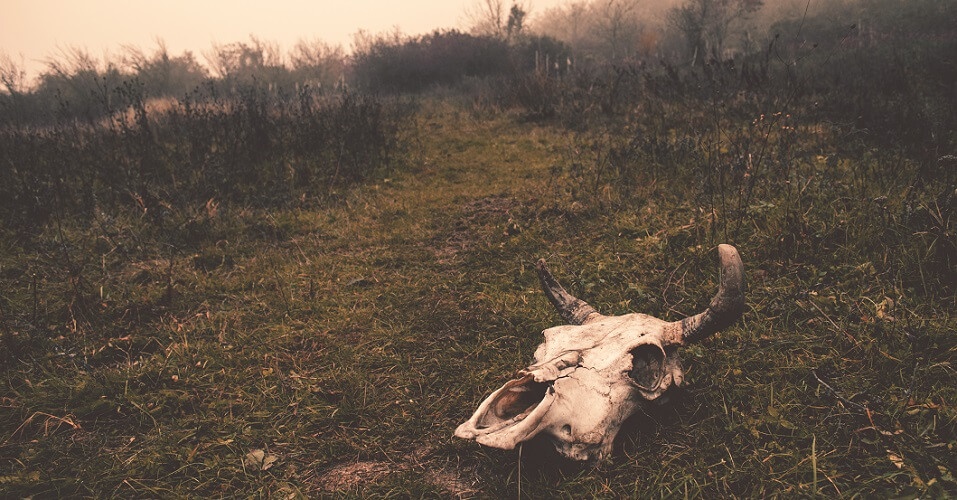
Cow Skull
A cow skull must have a heavy jaw to support strong mastication muscles – cows continuously graze and chew the cud. The frontal bone of a cow (or any animal with horns) also features an extra process called the cornual process. This paired process projects from the vaulted frontal bone.
The two cornual processes support the horns; keratin layers gradually lengthen and thicken to produce the long, pointed shape. Just like our hair and nails, horns grow continuously.
Horns are not to be confused with antlers; these are bone.
A horse skull is easy to distinguish from a cow skull. The frontal bone of the horse skull is narrower and the maxilla contains teeth; a cow only has lower incisors in the mandible.
If the teeth of an old herbivore skull have dropped away, you will still be able to see whether sockets are present. The maxilla of a cow has a dental pad without sockets.

Skull Fractures
When talking about skull fractures, we usually mean neurocranium fractures. Breaks in the viscerocranium are more likely to be called facial fractures.
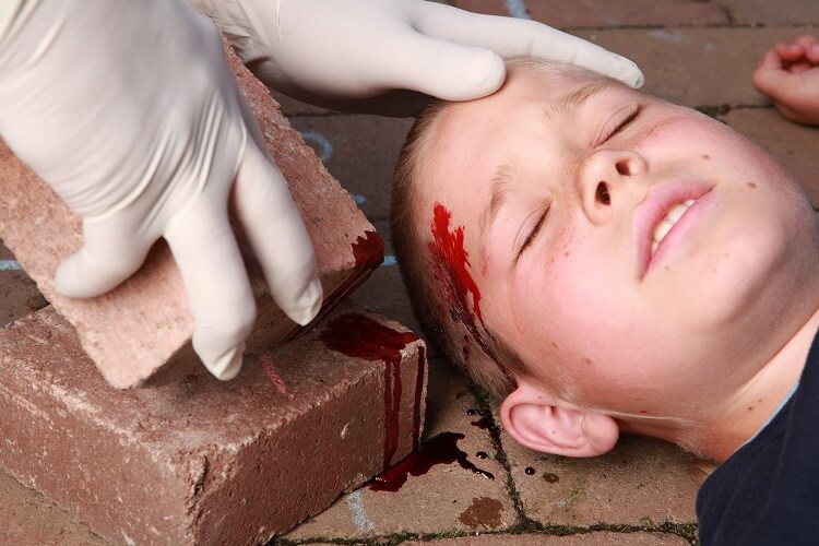
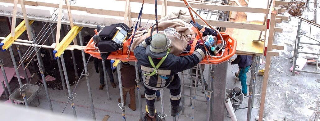
Most skull fractures are due to blunt force trauma caused by road traffic accidents, falls, contact sports, and assaults.
Skull fractures in very young children are relatively common as the sutures of the cranium take time to completely fuse.
One of the most dangerous types of neurocranium fracture is the basilar skull fracture or basal fracture. This is because the base protects both the brainstem and the cerebellum that control the autonomic nervous system.
Certain injuries are linked to specific basal fracture locations:
- Posterior cranial fossa: cervical spine injury and lower cranial nerve injuries. Often leads to hemi- or paraplegia.
- Middle cranial fossa: carotid injury and subsequent hemorrhage; injury to cranial nerves III, IV, V, and/or VI.
- Anterior cranial fossa: eye injury; cerebrospinal leaks through the nose; injury to cranial nerve I.
- Temporal bone fracture: carotid injury and subsequent hemorrhage; cerebrospinal or blood leaks through the ear; injury to cranial nerves VII and/or VIII.
A depressed fracture anywhere in the head or face can push into the delicate tissues of the brain and brainstem, the membranes surrounding the brain (meninges), blood vessels, and cranial nerves and their branches. Simple fractures without displacement are best treated conservatively.