Skin cells are the basic building blocks of the skin; a large, complex organ forms a protective barrier between our insides and the external environment. The most common type of skin cell is the keratinocyte, whose primary function is to form a tough, waterproof layer against UV radiation, harmful chemicals, and infectious agents.
However, the skin also contains highly specialized cells with important immunological, photoprotective, and sensory functions. The term ‘skin cell’, therefore, may refer to any of the four major types of cells found in the epidermis (or outer layer) of the skin.
Functions of the Skin
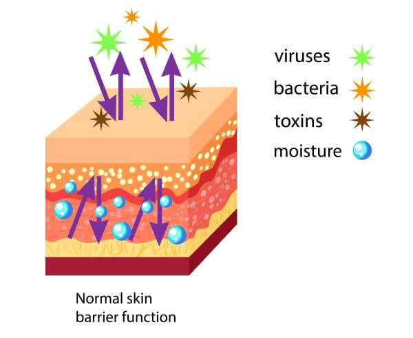
The skin is the largest organ of the human body and has a range of vital functions in supporting survival. The primary function of the skin is to form a physical barrier between the internal environment of an organism and the outside world. This protects internal organs and structures from injury and infection.
The skin also helps to maintain homeostasis by preventing water loss and regulating body temperature. It protects organisms from the damaging effects of UV light and helps to produce vitamin D when exposed to the sun. Finally, the skin functions as a sensory organ, allowing us to perceive touch, temperature changes, and pain.
The skin can perform all of these functions thanks to the highly specialized cells that make up the epidermis (the outermost layer of the skin).
Structure of the Skin
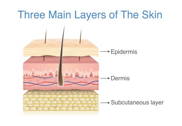
The skin consists of three major layers; the epidermis, the dermis, and the hypodermis (AKA the subcutaneous layer).
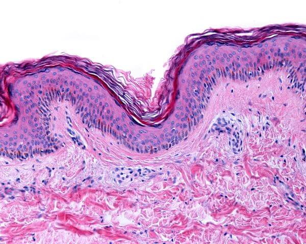
The epidermis is the outermost layer of the skin. This waterproof barrier protects the underlying skin layers and other internal structures from injury, UV damage, harmful chemicals, and infections by pathogens such as bacteria, viruses, and fungi. The thickness of the epidermis varies between different parts of the body. In the thin, delicate skin of the eyelids, the epidermis is only around 0.5 mm thick, whereas the more resilient skin of the palms and feet is about 1.5 mm thick.
The dermis is found directly beneath the epidermis and is the thickest of the three skin layers. This layer contains a complex network of specialized structures, including blood vessels, lymph vessels, sweat glands, hair follicles, sebaceous glands, and nerve endings. It also contains collagen and elastin, which are structural proteins that make skin strong and flexible. The main functions of the dermis are to deliver oxygen and nutrients to the epidermis and to help regulate body temperature.
The hypodermis (or subcutaneous layer) is the fatty, innermost layer of the skin. It consists mainly of fat cells and functions as an insulating layer that helps to regulate internal body temperature. The hypodermis also acts as a shock absorber that protects the internal organs from injury.
What is a Skin Cell?
The term ‘skin cell’ may refer to any of the four main types of cells found in the epidermis. These are keratinocytes, melanocytes, Langerhans cells, and Merkel cells. Each type of skin cell has a unique role that contributes to the overall structure and function of the skin.
Skin Cells of the Epidermis
Keratinocytes
Keratinocytes are the most abundant type of skin cell found in the epidermis and account for around 90-95% of the epidermal cells.
They produce and store a protein called keratin, a structural protein that makes skin, hair, and nails tough and waterproof. The main function of the keratinocytes is to form a strong barrier against pathogens, UV radiation, and harmful chemicals, while also minimizing the loss of water and heat from the body.
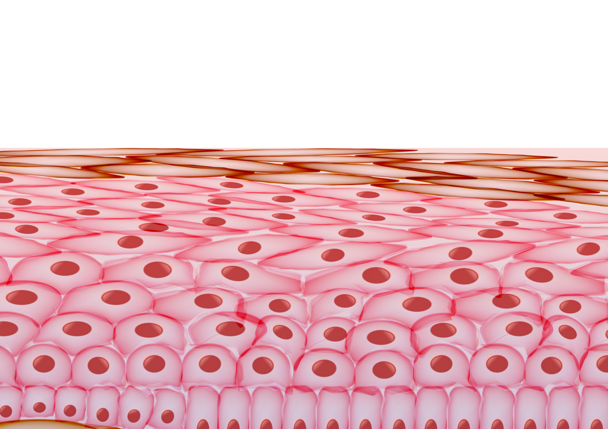
Keratinocytes originate from stem cells in the deepest layer of the epidermis (the basal layer) and are pushed up through the layers of the epidermis as new cells are produced. As they migrate upwards, keratinocytes differentiate and undergo structural and functional changes.
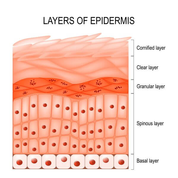
The stratum basal (or basal layer) is where keratinocytes are produced by mitosis. Cells in this layer of the epidermis may also be referred to as basal cells. As new cells are continually produced, older cells are pushed up into the next layer of the epidermis; the stratum spinosum.
In the stratum spinosum (or squamous cell layer), keratinocytes take on a spiky appearance and are known as spinous cells or prickle cells. The main function of this epidermal layer is to maintain the strength and flexibility of the skin.
Next, the keratinocytes migrate to the stratum granulosum. Cells in this layer are highly keratinized and have a granular appearance. As they move closer to the surface of the skin, keratinocytes begin to flatten and dry out.
By the time keratinocytes enter the stratum lucidum (AKA the clear layer), they have flattened and died, thanks to their increasing distance from the nutrient-rich blood supply of the stratum basal. The stratum corneum (the outermost layer of the epidermis) is composed of 10 – 30 layers of dead keratinocytes that are constantly shed from the skin. Keratinocytes of the stratum corneum may also be referred to as corneocytes.
Melanocytes
Melanocytes are another major type of skin cell and comprise 5-10% of skin cells in the basal layer of the epidermis.
The main function of melanocytes is to produce melanin, which is the pigment that gives skin and hair its color. Melanin protects skin cells against harmful UV radiation and is produced as a response to sun exposure. In cases of continuous sun exposure, melanin will accumulate in the skin and cause it to become darker i.e., a ‘suntan’ develops.

Langerhans Cells
Langerhans cells are immune cells of the epidermis and play an essential role in protecting the skin against pathogens. They are found throughout the epidermis but are most concentrated in the stratum spinosum.
Langerhans cells are antigen-presenting cells and, upon encountering a foreign pathogen, will engulf and digest it into protein fragments. Some of these fragments are displayed on the surface of the Langerhans cell as part of its MHCI complex and are presented to naïve T cells in the lymph nodes. The T cells are activated to launch an adaptive immune response, and effector T cells are deployed to find and destroy the invading pathogen.
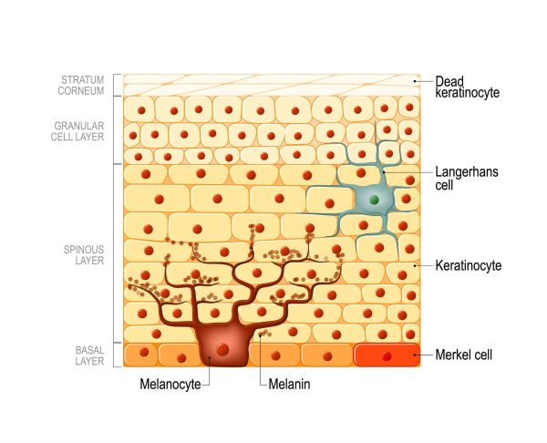
Merkel Cells
Merkel cells are found in the basal layer of the epidermis and are especially concentrated in the palms, finger pads, feet, and undersides of the toes. They are positioned very close to sensory nerve endings and are thought to function as touch-sensitive cells. Merkel cells allow us to perceive sensory information (such as touch, pressure, and texture) from our external environment.