Definition
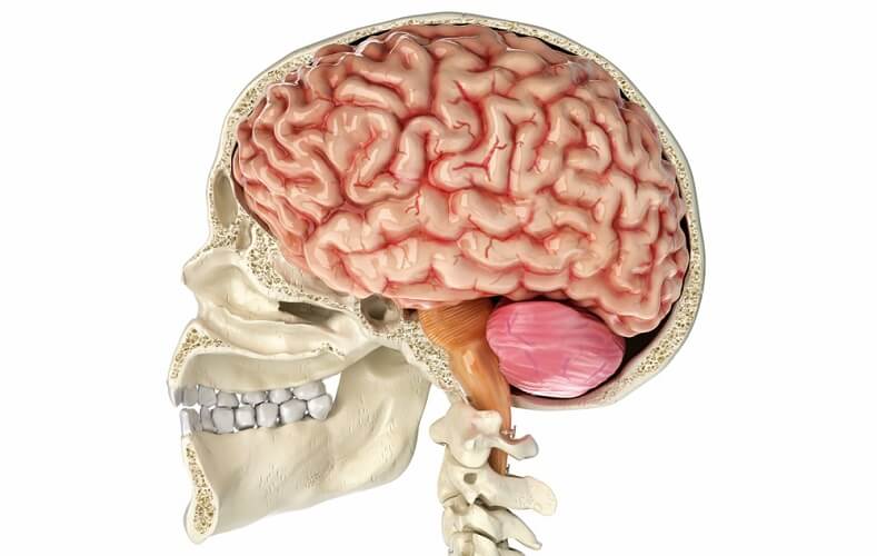
The reticular formation is a neuron network in the brainstem that enables consciousness, sensory and motor function, and endocrine and neurotransmitter regulation. This part of the central nervous system, spread in three main columns from one end of the brainstem to the other, is a core relay point that connects the nerves of the spinal cord with the brain via efferent and afferent neurons. Its full range of functions is not fully known.
Reticular Formation Function
Reticular formation function involves a broad range of autonomic, sensory, motor, behavioral, cognitive, and mood-based responses. It works together with other regions of the central nervous system to allow complex tasks such as the regulation of our state of consciousness, emotion processing, visual coordination, cardiovascular control, and posture. Approximately 100,000,000 impulses are received in the reticular formation (RF) every single second!
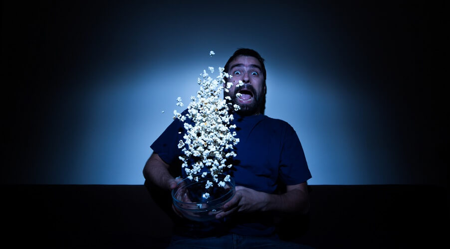
You can separate the functions of the reticular formation by looking at its two systems. These are the ascending reticular activating system (ARAS) that brings sensory messages from the RF to the brain cortex and vice versa, and the descending reticular system (DRS) that brings messages to and from the motor neurons of the spinal cord. You should see both systems as two parts of a single parallel system; they work at the same time, and the brainstem reticular formation modulates how many messages are processed. The two systems (ARAS and DRS) influence each other. That is why, if you are watching a really scary horror film, your muscles become tense – both your emotions and your muscles react. And when the scariest moment is over, you relax. The group term of reticular activating system or RAS is confusingly named, as it not only activates but deactivates associated neurons.
The ARAS is most commonly described as the regulator of consciousness and arousal; however, it relays information about lots of other processes including our respiration rate, cough response, cardiac rhythm, and the act of mastication (chewing). It is reported that chewing maintains cognitive function by stimulating the RF – absence of mastication is associated with dementia and disturbed sleep patterns. This shows how closely our muscle movements, cognitive functions, and consciousness are linked.
Reticular Formation Examples
A reticular formation example of activation and deactivation feedback would be someone dozing off during a long journey. Gradually, the person’s brain activity starts to fall and less information is sent from the resting brain cortex to the reticular system via the ARAS. When that person achieves rapid eye movement (REM) sleep, his or her muscle tone control rapidly falls to produce atonia via the DRS. If the head suddenly drops, signals are sent to the reticular formation from the suddenly-activated muscle spindles via the DRS. At the same time, the ARAS reacts and the nodding-off traveler suddenly wakes. This shows us the motor action of the DRS responding to and activating the ARAS and vice versa. Remember, the ARAS sends and receives sensory information, and the DRS sends and receives motor information.
Another reticular formation example is the action of general anesthetic medications used before surgery. First, a strong analgesic is administered that prevents pain sensations from traveling towards the RF. A sedative-hypnotic such as propofol reduces the firing rates of neurons in the brain cortex, thalamus, and reticular formation; this produces unconsciousness and halts the processes that form memory. Finally, a muscle relaxant in the form of curare inactivates the muscle spindles and spinal cord-regulated reflexes. The reticular formation arousal networks are depressed for the duration of the surgery by way of anesthetic gas and drugs, and a machine takes over the autonomic function of breathing as curare affects the skeletal muscle. The heart does not stop beating as specialized cardiac muscle cells act as pacemakers and the heart muscle is not skeletal muscle. However, the heart rate, which is influenced by midbrain reticular function, is affected.

Motor Reticular Formation
Receiving input via afferent (bringing towards) nerves that enter the reticular formation from the cranial nerves, we can move our facial and neck muscles. Remember, muscle movement is the result of motor nerves that are part of the DRS and also a response to the sensory stimuli that encourage muscle movement and travel via the ARAS. Just think of a horse twitches when a fly lands on its skin.
Involuntary smooth-muscle movements allow actions such as swallowing, coughing, and blood vessel dilation and constriction for blood pressure control. These messages are all relayed through the reticular formation. A chain of good motor reticular formation examples would be following a spoonful of food with the eyes as it travels to the mouth, chewing and swallowing the food, coughing if a crumb travels into the trachea, holding the breath during swallowing, and peristalsis in the digestive tract that pushes the food through and out of the body.
Voluntary motor function is also part of the reticular formation’s task, for example in our posture and equilibrium. Balance is not an involuntary act but learned, as we can see when watching a young child take its first steps. Through facilitatory and inhibitory pathways in the reticular formation, messages are sent to receptors in the joints and the associated muscle spindles. This muscle activity has been learned to the extent that we are not even conscious of these movements. Even so, posture control depends on complex physiological interactions, high levels of sensory processing, and the person’s goal, cognitive skills, and experience (motor memory).

Sensory Reticular Formation
The sensory functions of the reticular formation, guided via the ARAS but working together with the DRS, include how and when our bodies experience pain, how we balance, and – the most famous and studied of reticular formation roles – our levels of consciousness. However, the complete story of this small, anatomically unclear structure is still relatively unknown.
By forwarding sensory information to motor areas of the brain, the RF coordinates visual, auditory, vestibular, gustatory, olfactory, and tactile sensory input – sight, hearing, balance and movement, taste, and touch respectively – so we can perform and experience voluntary and involuntary physical and emotional responses.
Endocrine Reticular Formation
The endocrine function of the reticular formation does not mean that this part of the brainstem secretes or produces chemicals, but by relaying messages it does regulate hormone and neurotransmitter secretion. Probably the most famous endocrine system example of this particular reticular formation function is our stress response system. In the stress response system, the combination of memory and environment stimulates the RF to increase firing rates in the direction of the hypothalamus that encourage it to secrete corticotropin-releasing factor. This factor initiates the release of a cascade of stress hormones that make us alert, send more blood (oxygen and glucose) to the muscles and vital organs, supply less blood to non-vital organs, and so make the body ready to fight or run. Once the danger has passed, the relay-center of the RF modulates the sensory and motor messages that calm us back down.
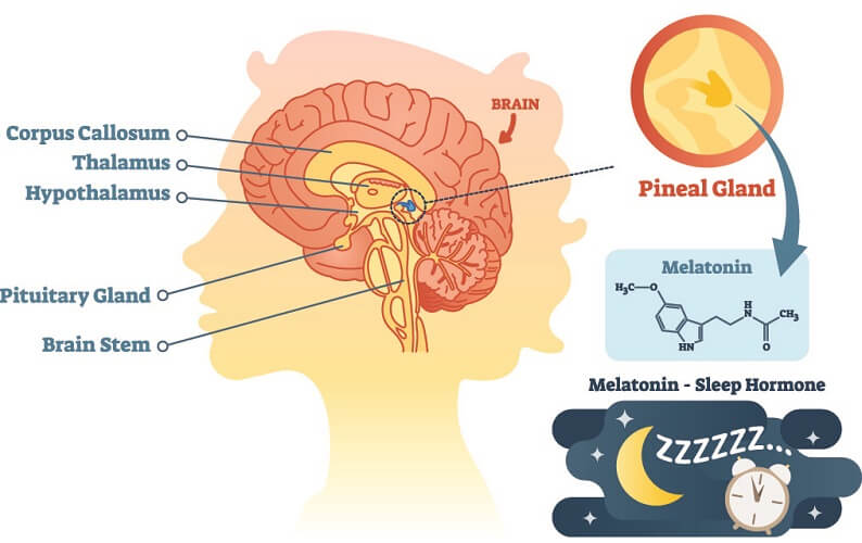
It is thought that the reticular formation transmits the information that controls the release and inhibition of an extensive range of hormones; this theory is supported by the fact that it lies extremely close to important neuroendocrine secretory organs such as the pineal, pituitary, and hypothalamus glands. As the pineal gland is responsible for melatonin production (where melatonin is a proven circadian rhythm regulator that helps us to fall asleep), this adds weight to the role of the reticular formation in our sleep-wake patterns.
Reticular Formation Location
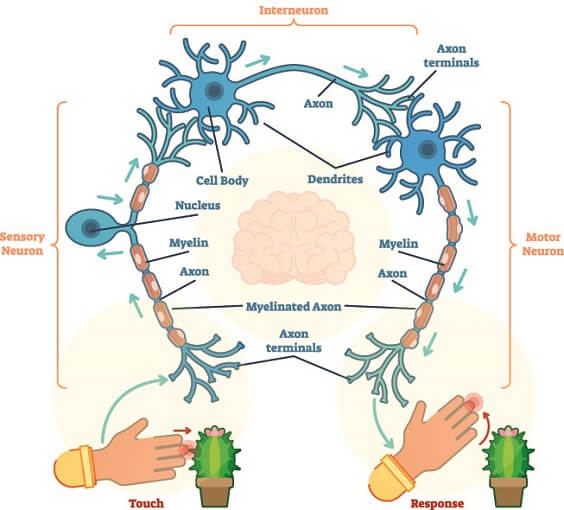
The reticular formation is located in the brainstem but extends into the spinal cord and thalamus; it passes through the medulla, pons, midbrain, and diencephalon. The RF does not completely fill the brainstem but is loosely split into three columns of nuclei (groups of nerve cells with their own set of functions) that run along its length. To simplify this quite diffuse structure, researchers split the RF into the median, medial, and lateral columns. While these areas are associated with their own range of functions, it is their response to specific neurotransmitters that makes them so different. This is because the reticular formation contains large numbers of interneurons with polysynaptic connections that connect directly or via other interneurons with a target cell. Interneurons are small versions of the reticular formation in that they are relay centers. The fit between two or more neurons and modulate how often and how effectively these neurons communicate. The interneurons of the RF are polysynaptic – this means that they do not only modulate messaging between two neurons but can relay information from multiple neurons, both sensory and motor, at the same time. A single RF nerve cell regulates multiple functions, so you should imagine the pictured interneuron below as connecting to many other neurons. These create a huge network of associated actions and reactions.
Median Column
The median column consists of a single, central column that runs through the midbrain. It is divided into three groups of nerve cells (nuclei): dorsal raphe nuclei, nucleus raphe pontis, and nucleus raphe magnus. You don’t need to know all of these names, but by grouping them we can get a better picture of this column’s known functions. The nerve cell networks in the median column contain groups of interneurons called raphe nuclei. The word raphe simply refers to the vertical midline seam where structures of the left and right side of the body join. This is why the nuclei in the medial column are all labeled raphe.
The dorsal raphe nucleus relays pain-control information. The nucleus raphe pontis connects to the cerebellum and is important for connecting involuntary sensory and motor information. The nucleus raphe magnus influences our perception of pain. All raphe nuclei primarily produce, regulate, and respond to the neurotransmitter serotonin (5-HT).
Medial Column
The medial column contains mixed medium and large nerve cells with synapses that primarily respond to, produce, and regulate the neurotransmitters gamma-aminobutyric acid (GABA) and glutamate. This column contains the gigantocellular nucleus, the ventral reticular nucleus, the oral pontine reticular nucleus, and the caudal pontine reticular nucleus. Again, you don’t need to learn these names off by heart.
The gigantocellular (big-cell) nucleus relays information that controls tongue movement. The ventral reticular nucleus is possibly linked to breathing and memory formation. The oral pontine reticular nucleus probably regulates how we enter and exit stages of rapid eye movement sleep; the caudal pontine reticular nucleus is associated with head and jaw movement. Probably and possibly are, unfortunately, the best we have right now. More research into the reticular formation is necessary before we can use more exact statements.
Lateral Column
The lateral column hosts at least six different nuclei, all of which mainly produce, regulate, and respond to the neurotransmitters noradrenaline and acetylcholine. The most studied of these nuclei are the parvocellular reticular nucleus, the nucleus locus coeruleus, and the pedunculopontine nucleus. These are associated with facial control and breathing, our physiological responses to stress, and our sensations of arousal, reward, movement, and attention respectively.
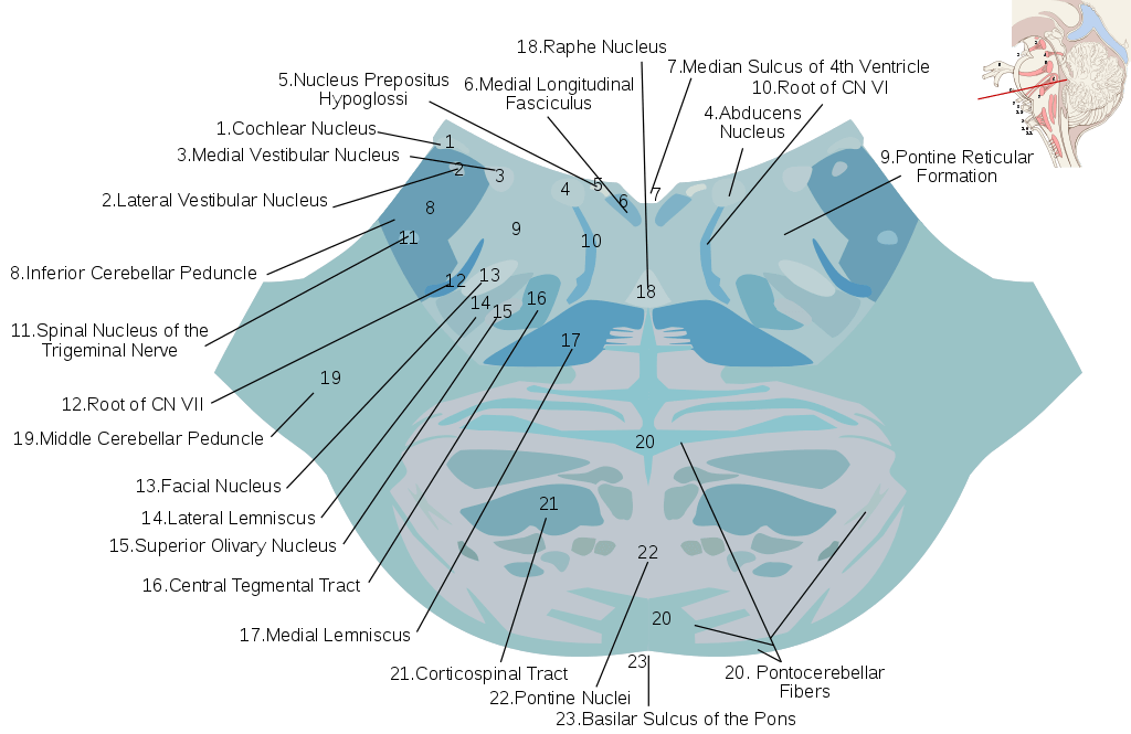
The image below gives a good indication of how various reticular formation nuclei are spread throughout the tissue of the pons.
Reticular Formation Pathways
Reticular formation pathways are split according to sensory and motor pathways (ARAS and DRS) and according to whether a nerve fiber or group of fibers enters or exits this part of the brainstem – in other words, whether the RF receives or transmits information. Connections bring messages to the reticular formation from the spinal cord and brain. Efferent pathways bring messages from the reticular formation directly or indirectly to other structures. Complex and simpler networks use the reticular formation as a central control or relay base.
Reticular Formation Afferent Pathways
When the reticular formation receives information from other regions, the routes these messages follow are afferent pathways. Messages travel via synapses from the spinal cord to the RF. These multiple, sensory pathways send us information about pain, temperature, crude touch, fine touch, vibration, and proprioception – the position and movement of our body.
Afferent pathways also arrive from the brain and cranial nerves. These bring information to the RF corresponding with eye movement, sounds, proprioception, and the presence of darkness and light that will, after being relayed through the RF, synchronize our sleep and wake patterns. A rather cruel study on cats in the late 1960s showed that the reticular formation has a lot of influence on how visual information accesses the brain.
Other cranial nerve and brain to RF pathways connect sounds to arousal, regulate hormone secretion, and adjust our levels of consciousness. When your alarm clock wakes you up in the morning, your ARAS is quickly stimulated through sound and your DRS opens your eyes and helps you to show that clock exactly what you think of it.

Reticular Formation Efferent Pathways
Efferent connections send information to other structures rather than receive it. In this case, efferent reticular tracts run out of the RF to the spinal cord or other regions of the brain – the cranial nerves, cerebellum, thalamus, and hypothalamus, for example. This information can be used to cause a response. Responses regulated via the RF are cognitive, sleep-wake, endocrine, emotional, and motor responses. The psychology definition of reticular formation function speaks of it being a regulatory center for sleep, alertness, fatigue, reward, and even different personality traits. The majority of responses to our inner and outer environments travel through the RF.
Reticular Formation Damage
Reticular formation damage may be the result of brainstem trauma, the aging process, tumors, and inflammation or infection. As the columns of specific nerve cells that run through the brainstem are so diffuse, the effects of minor lesions are not always predictable. Greater trauma to the site of the reticular formation is often fatal due to its central role in vital functions such as breathing and consciousness.
Low activity in the reticular activating systems produces unconsciousness and coma, while reticular formation damage in degenerative diseases such as Parkinson’s can lead to imbalance, tremors, and difficulty moving. Alzheimer’s disease is linked to lower levels of neurons that respond to acetylcholine throughout the central nervous system, including those nuclei of the reticular formation that respond to acetylcholine – like the cells in the lateral column of the RF.
Chronic reticular formation damage that dysregulates the messages leaving and entering the brainstem is known to produce REM sleep problems and the RF has even been found to be partly responsible for behavioral disorders such as schizophrenia. Other linked psychological effects are post-traumatic stress disorder and the relatively new diagnosis of chronic fatigue syndrome. Even personality traits such as introversion have been associated with RF abnormalities. As so many messages pass through the reticular formation, we should expect a long list of potential symptoms – from hormone regulation to motor responses, and from emotional effects to involuntary smooth and cardiac muscle control. After all, when the mail sorting office shuts down, all kinds of instructions and data is unable to get through.
Quiz