Definition
Pulse pressure reflects the difference between systolic and diastolic arterial pressure. This measurement is used with other diagnostic tests to evaluate arterial elasticity, atherosclerosis, and heart disease. Normal pulse pressure (PP) is 40 millimeters of mercury or 40 mm Hg.
What is Pulse Pressure?
Pulse pressure is the difference between systolic and diastolic blood pressure. In most circumstances, normal pulse pressure is less than 41 mm Hg. This measurement is used by cardiologists to predict heart health.
A wide pulse pressure (PP) of 60 mm Hg or higher is a risk factor for coronary artery disease in older populations. Atherosclerosis is the main cause of wide pulse pressure values.
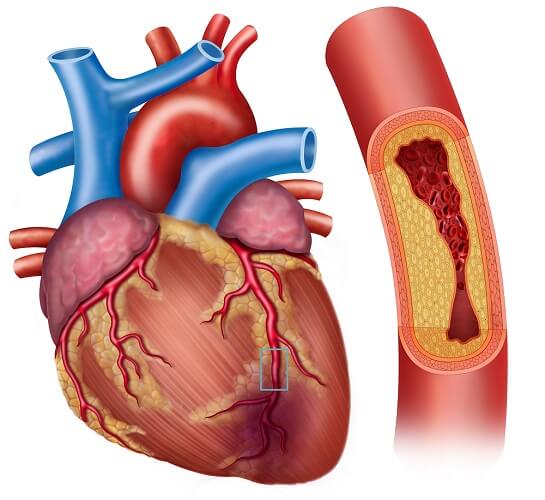
Systolic blood pressure gives the amount of pressure exerted by blood on the artery walls when the ventricles contract. This is always the higher number. Diastolic blood pressure also measures arterial wall pressure but this is the pressure exerted by the blood when the ventricles are relaxed.
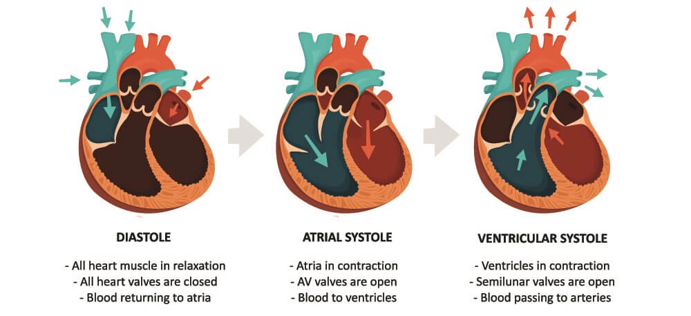
PP is the result of stroke volume and arterial compliance. Stroke volume is the volume of blood (in milliliters) pumped out of the ventricle with each heartbeat. Measurements are usually made at the left ventricle. The average stroke volume of a healthy young adult is about 80 ml.
Arterial compliance refers to how supple the main arteries are. When we are young and healthy our artery walls are elastic and can expand to accept blood pumped out of the heart. As we grow older, this elasticity is reduced.
Atherosclerosis – the buildup of fatty deposits on the inside of the artery wall – causes narrowing. This makes it harder for normal amounts of blood to travel through.

Another cardiaology term that helps us to understand PP is ejection fraction. This is a percentage value that shows the ratio of blood that leaves the ventricle every time it contracts compared to the amount of blood that remains inside.
A ventricle will never pump all of the blood it contains into the aorta or pulmonary artery. The amount it does pump out can be measured. Most cardiologists measure left ventricle ejection fraction (LV ejection fraction). A normal result is 55% or more. Results of between 50% and 55% are considered borderline. Reduced LV ejection fraction is any percentage under 50%.

The ventricles of a healthy younger person pump 55% or more of the blood they contain into the main arteries with every beat. This is approximately 80 ml. Healthy arteries close to the heart expand to accept this volume – they are compliant.
In an elderly person with heart disease and atherosclerosis the heart is usually enlarged. More blood pours into the ventricles from the atria because there is more space. However, the main artery walls are stiff and are spotted with fatty deposits that make them narrow. Even when the heart muscle is strong, these arteries cannot accept large volumes of blood at each stroke.
In this case, the ejection fraction will be lower. If the larger ventricle accepts 180 ml of blood but the arteries are only able to make space for 60 ml (stroke volume) of blood per beat, the percentage of blood left in the ventricle will be much higher than usual. The ejection fraction will be low – in the above example, just over 30%.
At the same time, low arterial compliance (low elasticity and atherosclerosis) increases blood pressure values. Even when the arteries relax they do not return to size as they have lost their elasticity. Diastolic pressure will be lower than usual.
A high blood pressure of 160/70 mm Hg gives a PP value of 90 mm Hg.
Low blood pressure can be caused by dehydration, sudden emotional or stressful situations, sudden changes in position, anemia, and blood vessels that remain dilated for longer periods.
Low blood pressure can also be the result of too-high doses of anti-hypertension medication. The root cause of high blood pressure is stiff and narrow arteries; it is uncommon to find low blood pressure in older individuals unless they are taking the wrong doses of anti-hypertensive drugs.
Low blood pressure is associated with narrow pulse pressure. In the elderly, blood pressure is lower due to widening of the arteries caused by medication. In addition, the arteries do not spring back as efficiently. An associated blood pressure of 90/70 mm Hg gives an abnormal PP of 20 mm Hg.
In healthy individuals with severe blood loss the arteries may function well but there is not enough blood to exert normal pressure on the artery walls. Pulse pressure in early hypovolemic shock is narrow. A blood pressure of 70/55 mm Hg would give a narrow PP reading of 15 mm Hg.

We can, therefore, say that abnormal PP reflects the difference between systolic and diastolic arterial pressure values due to problems with the artery walls, heart health and/or fluid levels.
Low and High Pulse Pressure
Low pulse pressure (narrow PP) is less than 25% of the systolic pressure. It is caused by multiple pathologies such as high levels of blood pressure-reducing medication, a rapid heart rate (tachycardia), and ascites (the build-up of fluid in the abdomen).
High pulse pressure (wide PP) is considered to be abnormal over 50 mm Hg. Normal pulse pressure range is between 30 and 50 mm Hg. Causes of wide pulse pressure are similarly broad; the most common of these are atherosclerosis, slow heart rate (bradycardia), low blood volume, and hyperthyroidism.
Although PP is not used to predict death, some studies find that high pulse pressure is common in elderly patients who die in hospital, no matter what their gender or whether they suffer from other chronic illnesses.

How to Calculate Pulse Pressure
Pulse pressure calculation – where the diastolic pressure is taken away from the systolic value – is easy. If your normal blood pressure is 120/80 mm Hg, your PP is 40 mm Hg – a normal result.
If the result is lower – narrow PP – these two values will be closer together. For example, 110/90 mm Hg gives a PP of 30 mm Hg. A wide pulse pressure – perhaps calculated with a blood pressure of 150/60 mm Hg would be 90 mm Hg.
Below is the pulse pressure formula:

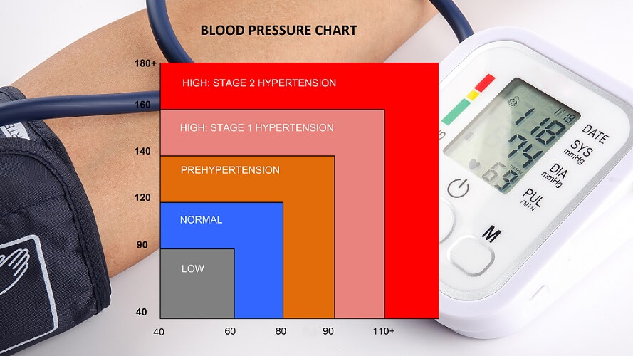
A pulse pressure chart is part of a medical blood pressure chart that shows whether a person might require treatment or follow-ups for hypertension.
Pulse Pressure Variation
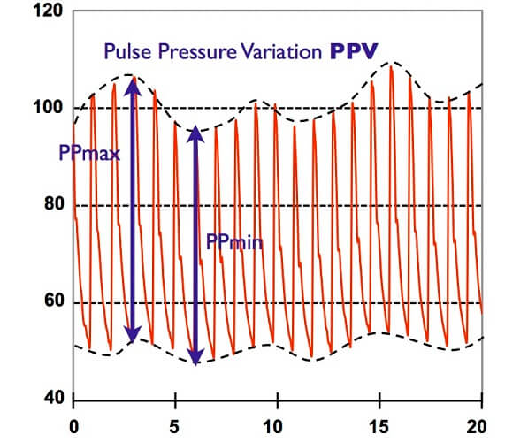
Pulse pressure variation is the slight change in arterial pressure that occurs when we breathe. It is most commonly measured during mechanical ventilation in the operating theater or intensive care unit.
PP variation is an important parameter for anesthesiologists and intensivists. Results specifically show changes in blood and pulse pressure when a patient is mechanically ventilated. This data helps the specialist to know how much fluid to administer via an intravenous line during surgery or assess risk of mortality in ventilated patients in intensive care.
