Definition
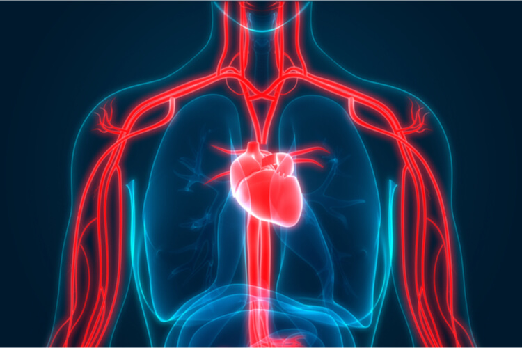
The pulmonary vein(s) are vessels carrying oxygen- rich blood from the lungs to the left atrium of the heart, which is critical for proper respiration. There are two major pulmonary veins for each lung, leading to a total of four major vessels. As these veins are critical for ensuring proper respiration, blockages and disorders at these vessels can quickly become serious and lead to broader conditions such as pulmonary hypertension and heart failure.
The Circulatory System
Before leading into the specific vessels of the cardiac and pulmonary systems, lets first look at the overall system and vessels found throughout the body.
All vertebrates (as well as a few invertebrate species) have a closed circulatory system, where blood flows throughout the body in a continuous loop by a series of vessels. The heart facilitates blood flow through the circulatory system by pumping to create a forceful pressure that maintains a viable flow rate. The major vessels involved in a closed circulatory system include arteries, capillaries, and veins.
Vessels
Arteries are thick- walled vessels that move blood away from the heart. They consist of mostly elastic fibers in addition to muscle fibers. The smooth muscle allows the arteries to contract, decreasing vessel diameter to slow down blood flow. However, the elastic fibers allow the arteries to return to a resting state so that diameter and blood flow can increase again. Very small arteries are called arterioles. These arterioles are needed to reduce blood pressure as blood prepares to pass through capillaries.
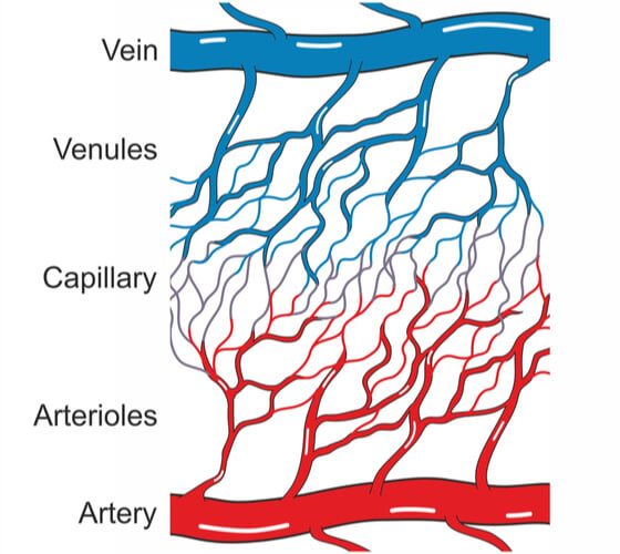
Capillaries are very thin vessels where gases and small molecules are exchanged from the blood to the tissues. These vessels are extremely thin, where the walls making up the vessels are only a single cell layer thick. Additionally, the capillary diameter is only wide enough to let red blood cells pass through one at a time. Large networks of capillaries are known as capillary beds. Once tissues use the oxygen and other molecules received, blood is brought back to the heart by flowing through venules and veins.
Veins are thin- walled vessels that move blood towards the heart. At this point, blood pressure is generally lower, allowing for veins to have larger diameters and thinner walls compared to arteries. Blood movement in veins is typically facilitated by skeletal muscle in the limbs. However, in large veins, valves are required to section the vessels into multiple segments, preventing blood from back-flowing. Very small veins are known as venules.
Sub-Circulatory Systems
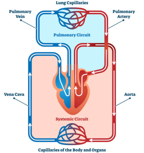
The circulatory system is broken down into two parts- the pulmonary circuit and the systemic circuit. The pulmonary circuit is the loop that moves blood from the heart and into the lungs (or gills for aquatic species), while the systemic circuit is the loop that moves blood from the heart and throughout the body. In fish species, gravity does not play a large role in maintaining blood flow, so blood pressure is consistently high throughout the body. However, in terrestrial vertebrates, gravity plays a larger role. Blood pressure must increase to overcome gravity while blood is traveling up towards elevated portions of the body. Therefore, separation of the two circulatory sub-systems allows for the overall system to remain balanced. Generally, the systemic circuit is high- pressured, while the pulmonary circuit in low- pressured. Specially, pulmonary blood pressure is only ~1/8th of systemic blood pressure.
The Heart
Chambers Within the Heart
The heart is broken down into at least two chambers in all vertebrates, where all species have at least one atrium and one ventricle. The atria (singular atrium) are responsible for collecting blood from the body, while the thick- walled ventricles are responsible for pushing the blood out of the heart. The number of atria and ventricles diversifies with species, where fish have one of each, while amphibians and many reptiles have two atria and one ventricle, and other reptiles, birds, and mammals (including humans) have two atria and two ventricles.

The pulmonary and systemic circuits are only partially separated in species lacking four fully developed heart chambers. Because of this, oxygen-rich and oxygen- poor blood can mix. In species with four fully developed chambers- such as humans- the two sub-circuits are completed separated. This prevents oxygen-rich and oxygen-poor blood from mixing, allowing for better cellular efficiency as tissues are able to receive more oxygen. The increased available oxygen is also needed to maintain higher body temperatures, which is why bird and mammal species are known as endotherms.
Blood Flow Through the Heart
The general movement of blood through the heart in species such as humans that have four fully developed chambers is as follows:
- Oxygen-poor blood enters the heart by flooding into the right atrium via the superior and inferior venae cavae (both of which are veins).
- The oxygen-poor blood then flows past the tricuspid valve into the right ventricle.
- The right ventricle pushes the oxygen-poor blood past the pulmonary valve and into the pulmonary arteries.
- The right and left pulmonary arteries lead into the right and left lungs, respectively, where the blood becomes oxygen-rich. This gas exchange occurs as the blood flows through the capillary beds of the alveoli within the lungs.
- The now oxygen-rich blood flows from the lungs back to the heart through the pulmonary veins, flooding into the left atrium. Each lung has two large pulmonary veins, where all four lead directly into the left atrium.
- The oxygen-rich blood flows past the mitral (or bicuspid) valve into the left ventricle.
- The left ventricle pushes the oxygen-rich blood past the aortic valve, into the aorta.
- The oxygen-rich blood then flows out of the heart and spreads throughout the arteries of the body.

Steps 1-3 consist of pulmonary circulation, while steps 5-8 consist of systemic circulation. Therefore, despite both vessels having pulmonary in their name, only the pulmonary artery is technically part of the pulmonary circulation. The pulmonary vein is actually part of the systemic circulation.
Location of the Pulmonary Veins
Multiple vessels are required to ensure blood flows efficiently through the pulmonary and systemic systems. Therefore, it is important to properly locate the pulmonary veins in relation to the rest of the system. All four of the pulmonary veins are located at the superior (topmost) portion of the heart. The right pulmonary veins stem directly off of the right lung’s root and run behind the right atrium and superior vena cava into the left atrium. Meanwhile, the left pulmonary veins stem directly off of the left lung’s root and run in front of the descending aorta, directly into the left atrium.
Diseases and Dysfunctions
Pulmonary Vein Stenosis
Pulmonary vein stenosis is a rare dysfunction where at least one- but usually multiple- of the four pulmonary veins become blocked. This blockage prevents the oxygen-rich blood from entering the left atrium of the heart from the lungs. This may occur when the walls of the veins thicken, causing the vessels themselves to narrow. Patients can undergo surgery to widen the veins. However, this is typically a reoccurring condition within a patient, so this solution is only short-term. If left untreated, this condition can lead to advanced pulmonary diseases, such as pulmonary hypertension and pulmonary arterial hypertension. Despite its rarity, clinical studies are ongoing to better understand this condition and applicable treatments.
Pulmonary Vein Thrombosis
Another rare dysfunction that can occur within the pulmonary veins is pulmonary vein thrombosis. Thrombosis- which is the formation of thrombi, or blood clots, within vessels- can decrease the available diameter of a blood vessel, thus restricting blood flow. When the thrombus is dislodged from its origin, it becomes a specific type of embolism (or intravascular mass) known as thromboembolism. The presence of these masses can lead to further pulmonary hypertension, blood clotting, and even sudden death.
Antecedent Heart Diseases
Additional antecedent heart diseases, such as mitral stenosis, can also increase blood pressure. As the mitral valve is on the left side of the heart separating the atrium and ventricle, pressure can buildup in the left atrium. The increase in pressure here can lead to increased pressure in the pulmonary veins pouring into the atrium. Similar to before, this can lead to pulmonary hypertension and pulmonary arterial hypertension. It is reported that the pathophysiology leading to mitral stenosis is analogous to pulmonary vein thrombosis.
Resulting Conditions
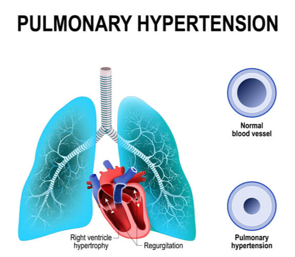
As mentioned in the previous two disorders, pulmonary hypertension is a broad condition defined by increased pulmonary pressure. Specifically, pulmonary pressure increases to 1/4th or more of systemic pressure. (Reminder: pulmonary pressure should only be ~1/8th of systemic pressure). This condition consists of several subgroups that are categorized based on the dysfunction cause. Typically, however, pulmonary hypertension results when surrounding arteries narrow (or if vascular blood flow increases). In order to push blood through these narrowed vessels, the heart must pump harder. Over time, this causes the heart to weaken, and the chances of developing heart failure increases. There can be multiple causes of pulmonary hypertension, with pulmonary vein stenosis and thrombosis being just a few of them. Pulmonary arterial hypertension is a specific type of pulmonary hypertension, but its root cause is at the small arteries of the lung specifically.
Conclusion
Pulmonary veins are critical vessels in the pulmonary system need to ensure proper respiration is dispersed throughout the body. These veins carry freshly oxygenated blood from the lungs to the heart via the left atrium, where oxygen-rich blood can spread to all subsequent tissues. The evolution of multi-chambered hearts in select species have allowed for the complete separation between the pulmonary and systemic circuits, preventing the mixing of oxygen-rich and oxygen-poor blood. Though disorders found directly in the pulmonary veins tend to be rare, they can contribute to larger conditions and diseases that can quickly become fatal. Research is ongoing to better understand some of these diseases and improve available treatments.
Quiz