Definition
The PR interval is found on electrocardiogram readings that measure the electrical activity of the heart. It is the first part of an electrocardiogram and indicates how well electrical signals pass from the atria to the ventricles. A depressed, short, or prolonged PR interval is associated with specific heart-related medical conditions.
What is the PR Interval?
PR intervals represent the first part of a heart beat and are measured in seconds or milliseconds. Each interval shows cardiologists how well the electrical impulses generated in heart pacemaker cells pass through the upper chambers of the heart (atria) and arrive at the lower chambers (ventricles). Any abnormal result will tell medical professionals that a problem exists within the atria.
A Short Introduction to the Electrocardiogram
Certain distinguishable waves and points show cardiologists how well the heart receives and sends electrical signals that control heart muscle movement.
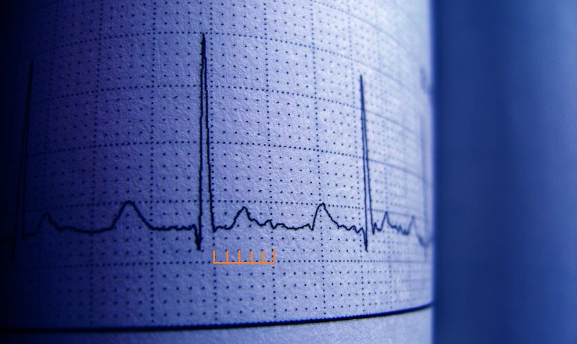
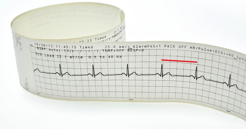
With electrodes placed on the skin to either side of the heart, impulses are transferred to a graph or screen. The typical shape of the electrocardiogram (ECG) is recognized all over the world.
Heart pacemaker cells produce their own electrical impulses to control heart muscle contraction. If the brain is damaged, the heart can continue to pump. The autonomic nervous system controls how often (heart rate) these impulses cause a reaction and regulates how strongly the heart contracts; it does not cause heart muscle cell contraction but regulates it.
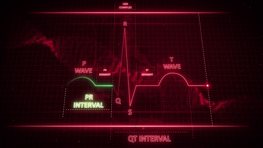
A normal ECG is composed of lettered components – P, Q, R, S, and T.
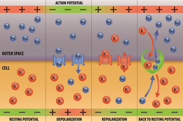
The P-wave (sometimes P-top) represents electrical depolarization in the atria. To find out more about how this electrical activity occurs have a look at the action potential article.
In short, depolarization means that an electrical signal is moving through a nerve or heart muscle cell membrane. In the heart, this causes the corresponding part of the muscle to contract.
The P-wave begins at the sinoatrial node of the right atrium where it passes into the right and left atria. The first part of the P-wave shows depolarization in the right atrium as this is closer; the latter part corresponds to the left atrium.
The Q-wave is the starting point of the QRS complex. This indicates the depolarization of cardiac muscle cells from the center of the heart and into the ventricles. As the ventricles are the main source of pumping power, this is the largest spike on an ECG.
- Q wave: depolarization of the interventricular septum
- R wave: early ventricular depolarization
- S wave: a downward deflection as this depolarization moves from the bottom of the ventricles upward, via the Purkinje fibers
Finally, the T wave is an upward deflection that occurs as the ventricles relax (repolarize).
As the space of time between each of these letters is important, an ECG is also split into intervals:
- PR interval: measuring the distance between the start of the P-wave and the start of the QRS complex
- QT interval: the distance between the start of the QRS complex and the end of the T wave
- ST interval: the distance between the end of the QRS complex and the end of the T wave
- TP interval: the distance between the end of the T-wave and the start of the next P wave
- RR interval: the distance between the peaks of two R waves that measure heart rate
A Short Introduction to the Heart Conduction Pathway
Heart muscle cells are unusual in that they do not need to receive signals from the central nervous system to contract. Instead, pacemaker cells continuously generate impulses inside the heart.
How quickly impulses are generated and how hard the heart contracts in response to them is controlled by the central nervous system; however, heart muscle contractions occur in direct response to pacemaker cells within the heart.
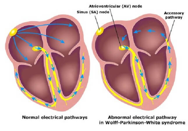
In a healthy heart, the sinoatrial node cells are the starting point for electrical impulse initiation. These action potentials begin at the top of the right atrium and spread through the right and left atria. This makes the upper chambers of the heart contract. The very slight delay as these impulses pass from the SA node means that the left atrium contracts a tiny bit later than the right atrium.
After passing through the atria these electrical signals arrive at the atrioventricular node that sits between the bottom of the right atrium and the intraventricular septum. At this point, there is a very short pause that gives time for all signals to arrive. Enough electrical energy must be present to make the larger ventricles contract.
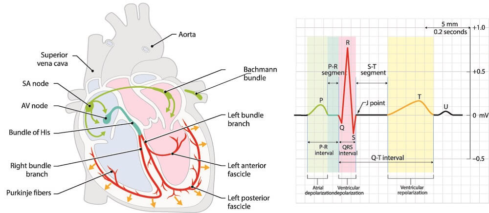
When enough signals arrive at the AV node, the signal continues into the bundle of His and travels down the intraventricular septum. From the bottom of the ventricles, Purkinje fibers travel upward to stimulate the entire ventricle to contract.
PR Interval Calculation
A PR interval calculation measures atrial depolarization time. It extends from the initial creation of electrical impulses at the sinoatrial node (SA node) of the right atrium and ends when these arrive at the ventricles. A slight delay at the atrioventricular node (AV node) is also responsible for the duration of a normal PR interval.
You can easily learn how to measure the PR interval when an ECG reading is printed onto graph paper. Every small square on an ECG represents 0.004 seconds. By multiplying 0.004 by the number of squares covered by the space between the start of the P wave and the start of the QRS complex, you can calculate the PR interval.
Doctors compare ECG results with normal ranges for the PR interval, QRS duration, QT interval, and RR interval.
Normal PR Interval
A normal PR interval is 0.12 to 0.2 seconds. This corresponds with four to five ECG printout squres. Any result of or between 120 to 200 milliseconds is considered normal. This shows that electrical activity is running smoothly from the sinoatrial node to the ventricles. This normal result does not necessarily mean that the atria are functioning perfectly, just that electrical signals are passing through as they should.
In tachycardia or bradycardia a PR interval can be normal; however, there may be an underlying problem with action potential generation. Even atrial arrhythmias can show normal PR intervals.
The result only tells us whether the electrical activity is moving through the upper chambers of the heart as it should. Other tests are required to indicate why an abnormal PR interval occurs.
Depressed PR Interval
A depressed interval, also called a depressed PR segment, is a downward-facing curve at this part of an abnormal ECG. As we already know, positive electrical activity during depolarization that moves from one electrode to the next gives a positive (upward) spike or curve. If this curve is upside-down it either means that the electrical signals are traveling backward or that there is insufficient electrical activity.
It is actually wrong to call this symptom a depressed PR interval as it is the space between the end of the P wave and the start of the QRS complex that shows anomalies. This part of the ECG is called the PR segment.
A depressed PR segment is found in the early stages of acute pericarditis where the membranes covering the heart become infected. Another cause is atrial damage – usually caused by coronary artery disease.
Short PR Interval

A short PR interval under 0.12 seconds or four squares can have more than one cause. The most common of these are:
- Junctional rhythm, where the AV node generates extra action potentials
- Enlarged heart
- Anxiety
- Stimulants
- Heart valve disorders
- Pre-exitation syndromes, where the ventricles respond too quickly to additional atrial signal routes
Even so, a short PR interval can be normal. There is usually no need for concern when the QRS complex is normal. Young athletes can have short PR intervals without any heart-related disorder.

Can anxiety cause a short PR interval? The answer is yes. Anxiety, stress, and even caffeine are known to cause shorter PR durations. Recent studies have also found a link between COVID-19 infection and PR interval shortening.
Short PR interval symptoms are often absent. Pre-exitation syndromes such as Wolff-Parkinson-White (where other action potentials bypass the AV node) can present with palpitations, dizziness, shortness of breath, anxiety, and fatigue. These symptoms are due to the heart’s inability to efficiently pump oxygenated blood to the body.
When the reason for the short interval duration is atrial electrical conduction and symptoms are present, short PR interval treatment includes cardioversion and radiofrequency catheter ablation. The former resets the heart rhythm, the second burns away bypass routes that generate abnormal electrical activity.
Medications causing short PR durations are limited. Drugs are more likely to cause the opposite effect – a prolonged PR interval.
Prolonged PR Interval
A longer than normal PR duration means that action potentials are not being efficiently conducted through the atria.
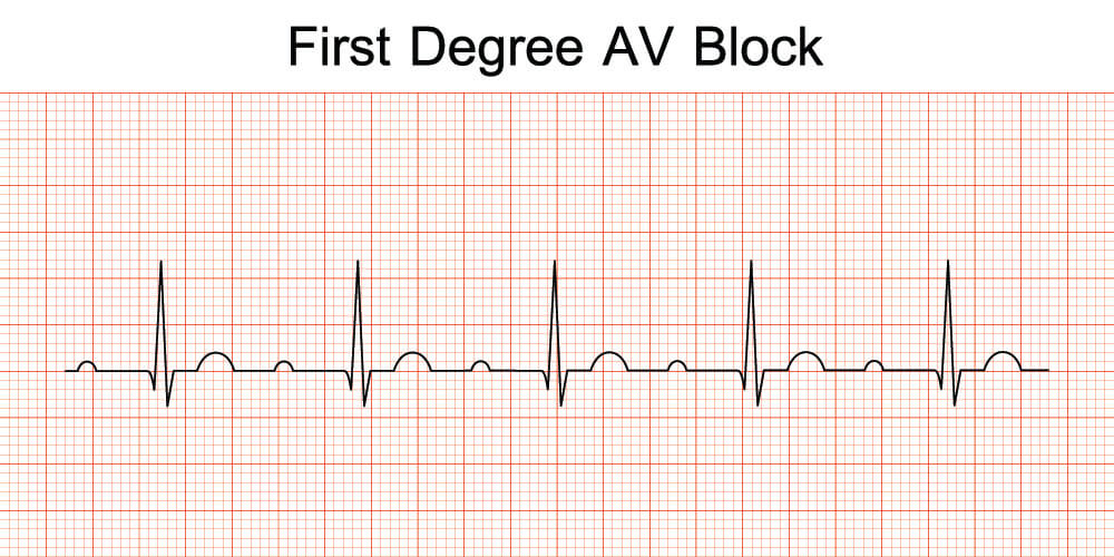
A long PR interval is most commonly caused by delayed conduction at the SA node. This is known as a first-degree AV block and usually presents with a PR interval of 200 to 220 ms. Intervals of over 300 ms are rare and anyone presenting with this duration will require a permanent pacemaker.
The older we become, the higher the risk of a prolonged PR interval. Over time, AV blocks can increase the risk for atrial fibrillation and heart-related death. A long PR interval means that certain medications may need to be reassessed – many drugs can lengthen the time it takes to send electrical impulses across the atria. One of these is digoxin or digitalis.

Another prolonged PR interval cause is untreated hyperthyroidism. When levels of thyroid hormone are too high the heart beats rapidly, pulse pressure increases, and both the PR interval and QRS duration lengthen. Someone with hypothyroidism who takes overly-high doses of thyroid hormone to compensate may present with these symptoms, too.