Definition
The palatine bone or os palatinum is a paired, flat, irregular facial bone. It forms part of the nasal cavity, oral cavity, and orbit of the eye. Composed of two plates, each bone sits between processes of the right or left maxilla bone and the single sphenoid bone. Paired palatine bones feature openings (foramina) that lead to the greater and lesser palatine canals. These canals provide space for blood vessels and nerves.
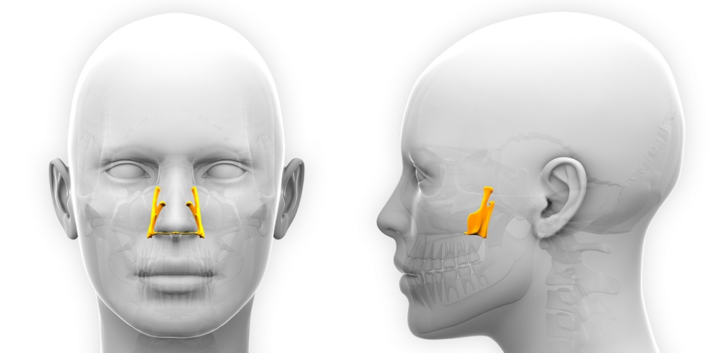
Palatine Bone Location
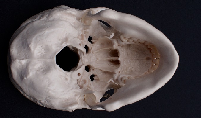
Part of the palatine bone location can be felt by running the tongue toward the back of the throat, stopping just before hard and soft tissue meet – this is the palatine surface of the horizontal plate (see palatine bone anatomy) that forms the back section of the hard palate (palate – palatine).
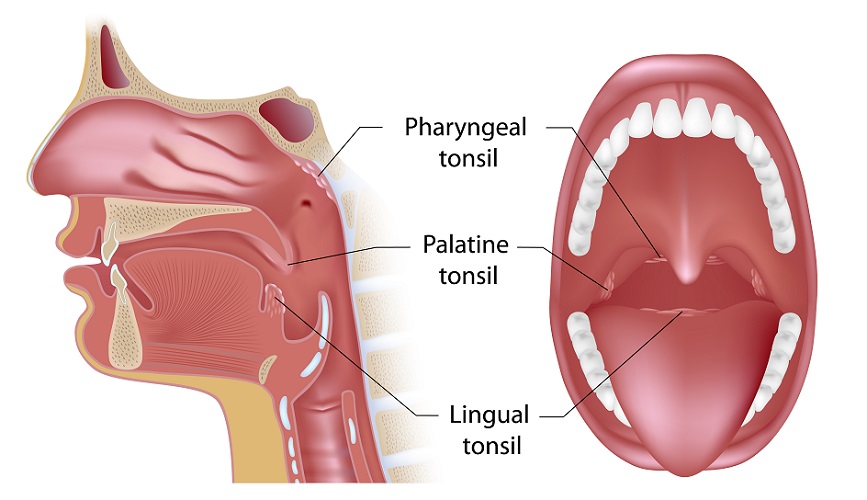
The tops of the palatine tonsils – lymphoid tissue masses on either side of the back of the throat – also indicate the location of the palatine bone base.
Each (facial) palatine bone forms an L-shape. Each bone is a mirror image of the other so that the horizontal lines (the horizontal plates) of the two L’s meet. The front section of each horizontal plate articulates with part of the maxilla. The back of the horizontal plates (posterior border) meets the cartilage of the pharynx.
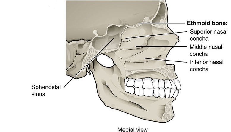
The perpendicular plate of the palatine bone is the vertical line of the L. This plate is located behind the nasal conchae. Both plates run up from the horizontal plate, articulating with the inferior nasal concha and sphenoid bone. You can see the positions of the three nasal conchae below.
Palatine Bone Anatomy
Palatine bone anatomy is usually split into two subcategories – the perpendicular plate and the horizontal plate; remember that these are paired bones – left and right.
The below image shows labeled palatine bones positioned as mirror images of each other. Imaging the soft tissue of the nose between them. The labels distinguish various anatomical structures; these are listed after the picture.
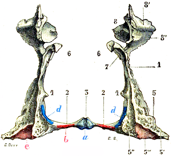
- 1 – General depiction of the perpendicular plate of the palatine bone
- 2 – General depiction of the horizontal plate of the palatine bone
- 3 – Articulation point of both palatine bones to form the nasal crest
- 4 – Conchal crest that meets the inferior nasal concha
- 5 – Various parts of the pyramidal process
- 6 & 7 – Sphenoidal process
- 8 – Various parts of the orbital process
- a & b – insertions of muscles that raise the uvula
- d – insertion for the superior pharyngeal constrictor muscle
- e – insertion for the medial pterygoid muscle
Perpendicular Plate of the Palatine Bone
The perpendicular plate – the vertical plate – has two functional surfaces. These are the nasal and maxillary surfaces.
The maxillary surface of the os palatinum meets the nasal surface of the maxilla bone (shaded pink at the upper nose in the image), as well as the pterygoidian process of the sphenoid bone (not pictured).
The irregular shape of the meeting of these bones means that space is made for the pterygopalatine fossa. The pterygopalatine fossa is a gap that provides space for important nerve tissue such as the maxillary nerve and pterygopalatine ganglion. These innervate various regions of the face, from the skin of the temples to the upper lip and teeth. The pterygopalatine fossa also provides space for the maxillary artery and various veins. Pterygo means wing-shaped.

At the top of the perpendicular plate is the orbital process. This part of the os palatinum forms part of the eye socket. A short distance under the orbital process is the sphenoidal process. This process not only forms part of the bony nasal cavity but also articulates with the pterygoid plates of the sphenoid bone; it surrounds a hole called the sphenopalatine foramen that connects the nasal cavity with the pterygopalatine fossa.
Other parts of the maxillary surface of the os palatinum form part of the maxillary sinus wall.
The perpendicular plates also features small holes or foramina. The greater palatine foramen opens into a groove that becomes a channel (canal) when the maxilla sits on top of it. Both greater palatine foramina (as we have two palatine bones) are found at the back of the hard palate. These open into the greater palatine canal. This canal protects the greater palatine nerve and its blood supply. The lesser palatine canals house the lesser palatine nerves. The lesser and greater nerves innervate the hard and soft palate, tonsils, and nasal cavity.
One set of greater and lesser palatine foramina are circled in pink (not red) below.
The nasal surface of the perpendicular plate – as its name suggests – faces the nasal cavity. This section of the perpendicular plate of the palatine bone provides structure at the back of the nose.
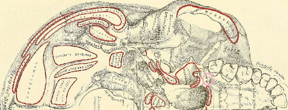
Between the meeting of horizontal and perpendicular plates is the pyramidal process of the palatine bone. It fits between two narrow plates in the sphenoid bone called the pterygoid plates to form the pterygoid (not the pterygopalatine) fossa. The pterygoid fossa provides space for masticatory muscles – muscles for chewing.
In the photograph, you can see the bases of the horizontal plates of the palatine bones and follow them across the pyramidal processes and down along the inside of each pterygoid fossa.
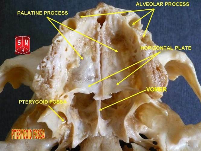
Horizontal Plate of the Palatine Bone
The horizontal plate can be felt in the back of the hard palate of the oral cavity. It also forms part of the bottom of the nasal cavity. This plate – just like the perpendicular plate – has two surfaces. However, these are the nasal and palatine (palate) surfaces.
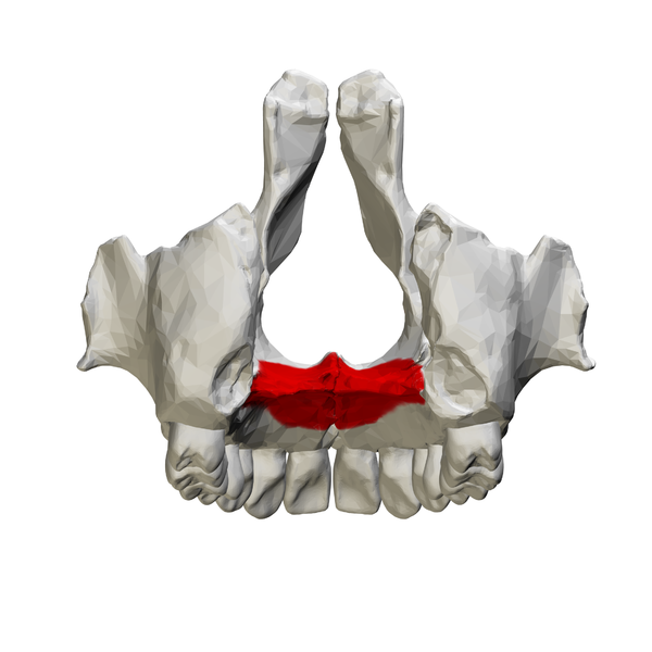
The meeting of each horizontal plate of the palatine bone to its mirror image rises is a little wider than the rest. The wider part forms the nasal crest and nasal spine.
The horizontal plate ends where it meets the pyramidal process of the perpendicular plate of the palatine bone – the pyramidal process is part of the perpendicular, not the horizontal, plate.
Palatine Bone Function
Palatine bone function is threefold. Firstly, each bone provides partial surfaces of the hard palate of the roof of the mouth, the nasal cavity, and the orbit bones.
The hard palate is formed by the palatine process of the maxilla and the horizontal plates of the palatine bones. It is the roof of the oral cavity and the floor of the nasal cavity.
The orbit bones are a group of seven facial bones (palatine included). The floor (bottom) of the orbit is composed of sections of the zygomatic, maxillary, and palatine bones. Look in the the eye sockets below – the many faint cracks indicate where these separate bones articulate.
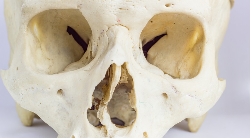
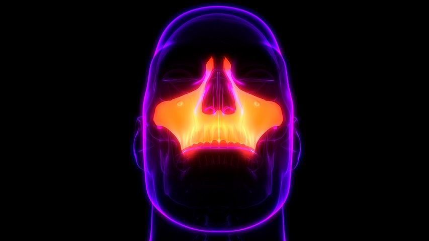
The nasal cavity warms and humidifies the air we breathe and traps potential pathogens and pollutants. Although we only have one nose, the cartilaginous septum (the rather collapsed structure in the above photograph) divides the nasal passage into two nasal cavities. The palatine process of the maxilla bone and the horizontal plate of the palatine bone provide the components of the nasal cavity floor.
The os palatinum is also an attachment point for facial muscles. Four muscles attach to the palatine bone:
- Medial pterygoid muscle (to the pyramidal process)
- Superior pharyngeal constrictor muscle (to the horizontal plate)
- Tensor palati (to the horizontal plate)
- Uvula muscle (to the horizontal plate)
Finally, the facial palatine bone also has three foramina that allow nerves and blood vessels to pass through. These nerves provide sensory and motor pathways to the hard and soft palate and nasal cavity.
Each bone has a set of three foramina:
- Greater palatine foramen: gateway for the greater palatine nerve and blood vessels between the pterygopalatine fossa and oral cavity
- Lesser palatine foramen: gateway for the lesser palatine nerve and blood vessels between the lesser palatine canal and the oral cavity
- Sphenopalatine foramen: gateway for the sphenopalatine artery and vein, nasopalatine nerve, and nasal nerves between the pterygopalatine fossa and nasal cavity
Palatine Process vs Palatine Bone
There is an easy answer to palatine process vs palatine bone – there are no palatine processes on palatine bones. Palatine processes are structures of the neighboring maxilla bones; these articulate with the palatine bones.
The palatine bone has three processes. When referred to, these processes should always include the name of the bone from which they originate (in this case the palatine bone) to avoid confusion. The three processes are:
- Orbital process of the palatine bone (forms part of the orbit bone)
- Pyramidal process of the palatine bone (forms part of the pterygoid fossa)
- Sphenoid process of the palatine bone (forms part of the pterygopalatine fossa)
When you see the word ‘process’ in a facial bone anatomy text, you know that this refers to an area or projection, and that this projection meets the bone after which it is named.
The addition of ‘of the’ – for example, the orbital process of the palatine bone – tells us that this process is part of the palatine bone and projects to meet the orbital bone. The frontal bone also has two orbital processes, so mentioning the bone of origin is important.
The palatine process of the maxilla bone, therefore, tells us that there is a projection from the maxilla that plays a part in the structure or function of the os palatinum.
Quiz