Definition
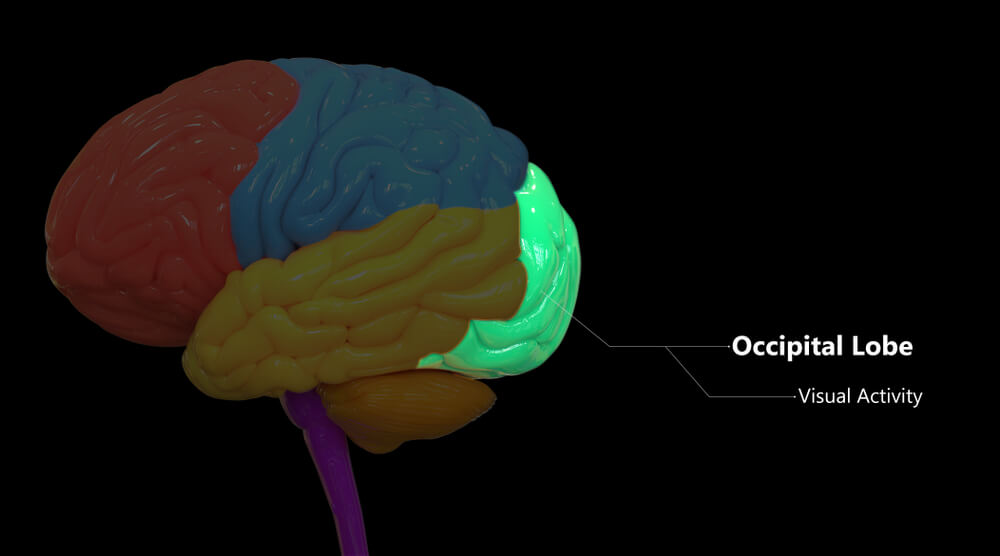
The occipital lobe, located at the back of the brain, is the smallest of the four lobes and enables visual processing and visual memory. Sitting behind both the temporal and parietal lobes, the occipital lobe is home to the primary and secondary visual cortices and is connected to the retinas of the eyes. Found in all vertebrates, this part of the brain is – evolutionarily speaking – the most recent.
Occipital Lobe Location
The occipital lobe is located at the back of the head and sits behind the parietal and temporal lobes, just above the cerebellum. Although furthest from the eyes, this part of the cerebral cortex is essential for vision. As with all of the lobes of the brain, the occipital lobe is divided between the two hemispheres. The right and left lobe communicate continuously so that we not only see flat images but full, three-dimensional scenes.
Primates have a well-developed occipital lobe but elephants do not; instead, they have disproportionately large temporal lobes (although their nerve tissue is nowhere near as dense as ours). Elephants are well known for their ability to communicate and depend on their sense of smell and hearing – all temporal functions. But their eyesight is very poor – an elephant can only see for distances of around twenty meters. Humans and primates have the best visual acuity (clarity of vision) of all mammals.
Occipital Lobe Function
Occipital lobe function involves receiving visual stimuli from the retinas of the eyes, processing this information, associating what is seen with our visual memory, and forwarding this processed data to other regions of the brain. While many of us think that images from one eye are sent to the opposite hemisphere of the brain, this is not correct. First of all, our vision is split into right and left hemifields – imagine cutting both eyeballs vertically in half. All the visual information from the left-hand side of both retinas is processed separately from the visual information processed by the right sides of both retinas.
The right hemisphere of the brain works with what is in our left-hand field of vision. For example, a tree to the left of your line of sight is captured by the right sides of both retinas. You can see that the eyeballs are split into green and red in the image below and that the path leading from the right sides of the retinas end at the right primary visual cortex, but take a look at the visual field – red is on the left and green on the right. They have swapped over.

The occipital lobe consists of different numbered regions – V1 up to and including V5 – that are shared between the primary and secondary visual cortices.
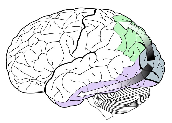
The primary visual cortex (V1) receives input from the retina via the optic nerve and thalamus. The secondary visual cortex consists of regions V2 to V5. Information is passed from the primary visual cortex to the secondary visual cortex via dorsal or ventral pathways. The dorsal pathway brings information about the location of a visualized object to the parietal lobe of the brain. The ventral pathway brings information to areas of the brain that deal with recognition and memory. The function of both cortices (primary and secondary visual cortex) is described in more detail below.
Primary Visual Cortex Function
The primary visual cortex is the largest area of the occipital lobe with a striped appearance that lends it another name – the striate cortex. Once visual information has arrived at V1, it is categorized and forwarded to the secondary visual cortex. The secondary visual cortices then send specific messages either to the parietal or temporal lobes by dorsal and ventral visual pathways.
If we break this down without digging too deep, images captured on the retina are sent via the optic nerve to the primary visual cortex. The primary visual cortex then figures out which information should be sent via the ventral visual pathway (that eventually ends up at the temporal lobe) and which information should be sent via the dorsal pathway to the parietal lobe. It can be compared to a mail sorting office. The ventral and dorsal pathways are also called the temporal (ventral) and parietal (dorsal) streams. You can remember which stream is which by thinking of the parietal lobe as a continuation of the line of the spine – a dorsal position.
Secondary Visual Cortex Function
The function of the secondary visual cortex is to further process the categorized visual information supplied by V1. The secondary visual cortex is also called the visual association area which perfectly describes its purpose – it gives meaning to the data provided by V1.
The dorsal visual pathway or dorsal stream brings input from the primary visual cortex (V1) to areas V5 and V3A. These areas process motion and shape information respectively. They also interact with the ventral visual pathway (just to make things more complicated), but mainly forward messages to the visual regions of the parietal lobe. This tells us that the dorsal visual pathway will end with some type of motor activity, like picking up an item. We know that the parietal lobe processes visual, navigational (proprioception), eye movement, and sense of touch information. This means that some scientists refer to the dorsal stream as the Where pathway.
The ventral visual pathway (the What pathway) is associated with memory and recognition. V1 data is forwarded to V2. V2 categorizes and passes this information on to areas V3 and V4 or directly to the parietal lobe. It is thought that V3 cells react to object orientation although very little is known about them. Visual area V4 might define how much attention is paid to an object and also help to form shapes and increase the contrast between different colors. From here, information passes into the part of the temporal lobe that produces complex images in combination with memory. This is particularly important in facial recognition. Prosopagnosia is the inability to recognize faces even when the eyes function well; this visual memory disorder is associated with temporal or occipital lobe damage.

Human Visual Development
Our cortical vision (vision perception in the brain) develops over time. Babies do not see well at all. Visual acuity (clarity) and contrast, color, shape, and motion sensitivity take months to reach optimal levels. Only binocular fusion (combining the information from two eyes into a single image), stereopsis (depth perception), and stereo acuity (clarity of a single image supplied by both retinas) develop relatively quickly, but at different times – between three months and three years of age. This means that early treatment can correct a cortical visual disorder before the visual cortices take on their mature form.
Interestingly, our cortical vision is not as influenced by our genes as it is by our environment. A rather cruel experiment that sewed one eye of newborn kittens shut produced permanent vision impairment throughout life, even in the presence of healthy eye and brain tissue. And, if raised only in the presence of horizontal lines, a kitten that becomes a cat will always be blind to vertical lines.

However, as with everything to do with the brain, things are not always so clear-cut. Brain plasticity or the ability of the brain to recover and reshape after damage has meant that some cortically blind people with early V1 lesions have experienced a sudden recovery of vision later in life. Another interesting point is that a one-sided occipital lobe lesion that develops when a fetus is in the mother’s womb does not necessarily result in one-sided cortical blindness after the baby is born. This is because the young tissue of the occipital lobe of the opposite hemisphere can learn to perform for both sides of the brain – embryos are notoriously adaptable. Another clue as to how complex our cortical vision pathways are is the unusual phenomenon known as blindsight, where a blind person can respond to visual stimuli even when cortically blind in both eyes; this tells us that other central nervous system structures are also at play. We still have a lot to learn.
Occipital Lobe Damage
Occipital lobe damage is usually the result of stroke, trauma, or brain tumors. You may have heard of occipital neuralgia, but this is caused by nerve injury close to the nape of the neck and is nothing to do with the occipital lobe – occipital simply means the back of the head.
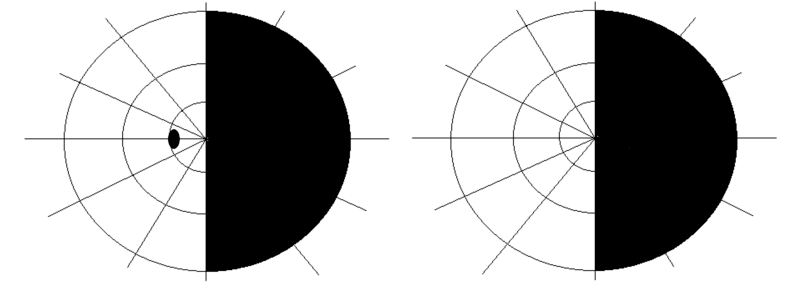
Early damage to the occipital lobe can lead to permanent cortical blindness even when other visual structures are unaffected. Unilateral (one-sided) occipital lobe lesions might cause visual field loss to the right or left vertical midline of an image (hemianopsia); a left-sided lesion will cause right-sided (contralateral) hemianopsia and a right-sided lesion produces left-sided hemianopsia. It is common to lose the same half of the visual field in both eyes – this is called homonymous hemianopsia.
Occipital lobe epilepsy (OLE) accounts for up to 10% of epilepsy forms; it is divided into idiopathic (unknown origin) and symptomatic (in the presence of a lesion) OLE. Usually beginning in early childhood, this type of epilepsy is often confused with ocular migraines (seeing spiked, shimmering rainbows) as both disorders produce the same type of hallucinations. Palinopsia (the recurrent visualization of an object that is no longer in the visual field), the presence of flashing lights, and temporary cortical blindness are also possible in OLE.
Anton syndrome is sometimes experienced by people who suffer from occipital lobe epilepsy. The affected person is unaware of a loss of vision and will believe hallucinated events and experiences are actual memories.
Riddoch phenomenon or statokinetic dissociation, also due to lesions in the occipital lobe, means a person can only see moving objects in the blind field; static objects are invisible. One patient with Riddoch syndrome described being able to see rain running down a window without being able to see through the window; the same person could see her daughter’s ponytail swinging but couldn’t see her daughter. The opposite of statokinetic dissociation is akinetopsia or motion blindness where static objects are visible and moving objects are invisible.
Color agnosia and cerebral achromatopsia are separate conditions that affect color processing and one’s ability to recognize color and perceive color respectively. This is also the result of occipital lobe damage.