Definition
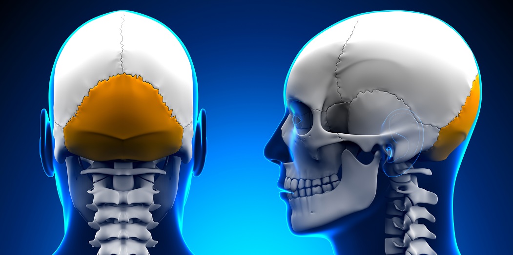
The occipital bone is a trapezoid-shaped single bone of the skull located at the back of the head. It forms immobile and mobile joints, contains grooves that protect certain cranial nerves and blood vessels, and hosts a number of raised or flat surfaces onto which muscles and ligaments can attach. The occipital bone is part of the calvaria or skull roof; it surrounds the foramen magnum through which the medulla of the brainstem, important arteries and nerves, and a protective membrane pass.
Where is the Occipital Bone Located?
Occipital bone location is the back of the head; the Latin term occiput specifically refers to this region. Also known as C0 because it forms one side of the atlanto-occipital joint between the cranium and first cervical vertebra (C1), this bone runs from the base to approximately halfway up the skull. The top (superior) part of the occipital bone is part of the calvaria or skull roof. The calvaria is composed of the superior parts of the occipital bone, frontal bone, and both parietal bones.
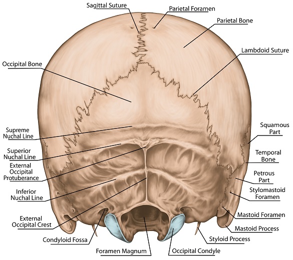
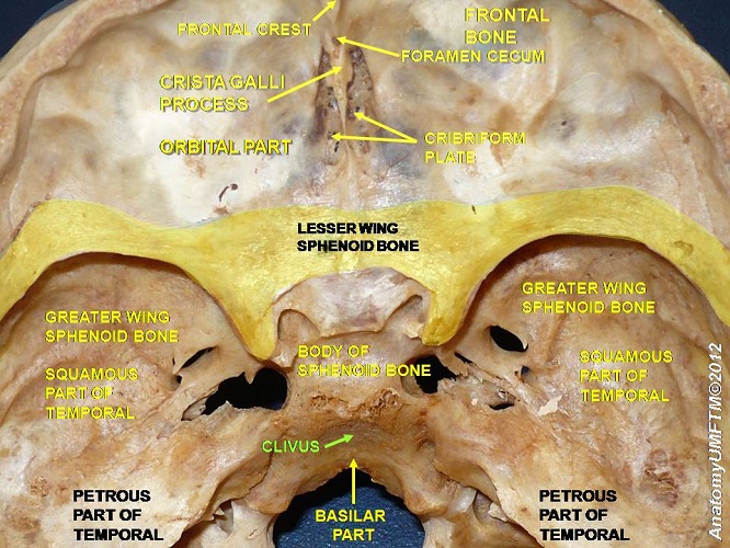
The occipital bone has borders with both parietal bones. Where they meet is known as the lambdoid suture. The ledges of both temporal bones are also connected to the occipital bone via the occipitomastoid sutures. The other connected skull bone is the sphenoid bone. This almost wasp-shaped piece of bone is extremely complex.
If you need to describe where the occipital bone is located in as few words as possible, it extends from the bottom of the skull to approximately halfway up the back of the head, forming joints with the parietal bones, temporal bones, and sphenoid bone, as well as with the first cervical vertebra.
Occipital Bone Anatomy
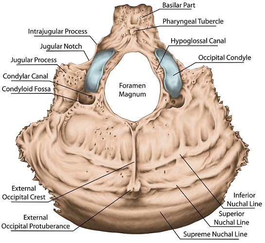
The occipital bone is trapezoid in shape. It surrounds the foramen magnum, a near-circular gap through which the medulla of the brainstem, nerves, arteries, ligaments, and tectorial membranes pass.
The occipital bone is composed of four parts: squamous, basilar, and two lateral parts. In the image the lateral parts are not labeled. They are composed of the areas around the occipital condyles. The squamous part is the largest area of this bone.
Squamous Part
The squamous part is a curved plate of bone that lies just behind the gap of the foramen magnum. The term squamous means flat, with flat bones primarily serving to protect internal organs and provide a surface for muscle and ligament attachments. The bone shape in this case is not flat but curved.
The squamous region of C0 has two surfaces – external and internal. The external (outside) surface has various ridges and protuberances that provide attachment points for the muscles of the cranium, neck, back, and shoulders, such as the superior, inferior, and median nuchal lines pictured above. Another of these external attachment points is the inion, a piece of bone that you can feel as an occipital bone bump in the middle of the back of your skull, just above the back of your neck. It is the origin of the nuchal ligament that holds up the neck. Giraffes have a particularly strong nuchal ligament for obvious reasons that inserts into the cervical vertebrae. It is such a thick ligament that it forms a visible hump at the bottom of the neck.

The internal surface of the squamous part features four fossa or hollows; the upper pair of hollows provides space for the occipital lobes of the brain and the lower pair for the two hemispheres of the cerebellum. A groove (sulcus) called the sagittal sulcus provides an open area where the top of the sagittal sinus can drain blood and cerebrospinal fluid into the circulatory system. Another indentation, the internal occipital crest is an attachment point for a folded membrane that separates the cerebellum from the cerebrum – the falx cerebri.
Basilar Part
The much smaller basilar section of the occipital bone or the basi-occiput lies in front of the foramen magnum. The surface of this wedge-shaped bone allows attachments for parts of the pharynx, neck muscles, and the anterior atlanto-occipital membrane, a thin sheet of tissue that connects the front of the first cervical vertebra to the front of the foramen magnum. The basilar part also has a sloped groove called the clivus of the occipital bone. This groove provides space for the medulla oblongata of the brainstem and a group of small veins that drain blood from the membranes that surround the soft tissue of the brain. You can see the wide indentation of the clivus in the photograph.
Lateral Parts
The pair of small lateral (side) sections are located on either side of the skull. The under surface contains two oval protuberances to the side of the foramen magnum that slot into the first cervical vertebra. Thanks to these protuberances – the occipital condyles – you can nod your head. In front of the occipital condyles lie two grooves called hypoglossal canals; these create space for the hypoglossal nerve (cranial nerve XII) to exit the cranium. Behind the condyles more indentations allow for venous drainage. The last section is the jugular tubercle. When you see the word tubercle, you know this means a sticking-out or thicker piece of bone with a rounded shape. This particular tubercle helps to protect the hypoglossal nerve by creating a wall to cover the hypoglossal canals.
Occipital Bone Function
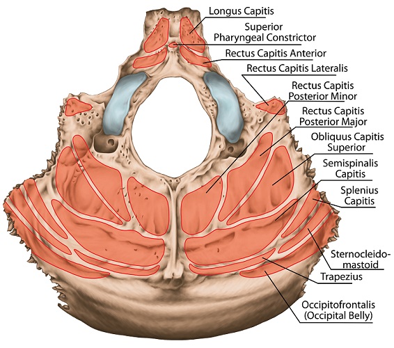
Occipital bone function is threefold. Firstly, it protects the delicate soft tissues of the cerebrum, cerebellum, medulla, and the nerves and blood vessels associated with them. The second function of the occipital bone is as one side of a two-bone joint. These mobile and immobile joints are described in more detail below. The final occipital bone function is its role as a muscle and ligament origin and insertion point. You can see the different muscles associated with the back of the skull in the image.
Occipital Bone Joints
The occipital bone forms joints with other bones either singularly or as a joint pair on either side of the skull.
The unpaired joints, in order of descending range of movement, are the atlanto-occipital joint between C0 (occipital bone) and C1 (atlas), and the spheno-occipital synchondrosis . Syn- is a Greek prefix that means together or united; the suffix –osis describes an abnormal state; chondr means cartilage. United cartilage abnormality. The abnormality in this case is the fusion of this cartilage to form a near immobile joint by the twentieth year of age (in humans). Because of this, forensic scientists and archeologists often use the spheno-occipital synchondrosis to determine the age of discovered remains. Boys tend to have a much slower joint-fusion rate than girls.
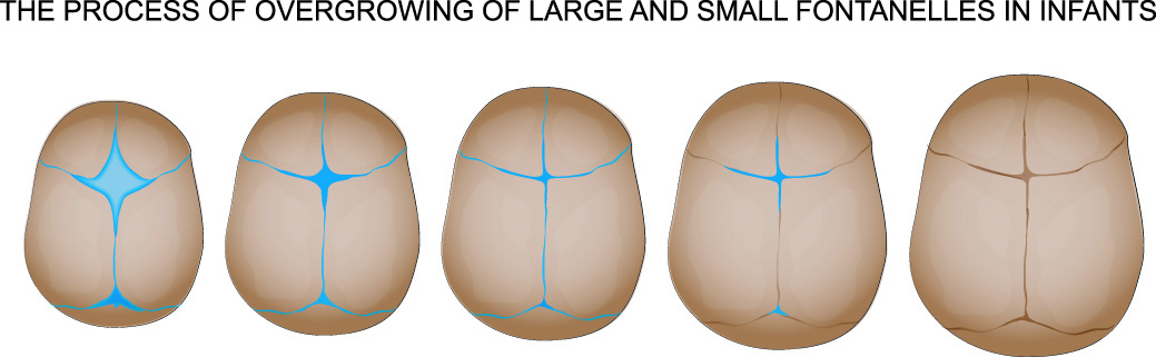
The paired joints are only mobile during infancy – we all know how delicate a young baby’s head is. This is because the skull must expand as the baby grows. The lambdoid suture between the occipital and parietal bones, and the petroclival and occipitomastoid sutures between the occipital and temporal bones, are found on both sides of the skull.
Occipital Bone Muscle Attachments
Occipital bone muscle attachments are multiple. As we have already seen, various protuberances and flat surfaces throughout the squamous, basilar, and lateral sections of C0 exist.
The occipitofrontalis muscle or epicranius muscle nearly covers the external squamous surface and is split into two parts (bellies) – occipital and frontal. It stretches from the eyebrows to the superior nuchal lines – imagine a rectangular, flat band of muscle that spans the skull from the eyebrows to the back of the head. When you raise your eyebrows or frown, you are using the occipitofrontalis.
There are a number of muscle attachments at the basilar part of the occipital bone. The external occipital protuberance is just one origin of the trapezius muscle of the upper back. The spinalis capitis muscle of the erector spinae attaches directly to the bottom of the skull and allows head, neck, and chest movement. The paired rectus capitis posterior minor muscles also play a role in keeping the head upright, inserting at the inferior nuchal line and underside of the occipital bone.
The rectus capitis major and obliquus capitis superior muscle pairs add head rotation to the mix; these also insert at the inferior nuchal line of the squamous part of the occipital bone. The rectus capitis lateralis muscle that allows us to flex our head from side to side attaches at the jugular process. Other muscles that enable neck flexion and also insert directly onto the occipital bone are the longus capitis and rectus capitis anterior muscles. While you do not need to know all of these names (one example is enough), the full range of muscle attachments for such a small bone is impressive.
Occipital Bone Damage
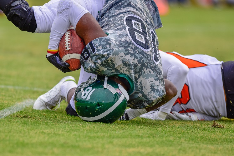
Damage to the occipital bone is usually the result of trauma. Falling on the back of the head is most likely to damage this part of the skull. When the skin above the bone is pierced, we tend to bleed profusely for various reasons.
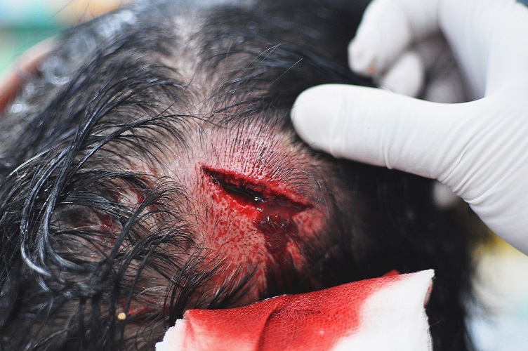
The main reason is the number of superficial and deep blood vessels in the skin of the scalp – the blood supply is so great, we can lower or raise our core body temperature when the scalp is exposed to extremes of temperature. If you have ever sat in the sun for a long time without a hat, you probably already know this. Also, the bigger the head or rather the bigger the head to body ratio – such as in young children – the greater the effect on body temperature. New parents are always advised to protect their babies’ heads from cold and heat.
Occipital Bone Fractures
Occipital bone fractures are not very common; high-energy injuries like whiplash can cause occipitocervical instability, as can certain genetic disorders such as Down’s syndrome. When the joint between the occipital bone and C1 becomes dislocated the result is often death. This is because the brainstem runs alongside the joint.
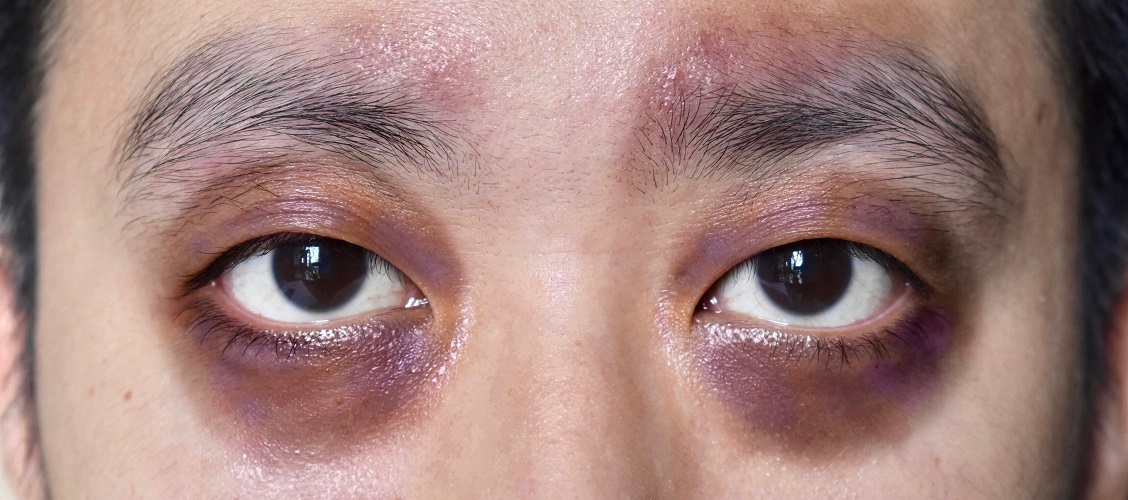
Any fracture around the foramen magnum is a medical emergency, although reparative surgery is complex. Stabilization of the neck and the surgical placement of drains to lower existing or potential high intercranial pressure are more likely to be used. If the basilar occipital bone is broken, Battle’s sign will commonly appear after one to two days. Battle’s sign is bruising behind the ear as a result of basilar trauma. It is sometimes accompanied by raccoon eyes and the presence of blood in the middle ear.
More serious fractures, as opposed to hairline fractures, can produce similarly serious symptoms. This is because of the proximity of blood and cerebrospinal fluid drainage systems, a number of cranial nerves, and tissues of the brainstem, cerebellum, and dorsal portion of the cerebrum. Even if there is no fracture, the brain knocking against the inner surface of the skull (concussion) can cause bleeding and swelling. The presence of blood and edema inside the brain means higher pressure as the skull does not provide extra space. If this occurs close to the occipital lobe, swelling in the rest of the brain may push the brainstem downwards. Damage to the brainstem can halt involuntary vital functions such as breathing and blood pressure regulation and is nearly always fatal.
Another example of occipital bone damage due to pressure inside the brain is sixth nerve palsy. Increased intercranial pressure – perhaps after a blow to the back of the head – causes swelling and pushes the abducens nerve (cranial nerve VI) close to the clivus of the occipital bone, trapping it. A trapped abducens nerve can lead to abducens palsy where one eye turns inwards.
The graphic autopsy pictures in this publication describing a calvarial bone fracture clearly shows a left occipital bone fracture in the first image. The brain has been removed to show the various internal grooves and projections.
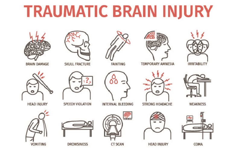
As trauma to the head can take time to cause observable symptoms – slow bleeds and limited edema sometimes continue for days or even weeks before the intercranial pressure increases and causes symptoms – an immediate clinical examination can be unreliable. Medical professionals are told to suspect brain damage after blunt trauma injuries if the patient presents with any of the following symptoms for up to four weeks after the event:
- Loss of consciousness
- Neck or occipital bone pain
- Impaired range of motion in the junction of head and neck
- Neurological deficit
Conservative treatment with neck and head stabilization are the chosen methods of treatment. If there is immediate danger to the soft tissue around the fracture, for example where a piece of bone becomes dislodged, surgical fusion of the bone using plates and screws will be necessary.
Quiz