Definition
The medulla oblongata is part of the autonomous central nervous system that directly connects the brainstem with the spinal cord. Medulla is Latin for middle; oblongata refers to this part of the brain’s elongated form. The medulla oblongata is located at the base of the brainstem and is essential for a broad range of somatic and autonomic motor and sensory functions. Practically all nerves signals pass through the medulla oblongata.
Medulla Oblongata Location
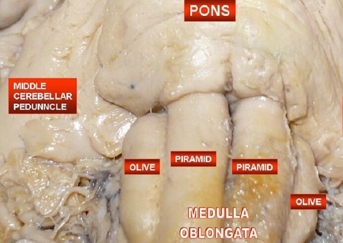
The medulla oblongata is located at the bottom of the brainstem, just under the pons, and merges with the spinal cord at the foramen magnum at the base of the skull. It contains both gray and white matter. The white matter is white due to the myelin sheaths of the many nerve axons that bring messages rapidly from the body to the brain and vice versa. Gray matter connects the medulla oblongata with four of the cranial nerves. These are the:
- Glossopharyngeal nerve (CN IX)
- Vagus nerve (CN X)
- Accessory nerve (CN XI)
- Hypoglossal nerve (CN XII)
Medulla Oblongata Function
Medulla oblongata functions are essential for nearly all nerve cell pathways. These functions are either specific to certain areas of the medulla or feature the medulla oblongata as part of a functional neural pathway or tract. One example of such a pathway is the medulla’s role within the reticular formation that controls vital functions such as breathing and heart rate. The medulla oblongata also plays an important part in our sleep-wake cycle or circadian rhythm.
In this article we will look at the function of the medulla oblongata (often shortened to medulla) according to specific pathways or tissue types. These are the cardiovascular and respiratory centers, the nucleus of the solitary tract (nucleus tractus solitarii in Latin, or NTS), the area postrema, the spinal trigeminal nucleus, the inferior olivary nuclei, the reticular formation, the pyramidal (motor) decussation, the cuneate nucleus, the gracile nucleus, the medial lemniscus, and the spinothalamic tract. You will not need to learn these very specific areas off by heart but will see how different areas of the medulla take part in complex pathways. These are described in more detail below.
Cardiovascular Center
The function of the medulla oblongata in the cardiovascular center is to regulate cardiac output. Cardiac output is the amount of blood that leaves the left ventricle of the heart per contraction (stroke volume) multiplied by the heart rate in beats per minute. Cardiac output regulation is made possible through information provided by baroreceptors and pH receptors in the major arteries.
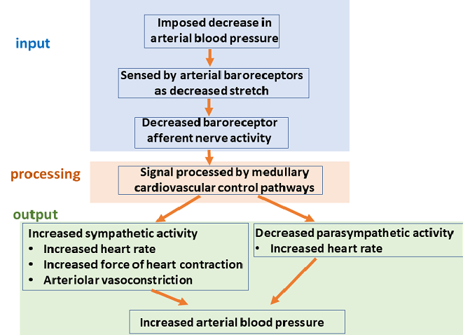
Chemoreceptors tell us about the acidity of the body – your blood becomes more acidic when carbon dioxide levels increase. They also tell the brain about the effects of hormones and neurotransmitters, most commonly in the form of epinephrine and norepinephrine. Baroreceptors send information about the amount of stretch in the major arteries, measuring the amount of pressure. If someone loses a lot of blood, for example, there is less pressure exerted on the blood vessel walls and the body will respond by constricting peripheral blood vessels and increasing the heart rate so that more blood reaches the vital organs.
The medulla oblongata responds to these signals by adjusting both the heart rate and stroke volume, which changes the cardiac output. This medulla oblongata function is part of the autonomic or involuntary nervous system and has three separate actions. The first is the cardio accelerator center that increases the heart rate and stroke volume (if there is enough blood) in reaction to the signals of the sympathetic (fight or flight) nervous system. The second is the cardio inhibitor center that slows the heart rate and stroke volume under parasympathetic (rest and digest) influence. The third system is the vasomotor center that regulates the constriction or dilation of arterial smooth muscle and so affects blood pressure and blood flow.
Respiratory Center
The medulla oblongata functions as a cyclic breathing center for the regulation of breathing. This is done through inspiratory neurons found inside the medulla that send motor impulses to the diaphragm and external intercostal (rib) muscles. Two cranial nerves – the vagus nerve and glossopharyngeal nerve – send data to the inspiratory neurons that has been collected from chemoreceptors. When stimulated, inspiratory neurons fire and cause the diaphragm and external rib muscles to contract. The pressure within the thorax becomes lower than the pressure outside the body and air is pulled into the lungs. This is inspiration.
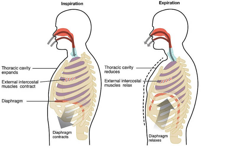
Expiratory neurons – also located in the medulla oblongata – send messages to the muscles of the abdomen and internal intercostal muscles. To create smooth respiratory motion, inspiratory and expiratory neurons slowly increase and decrease their firing rates. When the brain degenerates a few minutes before death, this is no longer possible and the result is gasping.
End of life gasping or agonal breathing is an involuntary medulla oblongata survival reflex that begins in the absence of oxygen. Some doctors think this may be an uncomfortable experience, others believe that the brain is no longer able to process thoughts of discomfort at this phase. You can look at the arguments in the Agonal Respiration and Suffering paragraphs of the free full-text PDF here.
Nucleus of the Solitary Tract
The nucleus of the solitary tract (NTS) or solitary nucleus (SN) refers to a group of medulla oblongata sensory cells that are part of the autonomic nervous system. Here, cardiovascular, visceral (certain internal organs), respiratory, gustatory, and orotactile information is received and forwarded. Orotactile information is especially important in newborns and young babies, where the sucking reflex is known to reduce pain and discomfort.
Efferent messages arrive in the medulla oblongata via chemoreceptors, stretch receptors, neurons connected directly to the visceral organs, and some of the cranial nerves (facial, glossopharyngeal, and vagus). These messages trigger a range of mammalian reflexes. They produce responses that may or may not be forwarded to other medulla oblongata functional areas, such as the reticular formation. All of these sensory functions are autonomic.
It is in the nucleus of the solitary tract that reflexes such as the gag reflex, cough reflex, and baroregulation reflexes (vasoconstriction and vasodilation), as well as gut motility and gut wall secretion mechanisms, are produced. Once these messages arrive, the NTS forwards them to other parts of the central nervous system to produce a response. For example, when stimulated, taste receptors in the NTS send a signal to the reticular formation that stimulates tongue and jaw muscle movement.

Area Postrema
The main medulla oblongata function in the area postrema is the vomiting reflex (not the gag reflex that is regulated by the NTS). Vomiting or emesis is the result of two separate medulla oblongata zones: the chemoreceptor trigger zone (CTZ) and the integrative vomiting center.
Messages arrive in the CTZ from the blood and cerebrospinal fluid when certain levels of toxins are present. These messages are forwarded to the integrative vomiting center. The integrative vomiting center is responsible for a reflex that is the combination of autonomic, visceral, and somatic motor pathways. The somatic motor pathways include respiratory and abdominal muscle contraction, while visceral pathways change how much our intestines move (peristalsis). Autonomic pathways increase salivation and sweating. The combination of these stimuli produces two vomiting reflex stages – the prodromal phase and the ejection phase. The first of these relaxes the muscles of the stomach and allows food that has traveled into the small intestine to move backward (retrograde peristalsis). This triggers the next phase – retching and vomiting (ejecting the stomach contents).
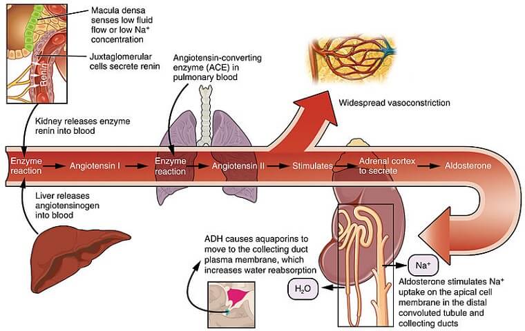
Other functions of the area postrema include chemoreceptor and osmoreceptor input from the major blood vessels and liver. Osmoreceptors tell the medulla oblongata about the fluid balance in the body. The area postrema is an important component of the renin-angiotensin-aldosterone system (RAAS). Newer research also shows that chemoreceptor information processed in the area postrema helps to regulate how forcefully we breathe in and out (respiratory drive) and contributes to an increase in appetite when the stomach is not sufficiently stretched.
Spinal Trigeminal Nucleus
Another group of sensory medulla oblongata functions occurs in the spinal trigeminal nucleus (STN). This area helps to provide our responses to temperature, touch, and pain – specifically of the face. This is because the sensory input comes from the trigeminal nerve, facial nerve, vagus nerve, and glossopharyngeal nerve. People with trigeminal neuralgia have symptoms that include a one-sided stabbing face pain. This might be the result of hyperactivity in the spinal trigeminal nucleus of the medulla oblongata.
Inferior Olivary Nuclei
The inferior olivary complex houses principle, medial accessory, and dorsal accessory olives that swap sensory and motor information between the spinal cord and the brain to enable learned actions. A lot of proprioceptive input (knowing where different parts of our body are at any time without needing to look) in combination with connections to the motor center of the brain and the eye allows us to practice and perfect learned movements. The medulla oblongata is, therefore, also crucial to our fine motor skills and coordination. In neurodegenerative disease, a damaged inferior olive can lead to the loss of previously perfected fine movement and a lack of coordination.
Reticular Formation
While not limited to the medulla oblongata, the reticular formation runs right through it. The reticular formation is a complicated network of nerve pathways. The entire structure involves parts of the medulla, pons, midbrain, hypothalamus, and thalamus. The function of the reticular formation is to regulate our states of consciousness and arousal and match sensory stimuli to motor, mental, and memory function.
General anesthetics administered before surgical procedures have a direct effect on the reticular formation; they lower consciousness, muscle tone, and responsiveness to external stimuli, stop memory formation (amnesia), and alter many autonomic responses. Anesthetic drugs reduce the effects of neurotransmitters such as adenosine, hypocretin, glutamate, GABA, and acetylcholine that are responsible for arousal and consciousness levels. If the medulla is damaged, the entire reticular formation can be negatively affected. If other areas are not working efficiently, less or more messages will pass through the medulla, causing excess or lacking stimuli and responses.
Pyramidal Decussation
The point at which the medulla oblongata meets the spinal cord is known as the pyramidal decussation. It is here that motor fibers from the medullary pyramids (paired vertical structures) cross over from one side of the brain to the opposite side of the spinal cord. Motor fibers that then continue into the spinal cord are renamed the corticospinal tract from this point on. The corticospinal tract is responsible for movement-related data transfer from the motor cortex of the brain to the spinal cord. It is because of the pyramidal decussation that damage to the left hemisphere of the brain leads to motor symptoms on the right side of the body and vice versa. Rather than a specific function, this part of the medulla oblongata is more of an anatomical marker. Various theories explain why decussation occurs in vertebrates. The most popular of these are the somatic twist and axial twist hypotheses.
Cuneate and Gracile Nuclei
The gracile nucleus (of Goll) and cuneate nucleus, both found in the medulla oblongata, have the same functions but serve different parts of the body. The gracile nucleus receives input from sensory neurons located in the lower body and sends this information to the thalamus. They provide proprioceptive (position), kinesthetic (movement), and epicritic (fine touch and temperature) information.
The cuneate nucleus has the same function but receives proprioceptive, kinesthetic, and epicritic data from the upper body before sending this data on to the thalamus.
Medial Lemniscus
The function of the medial lemniscus, located in the medulla oblongata, is directly related to the cuneate and gracile nuclei. The medial lemniscus, Reil’s band, or Reil’s ribbon starts at these nuclei, decussates (crosses over) at the lower part of the medulla, and then moves up to reach the thalamus. This is a major section of the dorsal column medial lemniscus pathway (DCML) that receives and forwards information about proprioceptive, kinesthetic, and epicritic data from the skin and joints of the body and head.
Damage to the medial lemniscus can be seen in cases of tertiary syphilis (Treponema pallidum infection). Symptoms are reduced proprioception and a lower sensitivity to fine touch. Damage to fine-touch pathways can be tested by way of a two-point discrimination evaluation. This test, where two slightly sharp points placed close together are applied to the skin, asks a person to report whether they feel one or two points. When the medial lemniscus is healthy and undamaged the test subject will know that two separate points are in contact with his or her skin; this is not the case when the medulla oblongata is damaged.
Spinothalamic Tract
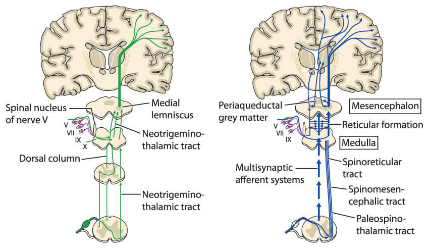
Finally, the spinothalamic tract gathers and forwards sensory information. The spinothalamic tract is a group of chain-reaction tracts that are individually called the anterior and lateral spinothalamic tracts, spinoreticular tract, and spinotectal tract. Don’t worry, you won’t need to know all the names.
The anterior and lateral tracts receive crude touch, pressure to the skin, pain, and temperature messages. The spinoreticular tract alerts us to these potentially harmful sensations, and the spinotectal tract turns our eyes to the source of the pressure or pain. All together, they provide a chain of information that can save our lives. The complete spinothalamic tract is the reason why we quickly pull our hand away when brushing it on the edge of a hot oven door, or ducking when something comes rushing towards our head.
The spinothalamic tract does not decussate at the brainstem but at the spinal cord. As with the medial lemniscus, the spinothalamic tract is not completely contained within the medulla oblongata, but the medulla oblongata is part of the spinothalamic tract and crucial to its performance.
This is the case with all medulla oblongata functions – it is not a single functioning unit but a central collection point for sensory and motor data that must pass from the central nervous system to the peripheral nervous system and vice versa.