Definition
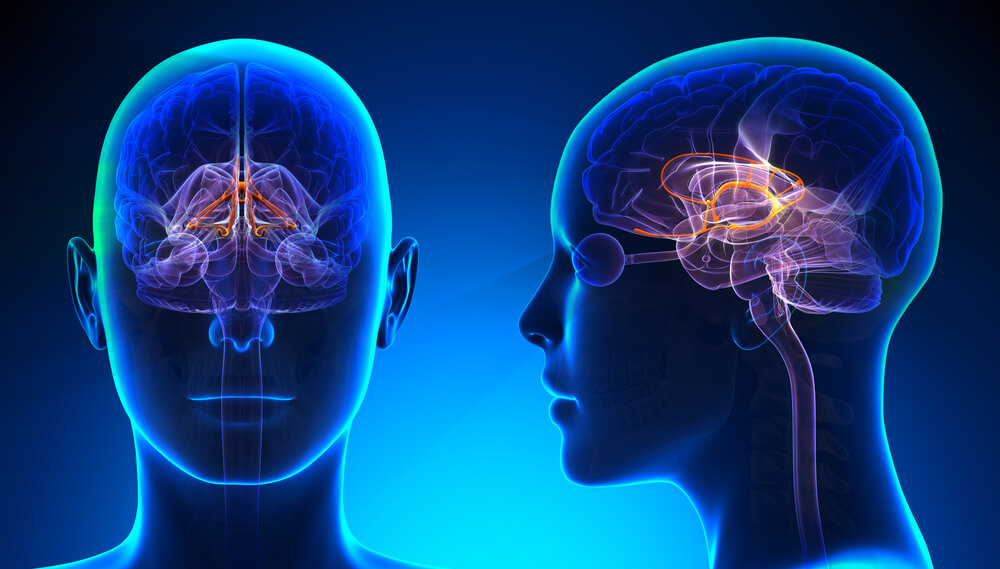
The limbic system consists of a series of brain structures responsible for processing senses and emotions to result in new memories and bodily changes. It processes these stimuli as electrical signals throughout the central nervous system, allowing for memory formation as well as autonomic and behavioral changes. Ultimately, the limbic system communicates with the endocrine system to allow for the release of hormones so that the body can properly respond to the stimuli presented.
Overview of the Limbic System
Why Is This Important?
Our emotions play a critical part in the way we behave, and it is believed that strong emotions- such as fear and anger- were utilized as an evolutionary mechanism for survival. For instance, when there was a stimulus in the environment that provoked a sense of threat, the limbic system would recognize this sense as fear and create a physical response in the body- such as increasing heart rate or releasing adrenaline into the blood stream- as a way to increase the chances of survival. The limbic system would then code this threat to memory, so the individual can quickly react if confronted again in the future.
How Does It Work?
The limbic system spans multiple regions across the brain, and the full functionality of the system is yet to be fully understood. Despite this, scientists have identified multiple structures that are important to the limbic system, such as the limbic lobe, hippocampal formation, amygdala, thalamus, and hypothalamus. These structures are able to communicate with each other by stimulating the structures’ individual cells, known as neurons. Eventually, connections are made between the neurons that allow for rapid processing from the initial stimulus to the final physical change.
Review of the Nervous System
The nervous system is made up of single nerve cells known as neurons, which are specialized by their ability to communicate with electrical signals at speeds of up to 120 meters/second. These high speeds are critical to the function of the nervous system, as it allows a single neuron to send information to the brain and back almost instantly.
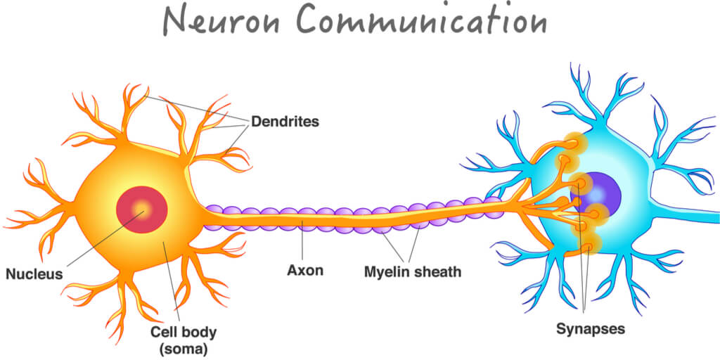
The neuron is made up of three main parts that work together to send these signals:
- Dendrites that convert and receive electrical signals
- The cell body (or soma) that contains the nucleus and processes the signal
- The axon that passes the signal on to the next neuron
At rest, neurons have a membrane potentialthat is determined by the concentration of ions on either side of the neuron’s membrane. When a stimulus is received, neurons create a quick and temporary change in this potential- now called an action potential. These action potentials rapidly change the polarization of the neuron, allowing for signals to be passed along between neurons.
Neurons form to create the entirety of the nervous system, which is broken down into two sub-systems: the central nervous system and the peripheral nervous system. The central nervous system (CNS) includes the brain and the spinal cord, while the peripheral nervous system (PNS) includes all other neurons distributed throughout the body. We will primarily focus on the brain of the CNS when discussing the limbic system, with slight mentioning of the PNS when discussing the hypothalamus.
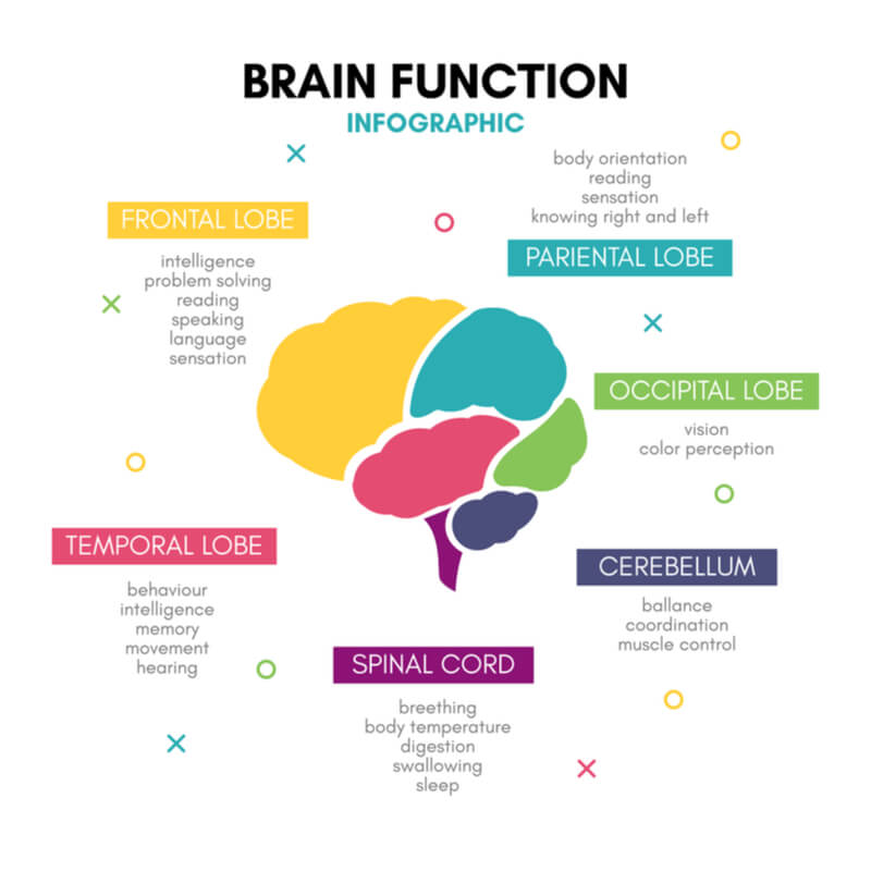
The brain is broken down into major structures, including the cerebrum, cerebellum, and brainstem. For the limbic system, the structure of focus is the cerebrum, which is split into two hemispheres that are connected by the corpus collosum. The cerebrum is further arranged into lobes, including the frontal, parietal, temporal, and occipital lobes. Each lobe is known for specific information processing and functioning, where specific structures tend to be localized within a specific lobe. The limbic system unites multiple structures and functions across these lobes to rapidly process new information and initiate appropriate responses throughout the body.
Structures, Connections, and Functions of the Limbic System
Limbic Lobe
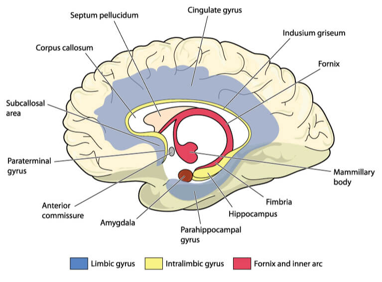
The first major region of the limbic system is the limbic lobe. The limbic lobe contains several important structures of the limbic system, but the entirety of the limbic system is not confined solely to this lobe. The significant structures found here include the cingulate cortex and the parahippocampal gyrus.
The cingulate cortex is an inner part of the brain that lies superior (above) and dorsal (behind) the corpus collosum. It consists of an anterior (frontal) and posterior (behind) section, which both receive information from the thalamus and hippocampal formation.
The primary function of this area is to regulate both autonomic function and conscious function. Autonomic functions include involuntary changes- such as an increase in heart rate to a threat in the environment- while conscious responses include voluntary choices- such as the decision to react to the threat (ex. taking a step back). The conscious response is also important when controlling emotions. In other words, instead of perceiving the emotion itself (ex. feeling pain), the cingulate cortex controls what the individual thinks of the emotion (ex. how you feel about the pain).
The parahippocampal gyrus is made up of several distinct regions in the middle section of the temporal lobe. The most significant of these regions includes the entorhinal cortex, which the parahippocampal gyrus uses to send information from the cortex to the hippocampal formation.
Hippocampal Formation
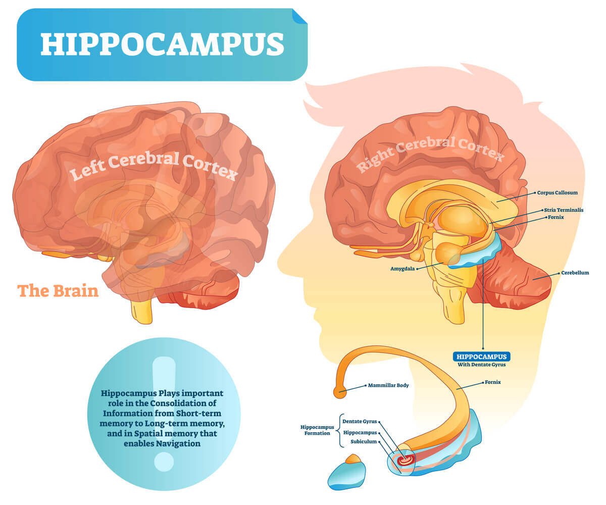
The hippocampal formation is found in the temporal lobe posterior to the amygdala and anterior to the corpus callosum’s splenium (the most posterior portion of the corpus collosum). The three main structures found in this area include the hippocampus (also known as Ammon’s horn, which is segmented into four groups of axons- CA1, CA2, CA3, and CA4), the dentate gyrus, and the subiculum region of the subicular complex.
The hippocampus is a horn- shaped structure that surrounds the lateral ventricles. The main function is to process new information and form long- term memories, which are then stored in alternate regions throughout the brain. If damaged, an individual is unable to create new long-term memories. However, the individual is able to remember long-term memories already stored.
The hippocampus most commonly receives information from the entorhinal cortex of the parahippocampal gyrus. When received, the information is typically mediated by the performant pathway. The performant pathway sends signals using fibers throughout the subiculum of the subicular complex and into the dentate gyrus, where fibers then extend from the dentate gyrus to the CA3 axons. Schaffer collaterals then connect with dendrites on the CA1 axons. The CA1 axons contain fibers that extend out of the hippocampus and into subsequent regions within the brain.
Amygdala
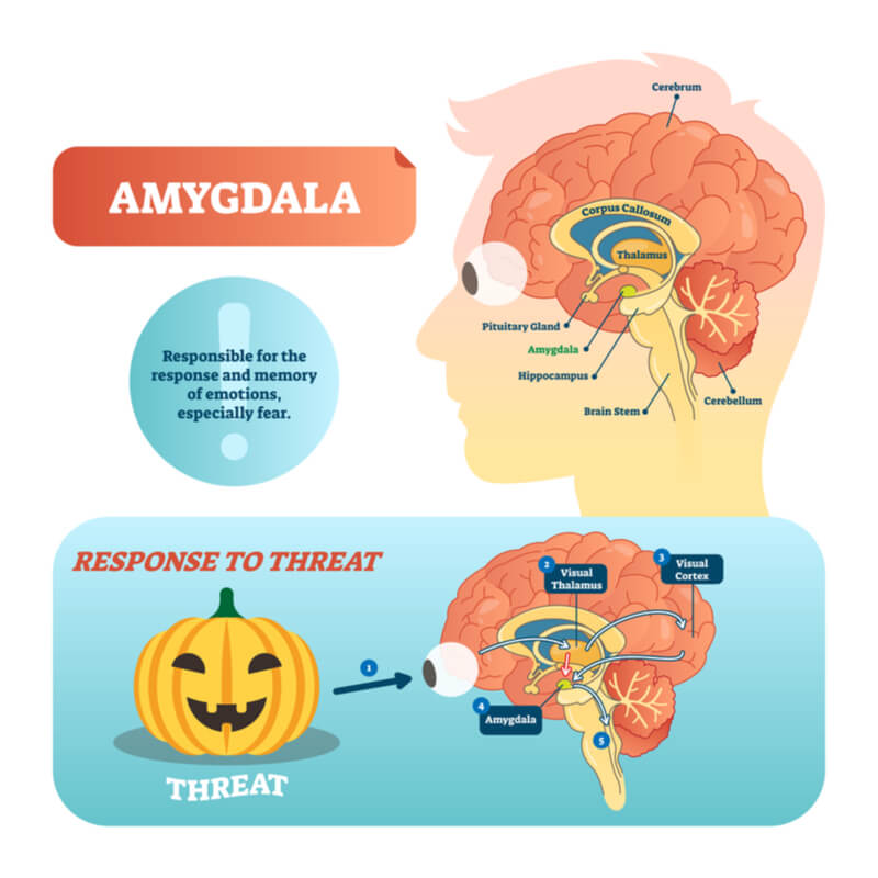
The amygdala is found in the anterior portion of the temporal lobe, directly in front of the hippocampus. The amygdala is responsible for coordinating voluntary, involuntary, and endocrine responses to anger, fear, and anxiety. Stimuli within an individual’s environment that are perceived as threatening are processed through the amygdala, allowing the individual to alter their behavior while exposed to the threat. This creates new behaviors that are associated to the threat, allowing the individual to quickly react when threatened in the future. This is, therefore, the structure that mediates survival, as fear and anger are critical emotions in alerting the brain of life-threatening situations.
The amygdala maintains two main pathways for information processing: the dorsal route and ventral route. Both routes send information to the hypothalamus, while the ventral route also sends information to the thalamus.
Thalamus
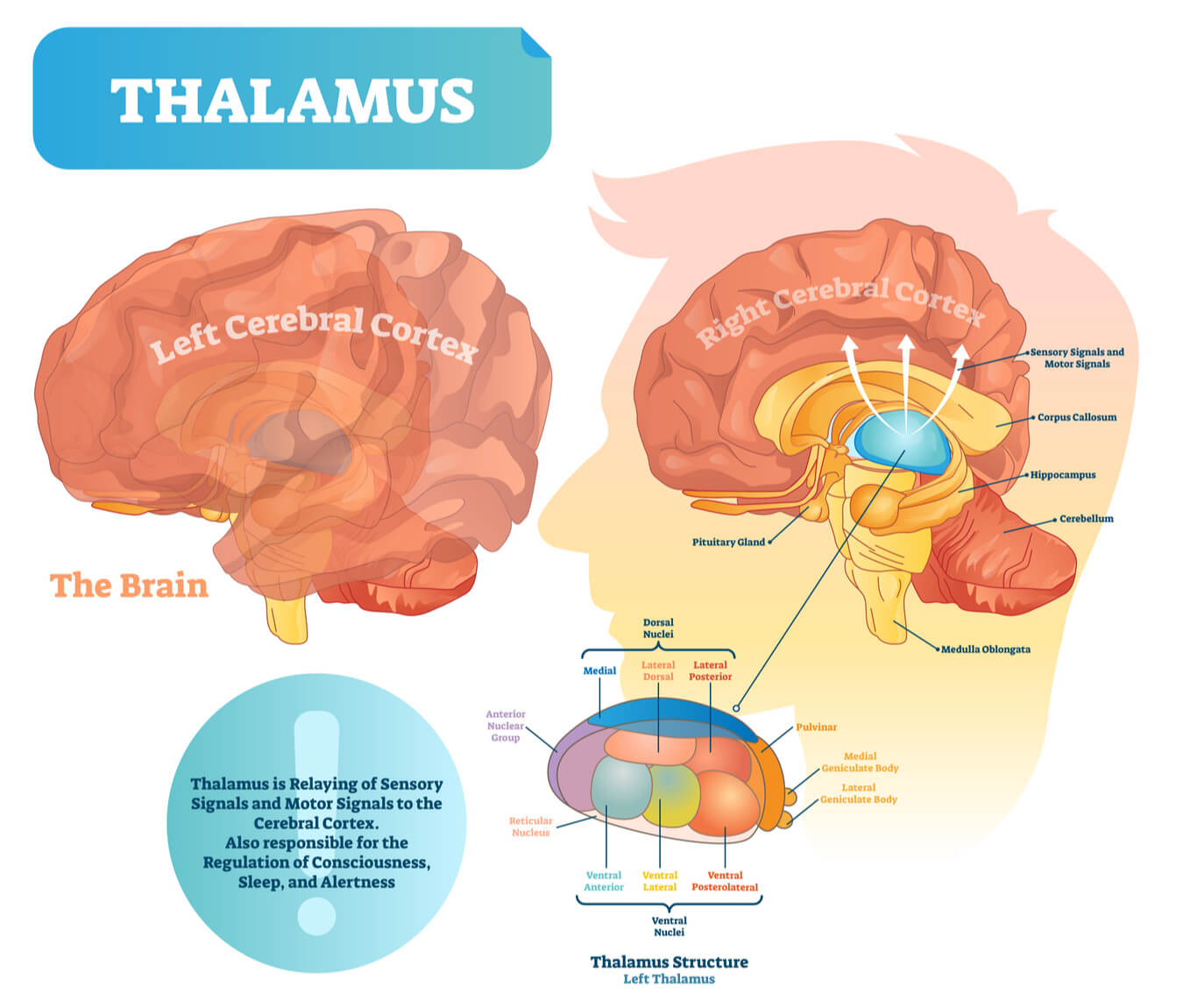
The thalamus is found superior and lateral to the third ventricle and makes up 80% of the diencephalon. The thalamus relays incoming information- such as senses including sight, sound, taste, and touch- and sorts it accordingly throughout the cortex. This allows information to be organized throughout the brain and sent to where it will create the most effective response. Ultimate responses include voluntary and involuntary responses, cortical arousals, learning, and memory formations.
*Note: Smell is not processed by the thalamus. Smell is receipted by the olfactory bulbs and is directly sent for emotional processing and memory formation.
Hyopthalamus
The hypothalamus is also located in the diencephalon, found inferior to the thalamus and both inferior and lateral to the third ventricle. The main function is in homeostatic regulation, as the hypothalamus has direct control over the autonomic nervous system (ANS). The ANS is part of the PNS that oversees involuntary actions and rhythms of the visceral organs, including heart rate, blood pressure, and digestive movement. The ANS encompasses two sub-systems- the sympathetic nervous system (“fight or flight”) and the parasympathetic nervous system (“rest and digest”).
The hypothalamus is the central structure that must be stimulated in order to create physical changes in the body. This is because the hypothalamus is directly connected to the pituitary gland, and thus immediatly influences the hormones released by the pituitary gland. This connection is a cross-talk between the nervous system and endocrine system, translating neural stimulations received by the brain into hormonal changes throughout the body.
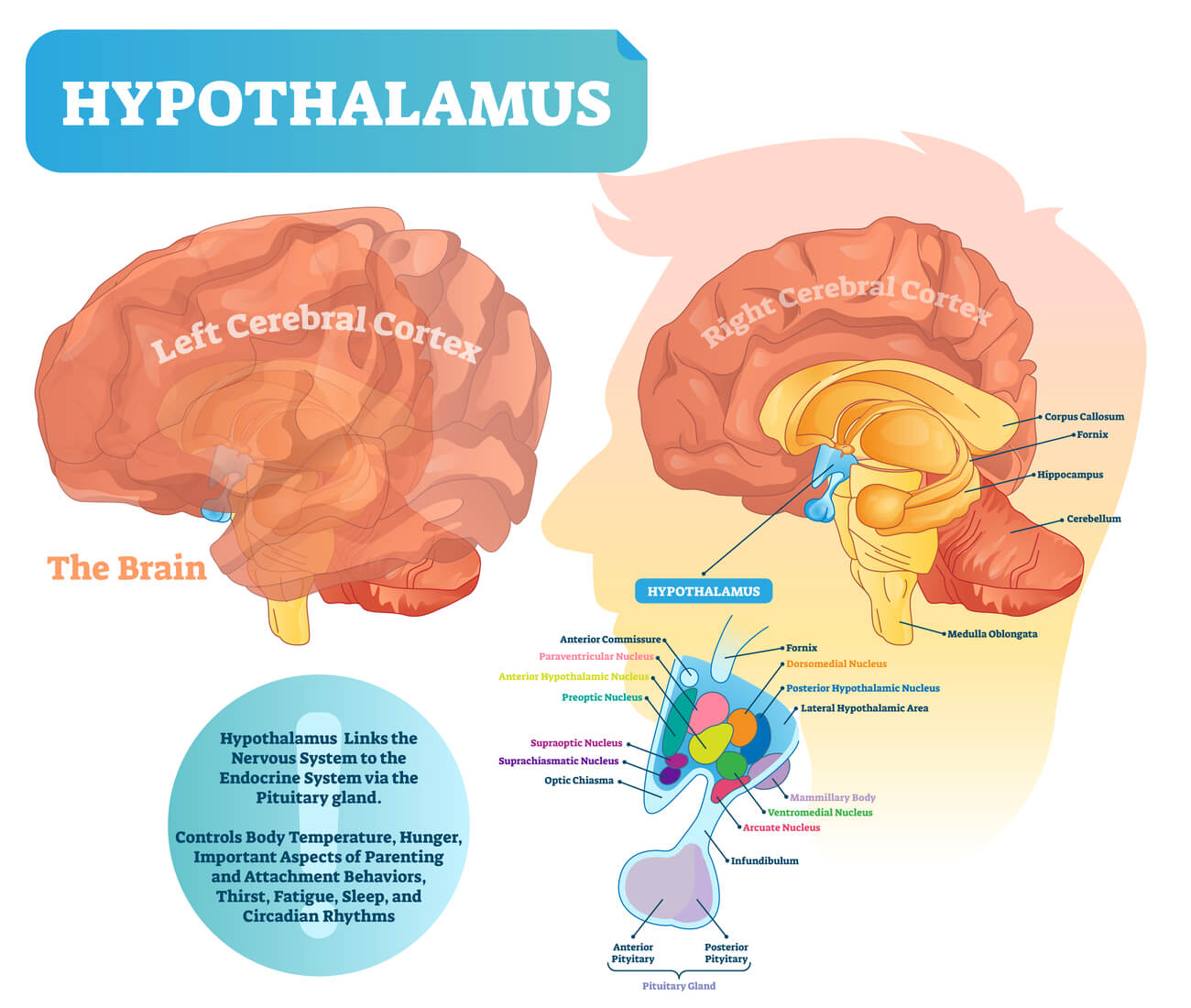
When stimulated, the hypothalamus directly releases corticotropin-releasing hormone (CRH) into the pituitary gland, thus activating the endocrine system. As the pituitary gland is made up of two parts- the posterior pituitary and anterior pituitary- this activation can be done in two ways. First, the hypothalamus can directly create and release neurosecretory cells (or neurohormones) into the posterior pituitary. The posterior pituitary then releases its own stored hormones- such as ADH and oxytocin- into the bloodstream to be spread throughout the body.
Alternatively, the hypothalamus can release neurosecretory cells into blood vessels that travel into and activate the anterior pituitary. The anterior pituitary then either increases or decreases the hormones distributed into the blood, depending on whether the neurosecretory cells from the hypothalamus are sent to stimulate or inhibit the specific hormone.
The portion of the pituitary that the hypothalamus interacts with- whether anterior or posterior- is contingent on the signal received in the brain and the bodily needs.
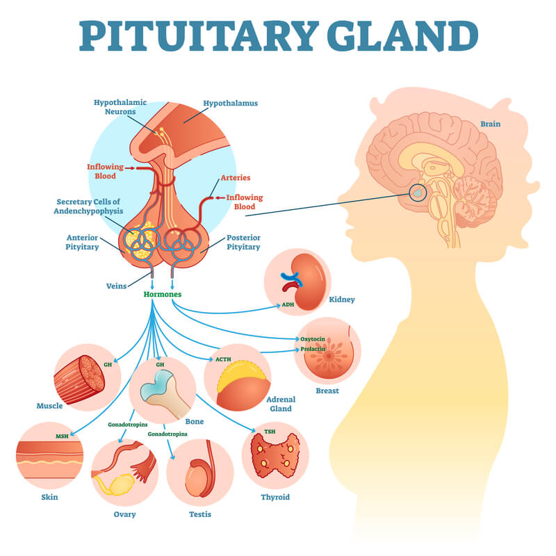
Example. During breastfeeding, the attachment of the infant to the mother’s nipple sends the sensory information from the nipple to the brain for processing. The hypothalamus is stimulated, and neurosecretory cells activate both the posterior and anterior pituitary gland. The posterior pituitary releases oxytocin while the anterior pituitary releases prolactin into the bloodstream, which both activate the needed bodily changes for lactation in the presence of the stimulus (the suckling infant).
Conclusion
The limbic system plays a critical role in processing senses and emotions throughout the brain to create bodily changes. It uses a highly interconnected system of structures and events to quickly create these changes as it serves as an important survival mechanism. While multiple brain structures have been previously defined, research is expanding to fully understand the potential of this system.
Quiz