Definition
The lacrimal bone or os lacrimale is a paired flat craniofacial bone that provides grooves for parts of the lacrimal apparatus (tear production) and a surface for muscle attachment. Both paired bones border with the maxilla, ethmoid, and frontal bones of the face and skull. The lacrimal bone is very small and quite delicate.
Where is the Lacrimal Bone?
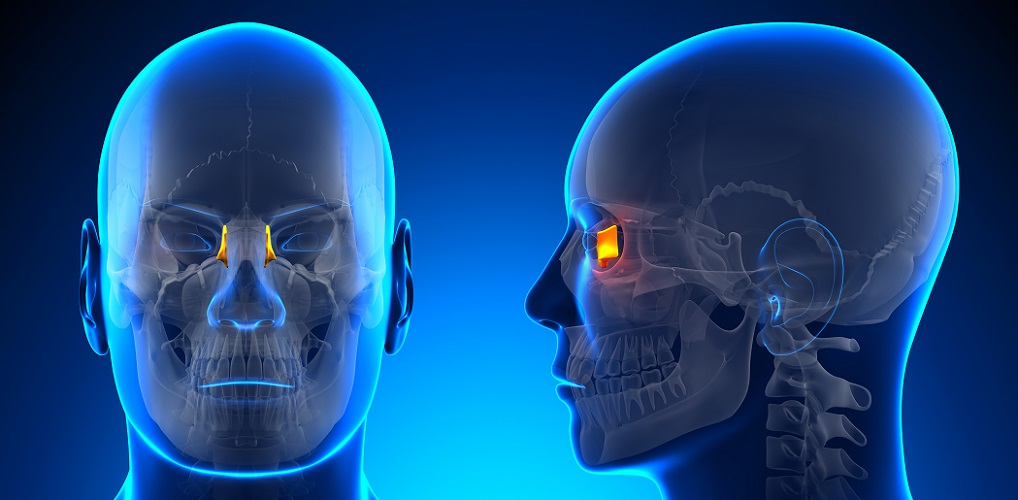
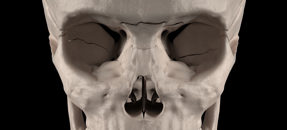
Where the lacrimal bone is located requires a little knowledge of the surrounding bones; however, its function as part of the lacrimal or tear-production system tells us that it is close to the inner corner of the eye. With a frontal view of the skull, both os lacrymal are nearly completely hidden behind the nasal bone.
When the nasal bone is removed, the front (superior) surface of each tiny bone can be seen in its entirety.
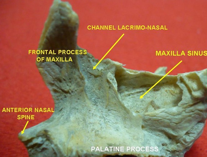
The above photograph shows the labeled lacrimal bone, with the anterior crest fitting into the frontal process of the maxilla bone and the top with the frontal bone of the forehead. The right-hand side of this picture of the left-hand side skull shows the bone fitting snugly into the eye socket.
Lacrimal Bone Anatomy
Lacrimal bone anatomy is relatively complex when we consider its small size in the human face – around one and a half centimeters in height and less than one centimeter in width.

If you wondering whether to label them as facial or cranial lacrimal bones, they are facial bones. In the face, the cranium ends at the frontal bone.
Both bones have two surfaces – the lateral (orbital) surface and the medial (nasal) surface. These surfaces contain grooves, ridges, and furrows that give the lacrimal bones an additional function. The two bones also have articulations with the frontal, ethmoid, and maxilla bones, and the nasal concha.
Lateral Surface of the Lacrimal Bone
The lateral surface of the lacrimal bone has four anatomical parts:
- Posterior lacrimal crest
- Lacrimal sulcus
- Lacrimal hamulus
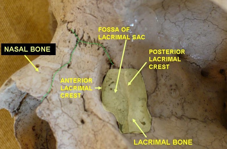
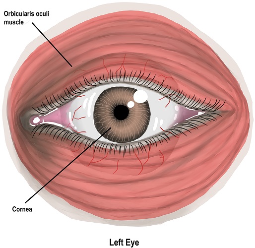
The posterior lacrimal crest divides the lateral surface into two sections. Lateral surface refers to the side surfaces of this bone. The posterior lacrimal crest is a narrow, vertical raised surface that creates a groove (the lacrimal sulcus) close to the eye. It is an attachment point for the orbicularis oculi muscle that closes the eyelids.
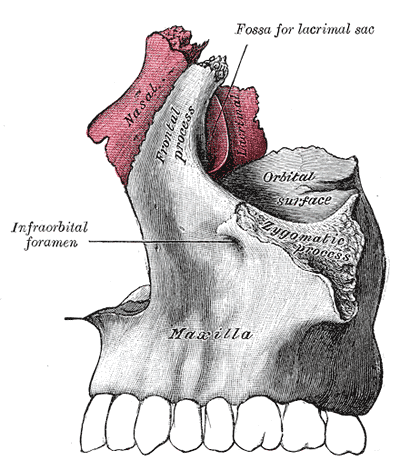
The lacrimal sulcus provides space for the soft tissues of the lacrimal sac and nasolacrimal duct. You can see this groove in the image, labeled as fossa for lacrimal sac. The crest ends in a small hooked shape called the lacrimal hamulus that produces a rounded orifice that houses the lacrimal canal.
The anterior lacrimal crest, mentioned in some articles as part of the lacrimal bone, is also considered to be part of the frontal process of the maxilla. This is the place where both bone surfaces join.
Medial Surface of the Lacrimal Bone
The medial surface of the lacrimal bone is the part that faces toward the midline of the body – the back surface. It has a long groove (furrow) along its length that runs in the same direction as the posterior lacrimal crest.
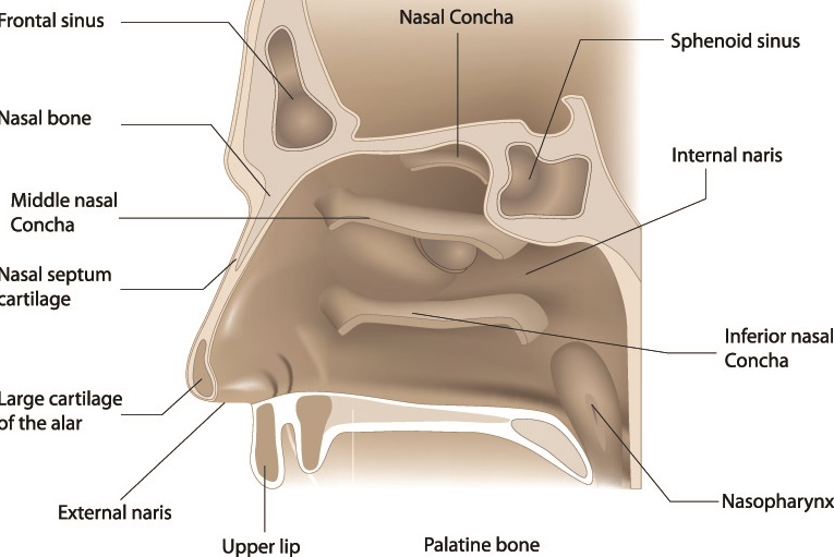
The medial surface provides a bony surface for the middle meatus of the nose that helps to support the front part of the middle concha (see image below).
Lacrimal Bone Borders
The lacrimal bone has four borders with other craniofacial bones called articulations. These articulations are found at the frontal, ethmoid, inferior nasal concha, and maxilla. As the lacrimal bones are some of the smallest bones in the body – and the smallest facial bones – they only articulate with larger bones at specific points.
Each bone borders the frontal process of the maxilla (see image) and the orbital plate of the frontal bone that forms the recessed area of the eye socket at the top of the eye socket.
The lacrimal bones also border with the eye socket-facing surface of the ethmoid bone. These bones are colored in shades of pink in the below image of the orbital bones.
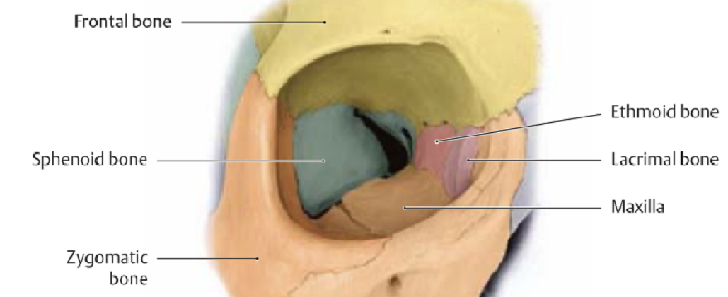
There is also a border between the lacrimal process of the inferior nasal concha.
These borders help us to pinpoint the lacrimal bone location.
Lacrimal Bone Function
The lacrimal bone has three functions. The first is to provide points of articulation between parts of the maxilla, ethmoid, and inferior nasal concha. Articulations rather than fixed sutures mean there is a very small range of motion when these bones move and, therefore, play a protective role.
The second lacrimal bone function is to provide an attachment point for the orbicularis oculi muscles. This circular muscle closes the eyelids but also helps with tear drainage – upon contraction, tears are pushed into the nasolacrimal duct.
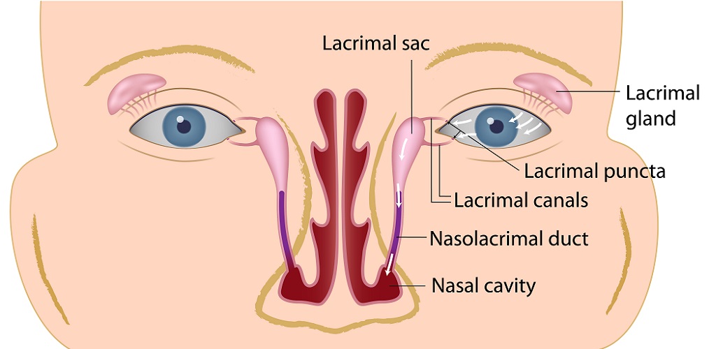
Finally, the shape of this bone means that soft tissue ducts can run along with it – the nasolacrimal duct that carries tears from the lacrimal sac to the inferior meatus of the nasal passage – the reason why tears come out of the nose when we cry. The nasolacrimal (or nasolacrimal) duct is very narrow and can become blocked.
Lacrimal Bone Fracture
Lacrimal bone fractures are part of greater facial traumas, particularly to the eye socket, nose, and forehead. An extremely fragile bone, fractures as a result of blunt upper facial trauma is not unexpected. Especially as the lacrimal and ethmoid bones are described as being of similar strength to an egg-shell when compared to other facial bones. This already delicate structure can be affected by osteoporosis ); there is a correlation between lacrimal bone thickness and general bone density throughout the body.
The lacrimal bones lie very close to the nasal airway and frontal lobe of the brain. Fractures in this region may cause airway obstructions and damage to the frontal lobe of the brain. Le Fort III fractures of the mid-face typically present with lacrimal bone fractures, in combination with complex mid-face fractures of the bones that surround the nose and eyes.
Maxillofacial surgeons need to restore the natural form of the face when treating such fractures. When the bones crack but do not shift, surgery is avoided. If the breaks have caused changes in position, they will need to be surgically treated. The tiny, fragile lacrimal bone is less likely to be fixed with plates and is fixed into place by attaching them to surrounding bones with wire. Where the orbicularis oculi detach from the posterior lacrimal crest, the tendon of this muscle is fixed into place using screw holes and sutures. In type III fractures – the most severe naso-orbito-ethmoid (NOE) fracture type – primary bone grafts are often required to replace lost or damaged bone.
Any lacerations to the tear ducts must be carefully closed to avoid permanent blockage of the nasolacrimal duct.
In cases of blockages due to congenital deformities, the ducts can be accessed via the inside of the nasal passage or via the corner of the eye. A dacryocystorhinostomy is the name of the procedure that opens the duct – sometimes a stent is placed to keep the duct open.
Quiz