Definition
The hyoid bone is the only bony structure in the larynx and the only bone in humans that does not articulate with other bones. Instead, it attaches to cartilage, muscles, and ligaments. Because of this, it is often said to be free-floating. It plays important roles in mastication (chewing), tongue movement, phonation, and swallowing. A broken hyoid bone can indicate strangulation.
What is the Hyoid Bone?
The hyoid bone, lingual bone, or tongue bone is a U-shaped or horseshoe-shaped bone. The Greeks named the upside-down letter U (upsilon) ‘hyodeides’ and this is where the tongue bone gets its most commonly-used name.
The lingual bone is part of the hyoid-larynx complex and is heavily involved in orofacial movement. It helps to keep the airway open between the oropharynx above and cartilaginous tracheal rings below.
Where is the Hyoid Bone Located?
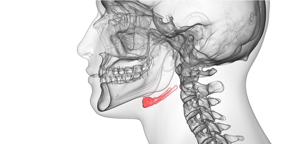
Hyoid bone location is between the chin and the cartilage of the thyroid gland at the level of the third or fourth cervical vertebra.
Moving upward along the midline of the neck from its base, the first cartilage you can feel is the cricoid cartilage. Above this is the thyroid cartilage, and above the thyroid cartilage sits the tongue bone.
The hyoid bone is located just below the submaxillary or digastric triangle – an anatomical landmark under the lower jaw (mandible).
Hyoid Bone Anatomy
Hyoid bone anatomy is not complicated but many different muscles depend on it as an attachment point. The lingual bone is part of the hyoid-larynx complex – the combined structures of the hyoid and its ligaments, and the thyroid, cricoid, and arytenoid cartilages and their ligaments.
The hyoid bone is unique because it does not have joints with any other bones. It is fixed into place by muscles, ligaments, and surrounding cartilage of the hyoid-larynx complex and ligaments of the temporal bones of the skull.
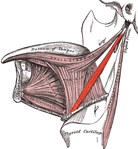
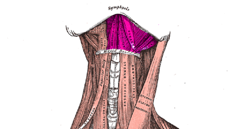
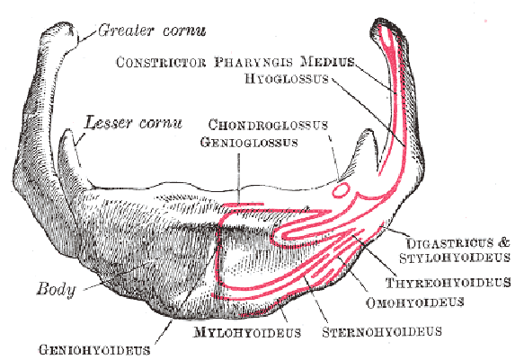
The tongue bone is suspended from the ends of the styloid processes of the left and right temporal bones by the left and right stylohyoid ligaments (in red). In the below image you can also see how the hyoid connects to the tongue.
Hyoid Bone Parts
The lingual bone is composed of a body and two horns that give it its distinctive shape.
The body is approximately 2.5 cm wide and one centimeter thick. The anterior surface that faces the front of the neck is convex and, when in a neutral position sits slightly forward and upward. A vertical mid-line ridge divides the body into two halves.
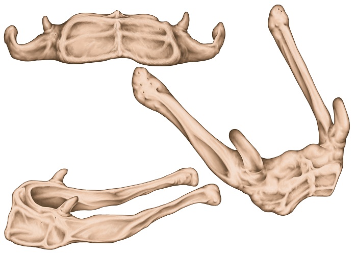
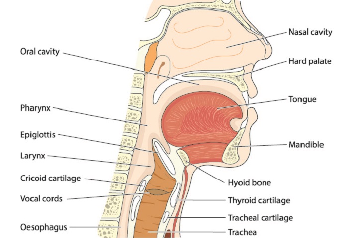
The posterior surface of the body that faces the trachea is concave and, in a neutral position, points slightly backward and downward. A membrane called the hyothyroid membrane separates the tongue bone from the soft tissue of the epiglottis.
The two paired horns are called the greater and lesser cornua.
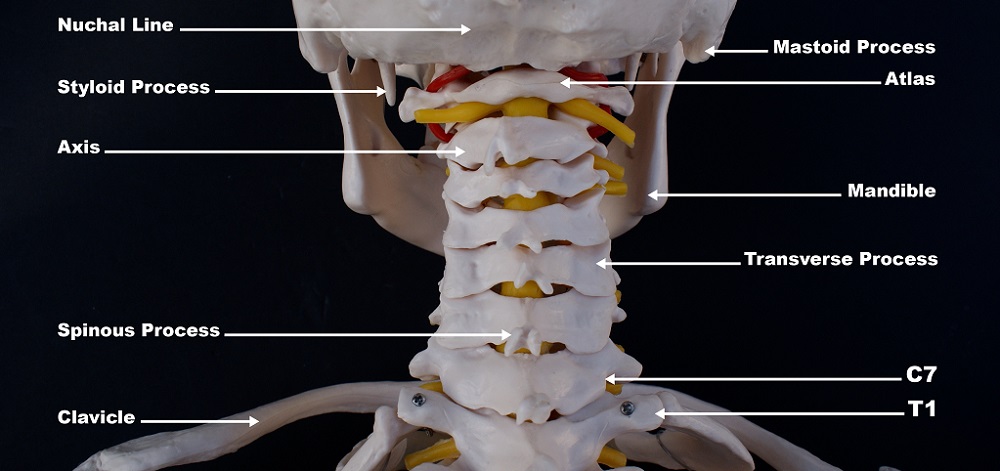
The greater horns of the hyoid bone protrude backwards and, like the body, are an attachment point for muscles. The shorter, stumpier lesser horns of the hyoid bone are an attachment point for the stylohyloid ligament. This ligament suspends the hyoid from the styloid processes of the temporal bones. You can see the styloid processes in the following image that shows the back of the skull and spine.
A clicking hyoid bone is a rare anomaly. It refers to a painful tapping sound in the throat. This is due to the tips of the greater horns knocking against the cervical vertebra that site behind them. Swallowing and neck movements can be painful. This paper includes some interesting photographs of a greater cornua resection during clicking hyoid bone surgery.
Hyoid Bone Muscle Attachments
Two groups of muscles – the suprahyoid (above the hyoid) and infrahyoid (below the hyoid) muscles – use this bone as a point of attachment.
Four pairs of suprahyoid muscles connect the lingual bone to the mandible, skull, and tongue. These muscles insert at the hyoid and have their origins above it; when the suprahyoid muscles contract, they pull the tongue bone up. The four muscles are the:
- Digastric muscle
- Stylohyoid muscle
- Mylohyoid muscle
- Geniohyoid muscle
When the digastric muscle contracts during speech, swallowing, and even breathing, the hyoid bone lifts. If other muscles prevent the hyoid from moving upward, the lower jaw will drop, opening the mouth.
Originating at the styloid process of the temporal bone of the skull, the stylohyoid muscle also lifts the tongue bone when we start to swallow.
The mylohyoid muscle lifts the lingual bone and tongue when we speak or swallow. Like the digastric muscle, it also helps to open the mouth. As most cartoons show extremely well, opening the mouth is the result of dropping the lower jaw or mandible; the upper jaw (maxilla) does not have such an exaggerated effect.

The geniohyoid muscle lifts the hyoid bone to open the airway. It is also involved in swallowing and mouth opening.
We also have four pairs of infrahyoid muscles that run from the hyoid bone to the shoulders. The lingual bone is the insertion point of these muscles and their origins are at the base of the neck and shoulders, meaning this muscle group pulls the hyoid down.
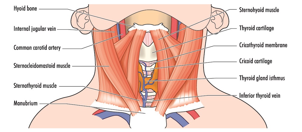
- Sternohyoid muscle
- Sternothyroid muscle
- Omohyoid muscle
- Thyrohyoid muscle

After the hyoid bone is lifted by the suprahyoid muscles, the infrahyoid muscles pull it back down. When the hyoid is elevated, the airway dilates.
When we swallow food, the airway must close to stop food particles from entering the lungs. Hyoid depression is also required when opening the jaw to speak, chewing, and in a lesser extent for certain head movements.
Two muscles that are not part of the aforementioned muscle groups have origins at the tongue bone. These are the hyoglossus and middle pharyngeal constrictor muscles.
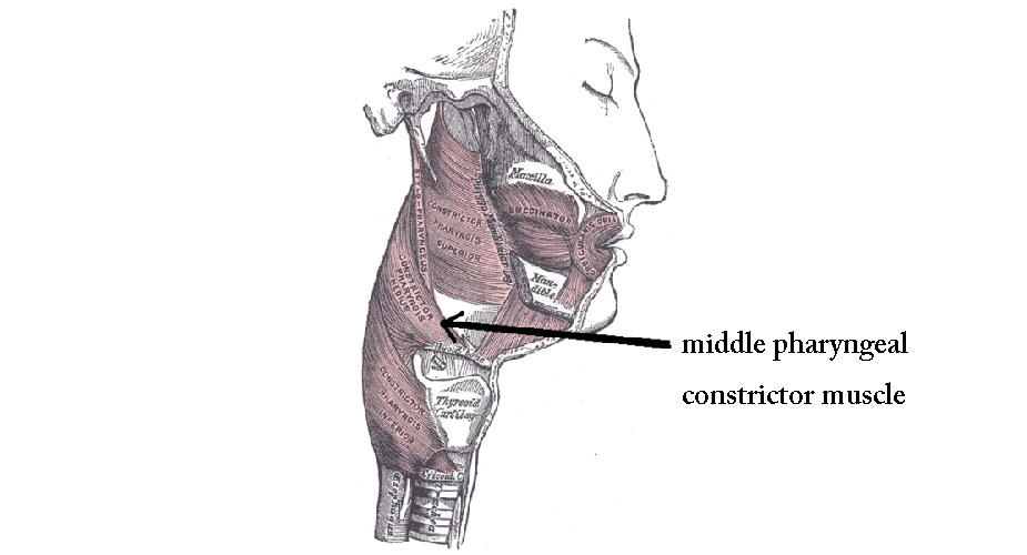
The hyoglossus depresses and retracts the tongue; the middle pharyngeal constrictor constricts around food that enters through the oropharynx (food bolus), pushing it down to the esophagus.
Hyoid Bone Ligamentous Attachments
The lingual bone is attached to the thyroid cartilage by way of a ligament called the thyrohyoid membrane. This tough membrane originates at the top of the thyroid cartilage and attaches to the back of the hyoid body and the greater horns.
As we have already seen, the stylohyoid ligament originates at the styloid process of the temporal bone and extends to the lesser horn of the hyoid bone.
The third ligament that attaches to the hyoid is the hyoepiglottic ligament. This connects the top of the hyoid body to the front (anterior surface) of the epiglottis.
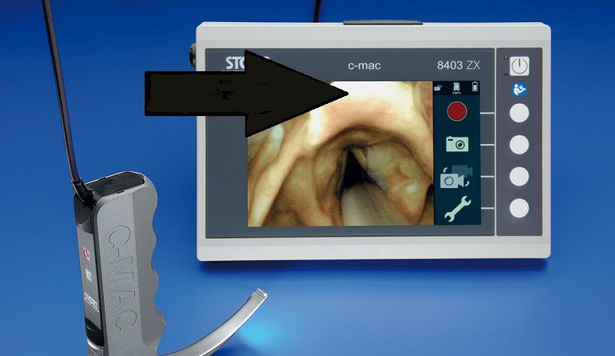
The epiglottis is a flap of soft tissue (indicated by the arrow below) that falls over the trachea when we swallow, protecting the airway. It can be seen during endotracheal intubation where a breathing tube is passed through the vocal cords (the edges of the triangular hole in the image) and towards the lungs.
Hyoid Bone Function
Hyoid bone function is structural – it holds up the tongue from below and keeps the larynx in place from above.
The lingual bone keeps the airway open between the oropharynx and tracheal rings and, therefore, plays a role in respiration. It is necessary when eating (chewing and swallowing) and speaking.
Other functions of the hyoid bone are its preventive action against regurgitation. It helps to control tongue movement and plays a minor role in head positioning.
Hyoid Bone Fracture
Hyoid bone fractures are a common clue during forensic autopsies as this small bone is often damaged during manual (with the hands) and ligature (hanging/garotting) strangulation. A hyoid fracture can also occur via blunt force trauma to the neck.
The most common cause of a broken hyoid is manual strangulation, as you will read in pages five to seven of this University of Mississipi Medical Center report.
Lingual bone fractures occur in approximately one third of strangulation victims; this type of fracture is more likely in older populations. As we age, the body and greater horns of the hyoid bone fuse – there is less give and these joints are more likely to snap. The fusion of bone joints is called ankylosis.
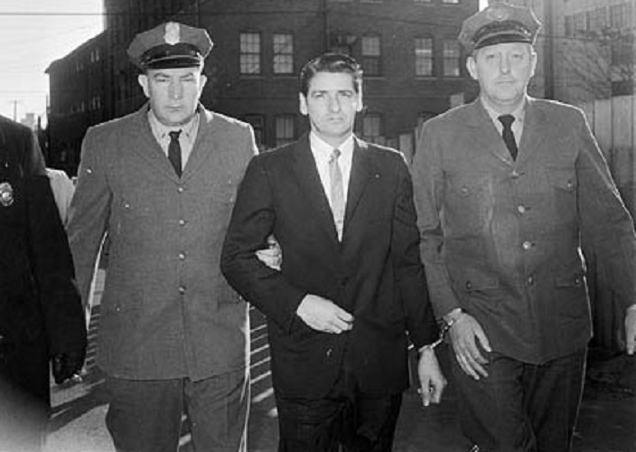
Various serial killers preferred to strangle their victims. This scientific report gives more insight into strangulation’s extremely unsettling effects.
As the greater horns protrude toward the back of the neck, they become compressed against the third or fourth cervical vertebrae during strangulation. With the superior horns of the thyroid cartilage attaching to the greater horns of the hyoid, pressure to the thyroid can also cause hyoid fractures and vice versa.
Hyoid Bone and Sleep Apnea
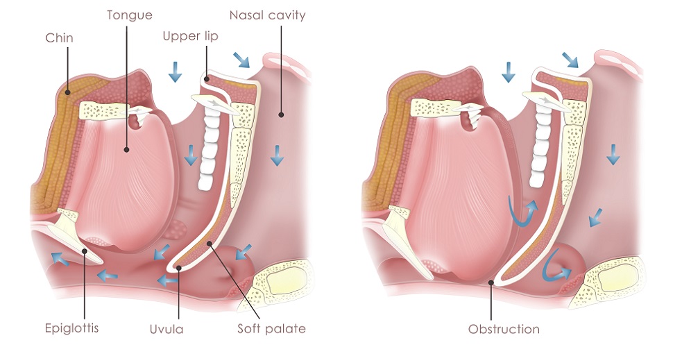
Obstructive sleep apnea syndrome (OSAS) is a chronic breathing disorder that occurs during sleep when the pharynx collapses.
As the tongue bone is an attachment point for the suprahyoid and infrahyoid muscle groups, and as it is these muscles together with muscles of the oropharynx that keep the airway open, it is easy to understand why this bone plays a role. When we sleep, many neurological stimuli fall away – this is particularly noticeable in our muscles. People who suffer from sleep paralysis will know exactly how unresponsive the muscles become.
In OSAS, relaxed muscles and pressure from fatty tissue (OSAS is usually associated with people who are overweight), cause the upper airway to close.
Hyoid position is a contributing factor to obstructive sleep apnea. When the two hyoid muscle groups are not working in sync, they contribute to pharyngeal collapse.
While OSAS is commonly treated with continuous positive airway pressure – the blowing of air into the airway under pressure – this chronic disorder can also be treated with surgery.
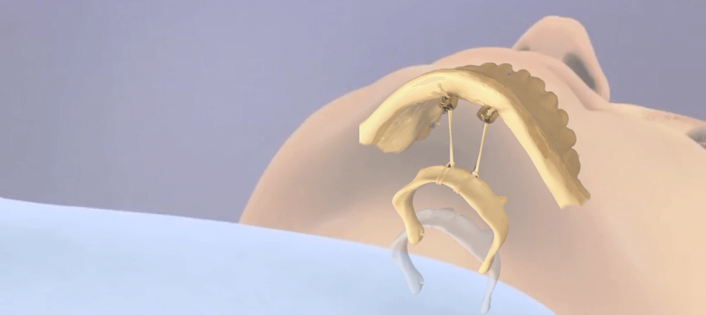
Surgical treatments typically involve the hyoid bone. Examples of sleep apnea surgery are hyoidothyroidopexy, hyoid suspension, and hyoid myotomy. The former stitches the hyoid to the upper portion of the thyroid cartilage. Hyoid suspension brings the bone forward by fixing it to the lower jaw (below). The latter surgery type (carried out with or without suspension) cuts into the muscles of the hyoid to create more room for air to pass through.
Quiz