‘Gram-positive’ and ‘gram-negative’ are terms used to broadly categorize two different types of bacteria. This distinction is made based on the structure of their cell walls, and their reaction to Gram staining.
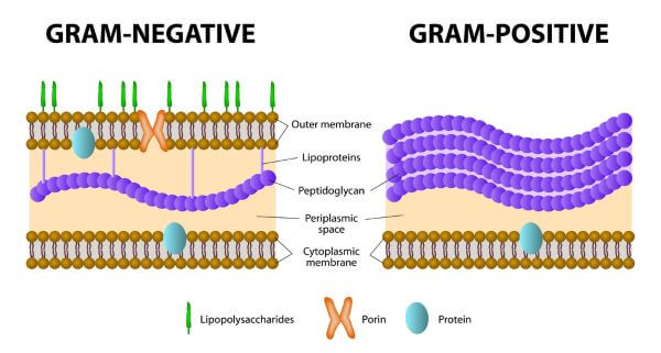
Gram-positive bacteria have cell walls made of a thick layer of peptidoglycan. The cell walls of gram-negative bacteria contain only a thin layer of peptidoglycan, but they also have an outer membrane that is absent in gram-positive bacteria.
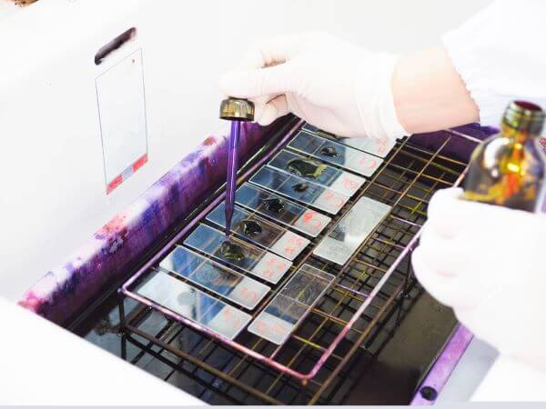
Gram staining is a technique that uses violet dye to distinguish between gram-positive and gram-negative bacteria. If the bacteria are gram-positive, the thick, peptidoglycan layer in their cell walls will retain the dye and they will stain violet. If the bacteria are gram-negative, the dye will leak out of the thin peptidoglycan layer, and the bacteria will stain red.
What Are Bacteria?
Bacteria (single: bacterium) are a type of unicellular, prokaryotic organism that inhabit almost every environment on Earth. Bacteria are very diverse and may be found living in soil, the oceans, hot springs, our houses, and even inside our own bodies. Many are harmless or even beneficial. For example, the bacteria that live inside the human gut (known as the gut microbiota) help with digestion and may even prevent or treat certain illnesses. Others (called pathogens) are not so friendly and are capable of causing diseases.
There are thought to be millions of species of bacteria on Earth, but they can be broadly divided into two categories based on a single characteristic: the structure of their cell walls.
Gram-Positive vs. Gram-Negative Bacteria
Gram-Positive Bacteria |
Gram-Negative Bacteria |
| Thick peptidoglycan layer in cell wall | Thin peptidoglycan layer in cell wall |
| No lipopolysaccharide membrane | Lipopolysaccharide membrane |
| Produce exotoxins | Produce endotoxins |
| Stained purple by gram-staining | Stained red/pink by gram staining |
Cell Wall Structure in Gram-Positive vs. Gram-Negative Bacteria
‘Gram-positive’ and ‘gram-negative’ are terms used to classify bacteria based on the structure of their cell walls.
Most bacterial cell walls contain a substance called peptidoglycan (AKA murein). Peptidoglycan is a large polymer made up of sugars and amino acids and is unique to bacteria. The cell walls of gram-positive bacteria are made up of a thick, mesh-like layer of peptidoglycan. Gram-negative bacteria have only a thin layer of peptidoglycan in their cell walls, but they also have an outer membrane containing lipopolysaccharides. This outer membrane is not present in gram-positive bacteria.
Toxins Produced by Gram-Positive vs. Gram-Negative Bacteria
Exotoxins and endotoxins are both types of toxins that are produced by bacteria. Endotoxins are also known as lipopolysaccharides and are found in the outer membranes of gram-negative bacteria. As gram-positive bacteria lack an outer membrane, they do not produce endotoxins.
Exotoxins are toxins that are secreted by bacteria and are produced by both gram-negative and gram-positive bacteria.
What is Gram Staining, and How Does it Work?
Gram staining is a common technique used to distinguish between gram negative and gram positive bacteria. It was named after Hans Christian Gram, a Danish bacteriologist who first developed the method back in 1882.
Gram staining involves staining a sample of bacterial cells with a crystal violet dye followed by a Gram’s iodine solution (containing iodine and potassium iodide). Next, a decolorizer (such as ethyl alcohol or acetone) is added to the sample. This dehydrates the peptidoglycan layer in the bacterial cell walls, causing it to shrink and tighten. In gram-positive bacteria, the crystal violet dye is trapped inside the thick, peptidoglycan layer. In gram-negative bacteria, the thin peptidoglycan layer is unable to hold onto the dye, and it leaks out of the cell wall.
Finally, the bacteria are viewed under a microscope. The color of the cells after staining indicates whether they are gram-positive or gram-negative.
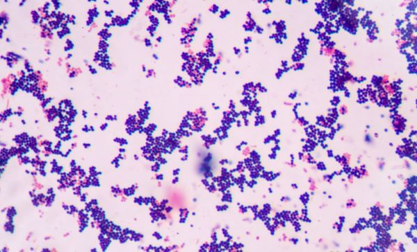
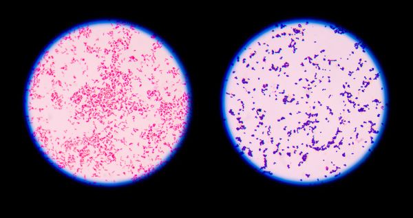
Gram Staining Results in Gram-Positive vs. Gram-Negative Bacteria
If bacteria are gram-positive, the Gram staining method will turn them violet. This happens because the thick, mesh-like layer of peptidoglycan in their cell walls retains the crystal violet dye. If the bacteria in the sample are gram-negative the dye will stain them red or pink, as their thin peptidoglycan walls do not retain the crystal violet dye during the decolorizing process.