Definition
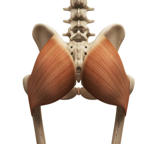
The gluteus maximus is one of three gluteal muscles that make up the buttocks. As the largest and most superficial of the three, it is the most prominent structure in which the buttocks and hips get their shape and function. The action of the gluteus maximus is to extend and laterally rotate the hip. Specific functions include maintaining body erectness, raising the body from sitting and bending positions, and ensuring balance when standing on one leg. While all three gluteal muscles originate at the ilium of the pelvis, the gluteus maximus specifically originates at the lateral surface of the iliac crest. It gathers to the side towards a tendon facing the body’s posterior and inserts on the greater trochanter of the femur. The gluteus maximus is innervated by the inferior gluteal nerve (L5-S2) and is vascularized by the superior and inferior gluteal arteries.
Background of Muscles
Muscle Tissues and Cells
There are three different muscle tissues that are found in the body: cardiac, smooth, and skeletal. Cardiac muscle makes up the heart and functions to circulate blood throughout the body. Smooth muscle is found in many bodily systems that undergo involuntary movement, such as the digestive tract, respiratory system, urinary tract, and reproductive system, as well as along blood vessel walls. Skeletal muscle is found throughout the body and has many functions. Primarily, skeletal muscle is used to voluntarily move body parts. It is also found at the openings and closings for the digestive tract, urinary tract, and respiratory system.
The gluteus maximus is made up of skeletal muscle tissue.
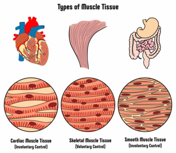
All three types of muscles are made up of individual cells known as myocytes, where the cells for each tissue type slightly differs. Cardiac muscle cells are short, branched, and striated, and are connected to each other by intercalated discs. These cells usually only have a single nucleus. Smooth muscle cells are also short but are instead spindle-shaped and non-striated. There is only one nucleus present per cell. Skeletal muscle cells are both long and striated with a cylindrical shape. These cells contain multiple nuclei. Cardiac and skeletal muscles can also be referred to as muscle fibers.
Skeletal Muscle Parts and Properties
Several general properties are associated with muscles, such as having high elasticity. This means that the tissue can stretch, extend, and recoil back to its original shape and size. As muscles utilize a large amount of energy in the form of adenosine triphosphate (or ATP), muscle cells contain a significant number of ATP-producing mitochondria. Additionally, muscle cells are “excitable”, similar to nerve cells when stimulated by action potentials. This feature is critical for muscles to contract.
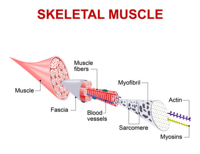
Muscle cells have similar properties, structures, and organelles to other cell types, but also have their own specific terminologies and differentiating properties. Generally, all muscle fibers have a cell membrane that is called the sarcolemma. Additionally, muscle cells contain sarcoplasm, which is the intracellular equivalent to cytoplasm.
To accompany muscle function however, these cells contain transverse tubes that pass along contraction signals throughout the fiber. Myofibrils are responsible for extending the length of the cell when muscles expand. These myofibrils contain repeating sarcomere structures, which contain thick (myosin) and thin (actin) filaments that increase tension to shorten the muscle during contractions. The myofibrils are surrounded by the sarcoplasmic reticulum (comparable to the endoplasmic reticulum), which is important for storing calcium ions (Ca2+). Branches at the end of the sarcoplasmic reticulum come together to form the terminal cisternae, which is used for calcium ion uptake following muscle contractions.
Neuromuscular Junction
Muscle contractions occur when fibers are stimulated by neurons at the neuromuscular junction (also known as the myoneural junction). For each skeletal muscle fiber, there is a nerve cell called the motor neuron. The motor neuron releases acetylcholine– a chemical messenger- that excites the muscle fiber. The neuromuscular junction is the space where the muscle fiber and motor neuron meet.
Neurons are made up of three main parts: the cell body (or soma) which contains the cell’s nucleus, several short and branching dendrites, and a long axon that generates action potentials. An action potential is the electrical change that occurs when cell membranes depolarize (or experience a sharp and sudden increase in membrane charge due to fluxing ions). The end of the axon contains the synaptic terminal. Located in the synaptic terminal are synaptic vesicles that contain acetylcholine. The synaptic vesicles release the stored acetylcholine into the small space between the terminal and muscle fiber sarcolemma- which is known as the synaptic cleft.
(Note: Acetylcholinesterase- which is an enzyme- is also released into the synaptic cleft to prevent the muscle fiber from becoming overstimulated.)
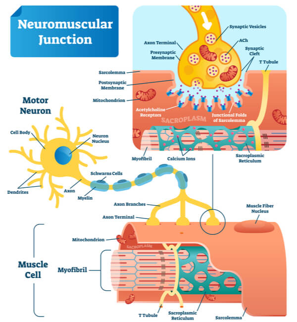
Once at the muscle fiber, the released acetylcholine binds to receptors on the sarcolemma. This binding creates an action potential that “excites” the muscle fiber and spreads contraction signals throughout the cell. Once excited, calcium ions from the sarcoplasmic reticulum are released into the sarcoplasm, which then bind to troponin (a protein found on the muscle fiber). This binding removes the pre-attached tropomyosin to expose active sites on G- actins for myosin heads to attach. The attached myosin heads then “walk” along the action, causing the thin filaments to slide and thus shorten the sarcomere. As this occurs in multiple sarcomeres, myofibril length decreases and the overall muscle contracts. Following muscle contraction, the released calcium ions are actively transported out of the sarcoplasm and into the terminal cisternae.
The motor neuron that innervates the gluteus maximus is the inferior gluteal nerve.
Muscle Actions
When referring to muscles, there are multiple terms used to indicate specific actions. A few of the general body terms and their associated actions are as follows:
Abduction: Moving a body part away from the body’s center
Adduction: Moving a body part towards the body’s center
Elevation: Moving a body part up
Depression: Moving a body part down
Flexion: Decreasing the angle of a body part (ex. bending an elbow or tilting your head towards your chest)
Extension: Increasing the angle of a body part (ex. straightening your legs to stand after sitting in a chair)
Rotation: External- Moving a body part out away from the body’s axis (ex. rotating your hip and leg so that your foot points out to the side while your body still faces forward)
Internal- Moving a body part in towards the body’s axis (ex. rotating your hip and leg so that your foot realigns to point forward while your body still faces forward)
Note: The head can also be rotated in either direction of the body’s axis
The action of the gluteus maximus is to both extend and laterally rotate the hip.
Gluteus Maximus
The gluteus maximus is one of three gluteal muscles that form the buttocks, along with the gluteus medius and gluteus minimus. As the largest and most superficial of the three muscles, the gluteus maximus makes up most of the buttock’s and hip’s shape. The major actions are to extend and externally rotate the hip and thigh. Additionally, the gluteus maximus also plays a major role in keeping the body erect. Combining these functions, this muscle is highly used when completing many simple actions, including rising after sitting in a chair, raising the upper torso after bending over, and walking up stairs. Furthermore, the gluteus maximus supports the pelvis when the body is balancing on one leg, as well as steadies the femur through its deep connections.
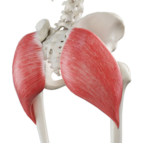
As a large muscle, the gluteus maximus originates at several points of the pelvis. Specifically, it originates at the iliac crest of the pelvis, the inner upper ilium, the lower part of the sacrum, and the coccyx. Meanwhile, it inserts into two points: the superficial fibers of the greater trochanter and the fascia lata of the thigh. It also connects to deep fibers that insert into the gluteal tuberosity found between the adductor magus and vastus lateralis (both found on the thigh). While the gluteus maximus runs both inferior and laterally, it is further split into superior and inferior portions. The superior portion inserts at the fascia lata’s iliotibial tract, while the inferior portion inserts at the femur’s gluteal tuberosity.
Additionally, the gluteus maximus is vascularized by the superior and inferior gluteal arteries.
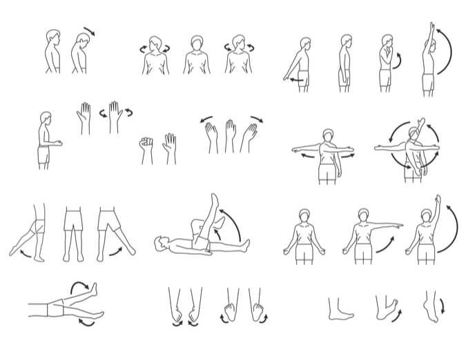
Gluteus Maximus Exercises
The gluteus maximus is regarded as the primary muscle responsible for hip extension. This is especially evident in exercises that utilize simultaneous hip and knee movement, such as with squats and leg presses.
Electromyography (EMG) is a common technique used by scientists and medical professionals to measure electrical potential fields produced during sarcolemma depolarization. As a result, the EMG can sum together the action potentials of motor neurons to indicate muscle activation levels when specific actions are monitored in controlled conditions. This thus allows exercise physiologists to identify effective movements for strengthening specific muscles, including the gluteus maximus.

Ultimately, it was found that step-up exercises- including associated variations such as lateral, diagonal, and cross- over step-ups – yield the highest gluteus maximus activation levels. Bilateral exercises- such as hip thrusts, squats, lunges, and deadlifts- can also lead to high levels of gluteus maximus activation. It is important to note however, that individual differences in exercise methodology can lead to varying activation levels. Additionally, outlying factors- such as exercise kinetics, movement velocity, motion range, fatigue levels, and mechanical complexity- can also lead to varying results.
Gluteus Maximus Associated Pain and Weakness
The neuromuscular system is biologically optimized to provide movement in spite of muscle dysfunctions. The gluteus maximus is such a case, where internal muscle coordination can change, and biochemical overload injuries can occur despite dysfunctions appearing without physical symptoms. This is especially evident when one muscle continues to perform while a corresponding muscle is compromised, thus leading to multiple potential injuries. For example, gluteus maximus weakness can follow knee pain, lower back pain, hamstring stress, ankle sprains, and other preliminary injuries. Reduced gluteus maximus activation following these injuries is thought to benefit short- term muscular heath and prevent injuries following large movements.
Given the evolutionary importance of the gluteus maximus to maintaining erect postures and proper movements, certain lifestyle choices can weaken this muscle and eventually cause pain. Notably, prolonged sitting can lead to gluteus maximus weakness and increased cell atrophy. Secondary hip extensor muscles- including the hamstrings– are more heavily used when sitting due to the body utilizing more energy efficient motor routes. Thus, these sitting positions expense the energy demands of the less used muscles, leading to further gluteal area pain. Additionally, the hip flexor muscles can tighten over a prolonged time period, causing the gluteus maximus cells to elongate.
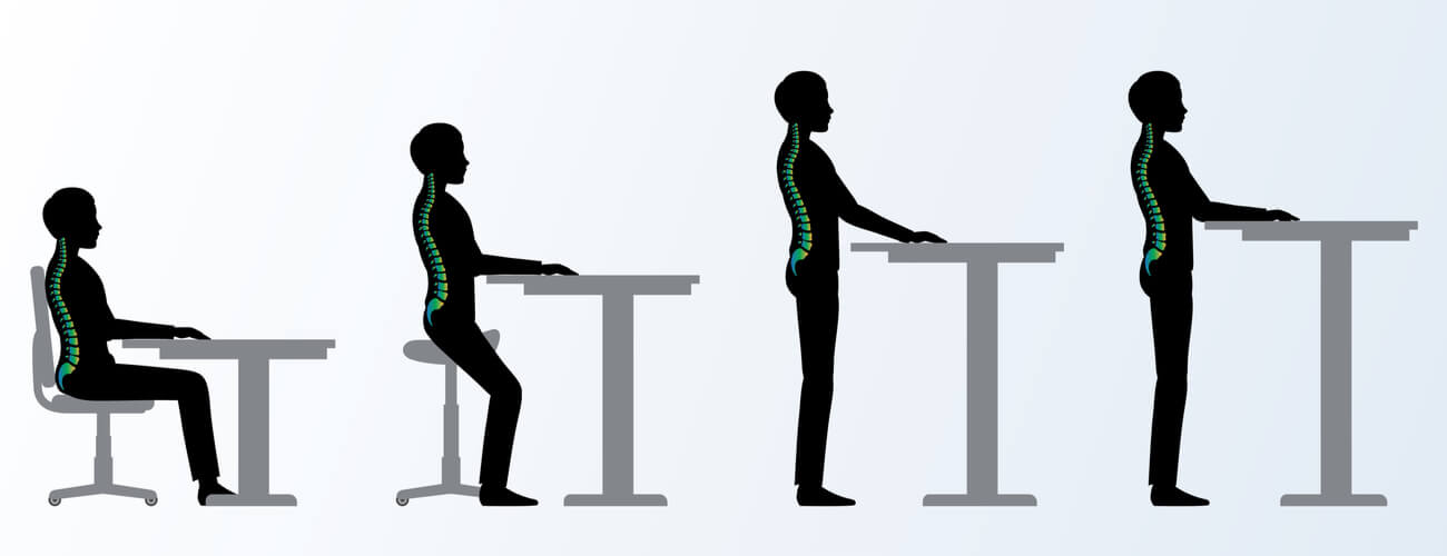
Treatments to overcome gluteus maximus weakness and pain are case dependent, but can include the following: treating the initial injury that led to the gluteal weakness, undergoing physical therapy, using inhibitory injections- such as botulinum toxin type A-, increasing exercises surrounding the gluteus maximus, correcting sitting and standing body postures, and reducing sitting periods. For pain related to muscle tightness, self-massage techniques using a foam roller can increase overall motion and performance. Additionally, all exercises undertaken should be properly followed to ensure the correct muscles are being targeted. These exercises should replace alignment of the muscles incorrectly targeted in the past (whether it was due to poor sporting behaviors or sitting postures) with the intended targets instead.
Conclusions
The gluteus maximus is the largest and most superficial of the three gluteal muscles that constitute the buttocks and hips. Innervated by the inferior gluteal nerve, the action of the gluteal maximus is to extend and laterally rotate the hip, which allows the body to maintain body erectness and recover from sitting and bending positions. Improper sitting postures and additional injuries in the surrounding areas can lead to gluteus maximus weakness and pain, where step-up and bilateral exercises can aid in strengthening the muscle.
Quiz