Definition
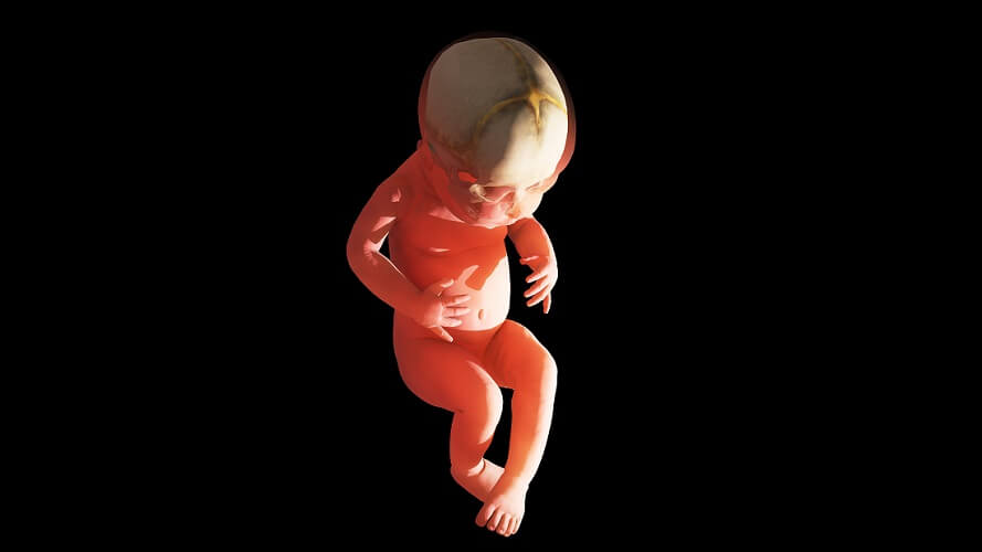
A fontanelle, fontanel, or soft spot is an anatomical feature of a baby’s skull. A baby has six fontanelles that consist of membranous tissue in the areas where certain adult skull sutures are found. Fontanelles allow the skull to pass through the birth canal and also provide a means of expansion as the brain grows.
The Baby Fontanelle
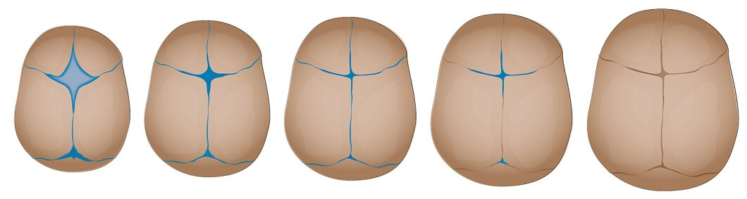
Six baby fontanelles close at different stages, from early babyhood until around the age of eighteen months. This period can vary slightly from child to child.
The fontanelle is quite delicate and should be protected from trauma – only thick membranous connective tissue protects the delicate structure of the underlying brain. Even so, many new parents are overly cautious concerning this part of the newborn anatomy.
The six fontanelles are the:
- Anterior fontanelle x 1
- Posterior fontanelle x 1
- Mastoid fontanelle x 2
- Sphenoid or sphenoidal fontanelle x 2
Anterior Fontanelle
The anterior fontanelle is the largest. This diamond-shaped soft spot is located between the frontal and parietal bones. At this point in life, the frontal bone is not a single bone but consists of a left and right bone that must grow in size and fuse as the brain increases in mass.
The position of this largest soft spot can be easily seen on a newborn; in adults, the location of the now-fused fontanelle is at the junction of the frontal, coronal, and sagittal sutures of the skull. Where these sutures intersect is called the bregma – Greek for the top of the head. An anterior fontanel is an early form of the bregma.
This soft spot usually closes by the age of eighteen months.
Posterior Fontanelle
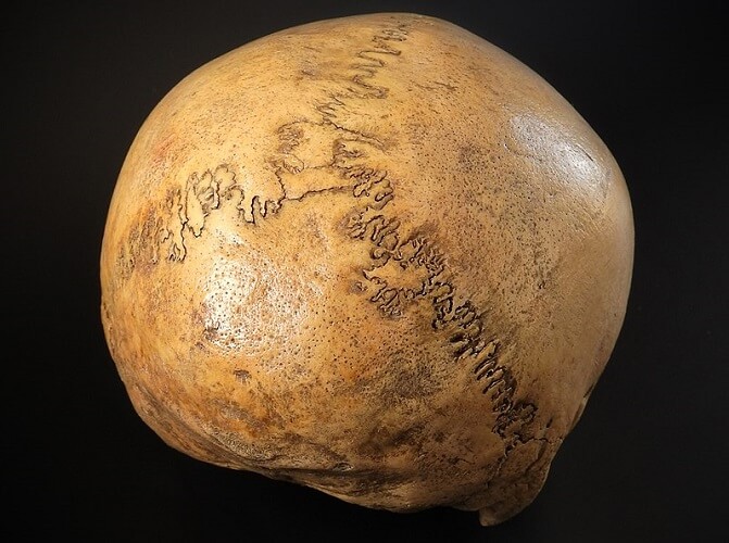
The posterior fontanelle is triangular and closes much more rapidly – at around the eighth postnatal week. It is found where the parietal and occipital bones eventually meet at the junction of the sagittal and lambdoid sutures. Where these sutures intersect is called the lambda. A posterior fontanelle is an early form of the lambda.
Mastoid Fontanelle
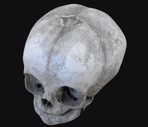
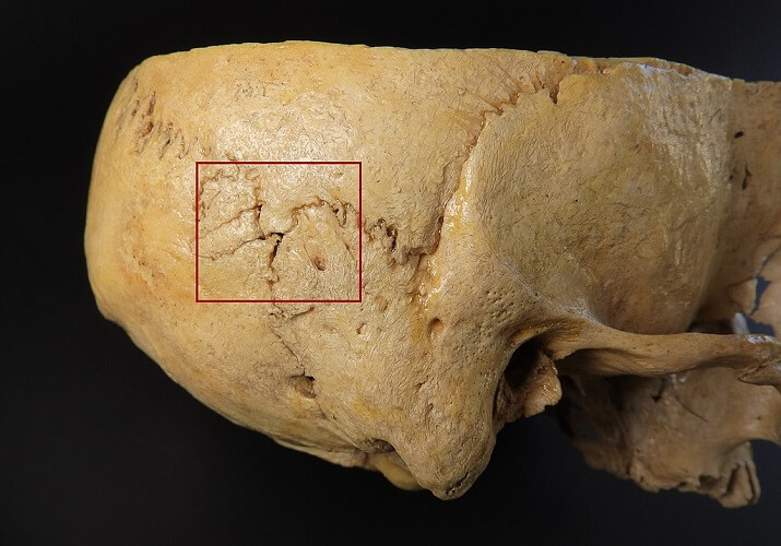
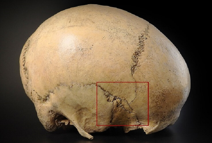
The paired mastoid fontanelle or posterolateral fontanelle – left and right – is located between the temporal, occipital, and parietal bones, behind the ear. In the photograph below, you can see the entrance to the acoustic meatus behind the zygomatic process of the temporal bone.
The mastoid fontanel marks the position of the junction of the parietomastoid, occipitomastoid, and lambdoid sutures at the side of the head. This point is called the asterion. A mastoid soft spot is an early form of the asterion.
It closes between the sixth and eighteenth postnatal month.
Sphenoidal Fontanelle
The paired sphenoid fontanelle or anterolateral fontanelle is located between the sphenoid, parietal, temporal, and frontal bones, at the temple. It marks the position of the junction of the coronal, sphenofrontal, sphenoparietal, sphenosquamosal, and squamosal sutures. This area is known as the pterion – the weakest part of the adult cranium. A sphenoidal fontanelle is an early form of the pterion; it closes in or around the sixth postnatal month.
The Third Fontanelle
In rare cases, a third soft spot is found. This indicates a disorder – most commonly Down syndrome or congenital hypothyroidism (low levels of thyroid hormone). This abnormal fontanelle is located in front of the posterior fontanel.
Other Disorders
Normal soft spots close at slightly varying rates; however, abnormal sizes may indicate underlying disorders.
Overly-large soft spots are associated with:
- Chromosomal disorders such as Down syndrome, trisomy 18, and trisomy 13;
- Genetic disorders including achondroplasia (dwarfism) and Beckwith-Wiedemann syndrome (abnormal growth);
- Hypothyroidism;
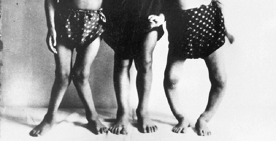
- Malnutrition, including rickets (prolonged vitamin D deficiency);
- Problems during pregnancy such as fetal hydantoin syndrome (when a pregnant mother takes a certain type of epilepsy medication);
- Increased pressure in the brain, most commonly due to hydrocephalus (water on the brain).
When the brain is unable to drain away excess cerebrospinal fluid or leaked blood, fontanels can save a baby’s life. While an adult skull cannot change shape once fused, soft spots allow a young baby’s skull to expand. As a result, the skull can become misshapen if the cause of the brain’s swelling is left untreated.
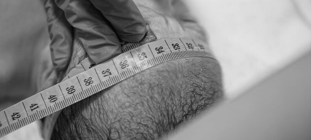
Very small fontanelles can be caused by hyperthyroidism (too much thyroid hormone production), infection, and genetic disorders that cause microcephaly (small head), and craniosynostosis. In craniosynostosis, the bones of the skull fuse too early. If this only occurs at one suture, the other sutures will expand to allow the brain to increase in size and the head will be misshapen. If all of the sutures are affected, immediate surgery is required to relieve the resulting high intracranial pressure.
Fontanelle Closure
Fontanelle closure, if not affected by the above-mentioned disorders, is relatively predictable.
- Anterior closure: 18 months
- Posterior closure: 8 weeks
- Mastoid closure: 6 – 18 months
- Sphenoid closure: 6 months
Up to the time the soft spots have disappeared by way of a process called intramembranous ossification, these areas of the skull are susceptible to damage. If the skin is broken, bacteria and/or viruses can potentially access the brain.

Normal Fontanelle vs Sunken Fontanelle
A normal fontanelle is flat or very slightly sunken and can easily be felt with the fingertips. Many new mothers are likely to panic when they feel it bulging when the baby cries (as you can see below) – this is normal. When the baby stops crying, the bulge disappears.

A normal fontanel can also pulsate, as only thick membranes separate the skin from the blood supply to the brain.
A sunken fontanelle that has a visible dip, however, is a sign of dehydration. Reasons for dehydration are often uncomplicated and easily treated – diarrhea, vomiting, fever, or overheating.
If a young baby stops producing wet nappies or cries without tears, this is a medical emergency. Once fluid levels improve, a sunken soft spot will return to a more healthy form.

Bulging Fontanelle
A fontanelle bulge can occur when a newborn is crying or even when frustrated and angry; however, a soft spot bulge that remains when the crying has stopped could point to a variety of pathologies.
The list of possible illnesses is long and includes neurological complications, cardiovascular problems, infections, blood disorders, endocrine and metabolic imbalances, toxicity, and trauma. It is vital that a bulge at any of the soft spots (that is not only present when the baby is crying) is seen by an experienced pediatrician, preferably when the baby is calm and happy.
A pediatrician will test for the Macewan sign or “cracked pot” sign – this is the sound the fontanel makes when gently tapped. Alternatively, the doctor may use his or her stethoscope to listen for a bruit – this rushing sound may indicate an arteriovenous malformation or AVM. An AVM is tangled mix of arteries and veins that lowers circulating oxygen levels.