Definition
The ethmoid bone or os ethmoidal is a singular (unpaired), irregular, highly perforated bone of the cranium located between the eye sockets and running back to the frontal lobe of the brain. Ethmos means sieve in Greek which indicates this bone’s many perforations. Grooves allow nerve branches to pass from brain to nasal passage; air chambers make this bone part of the paranasal sinuses.
Ethmoid Bone Location
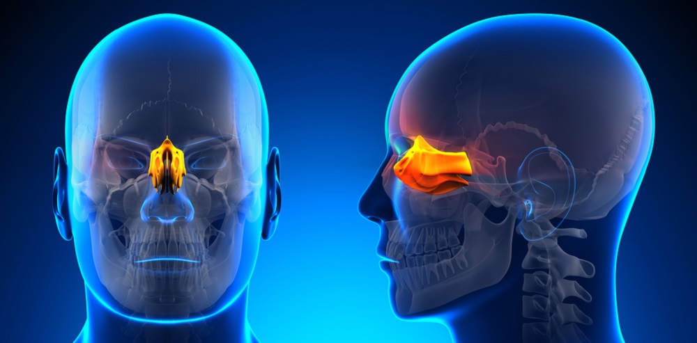
The ethmoid bone is located between the orbital bones of the eyes, at the top of the anterior cranial fossa. It sits above the sphenoid bone and behind the nasal bone. A spongy, lightweight cube-shaped bone, it contributes to the paranasal sinuses and presents with characteristic channels and grooves.
The lightweight form and many perforations mean this bone is relatively fragile. Ethmoid bone location, set between many other bones of the face, means that it is rare for it a single structure to be damaged. Blows to the face (assault) or falls directly onto the face can lead to naso-orbito-ethmoidal fractures – combination of fractures to the nasal bone, orbital bones, and ethmoidal bone; the most common cause of this complex fracture is the motor vehicle accident, when the victim’s forehead hits the dashboard.
The below image shows a labeled ethmoid bone surrounded by many other bony structures. It is extremely well hidden..
Ethmoid Bone Anatomy
Ethmoid bone anatomy is quite complicated. It is one of eight cranial bones, two of these – the parietal and temporal bones – are paired; the others – the frontal, occipital, sphenoid, and ethmoid bones – are unpaired.
There are four main ethmoid bone parts. These are the cribriform plate, two ethmoid labyrinths, and a perpendicular plate. Each part has its own set of functions.
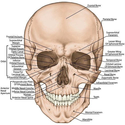
When we look at ethmoid bone anatomy from the front (anterior view), most of the bone is hidden by the orbital cavities of the eyes, the frontal bone, and the short nasal bone. If we remove these we see a narrow piece of bone with a central protuberance called the lamina perpendicularis, vertical plate, or perpendicular plate (see the labeled ethmoid bone anterior view a little further on).
Perpendicular Plate of the Ethmoid Bone
The perpendicular plate of the ethmoid bone runs vertically and ends to form the first two-thirds of the nasal septum. It provides an attachment point for the septal cartilage we can feel between our nostrils. The perpendicular plate begins just under the cribriform plate.
The top of the ethmoid bone’s perpendicular plate has many grooves and channels (canals). These also run from the cribriform plate and into the nasal passages and provide a protected surface for the olfactory nerves.
Cribriform Plate of the Ethmoid Bone
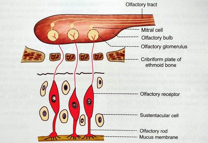
The cribriform plate or horizontal lamina fits into a groove at the underside of the frontal bone. This ethmoid bone part provides a roof for the nasal cavity and a floor for the olfactory bulb. The cribriform plate of the ethmoid bone is also perforated to allow olfactory nerves to travel along the vertical perpendicular plate and mucous membranes of the nasal cavity. Another nerve that passes through the cribriform plate is the nasociliary nerve.
If the cribriform plate of the ethmoid bone is the flat roof of a house, the perpendicular plate is that house’s very narrow front façade. The roof has a chimney right at the back – the crista galli of the ethmoid bone. Crista galli is Latin for a cockerel’s comb; it rises vertically from the cribriform plate. The back of this protuberance serves as an attachment point for the falx cerebri – a major fold in the dura mater membrane of the brain that separates both brain hemispheres. The front of the crista galli ‘chimney’ is a point of articulation with the frontal bone.

Ethmoid Labyrinths
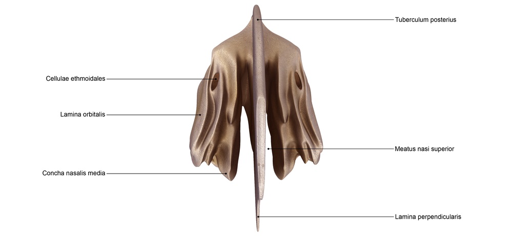
The posterior view of the ethmoid bone, labeled above, shows the flat roof of the cribriform plate. On either side are two ‘wings’ – the sidewalls of the house example. If you compare the posterior view of the wings with the anterior view, you will see that the fronts of the both bones are grooved. These twin bones are the ethmoid labyrinths or lateral masses.
Ethmoid labyrinths have two important structures. The first is their inner surfaces that form the superior and middle conchae. The inferior concha is a separate structure and not part of the ethmoid bone.
Conchae or turbinates are covered with ciliated epithelium and make a scroll-like shape on either side of the nasal passage. They increase the total volume of air within the bony structures around the nose. This means that the air we breathe in will be slightly warm and a little humid.
The most interesting part of ethmoid bone anatomy is the ethmoid air cell. Multiple air cells are found in both of the labyrinths and are also referred to as ethmoid sinuses. Ethmoid air cells are not eukaryote cells but air chambers and part of the paranasal sinus network. As we grow older, more ethmoidal air cells appear. Young children have up to four; an adult can have more than ten; these will also be larger.
Ethmoidal air cells get bigger over time. This process is known as pneumatization. Sinus pneumatization is a continuous process that causes the paranasal sinuses to increase in volume. The ethmoid sinus is the first of the paranasal sinuses to initiate pneumatization. In one study, the ethmoid air cells of all 46 newborn study subjects already showed signs of early pneumatized ethmoidal sinuses. The other three sinuses can take months or years to initiate pneumatization. Why this occurs in the ethmoid bone sinus first is not yet understood.
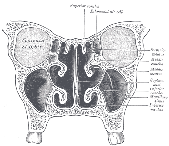
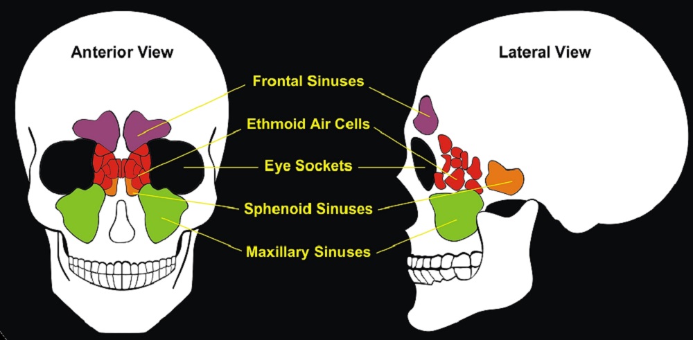
There are three ethmoidal sinuses – anterior, middle, and posterior. The ethmoid bone cavity network is often described as honeycomb-like. These networks of air chambers open – as do all paranasal sinuses – into the nasal cavity. Sinus to nasal cavity openings are referred to as ostia (singular: ostium).
All paranasal sinuses – frontal, sphenoid, maxillary, and ethmoid – increase the volume of the nasal cavity. These aerated channels are lined with ciliated pseudostratified epithelium and goblet cells.
Ethmoid Bone Function
Ethmoid bone functions are quite easy to figure out when you have looked at the anatomy, although some functions may be surprising. With perforations and grooves for olfactory nerves and the ethmoid bone’s proximity to the olfactory organs, it is obvious that it plays a role in our sense of smell. However, this small cranial bone does have other uses.
Face Stabilizer
The ethmoid bone is one of the most complex bones in the human body. Its small size and light weight might make it an unlikely candidate for increased facial stability; however, the ethmoid forms a strong and supportive border with the orbital cavities and the nasal cavity. The form of the ethmoid air cells also provides a higher level of structural stability.
Naturally, any bone protects the soft tissue that lies underneath. The ethmoid protects part of the frontal lobe of the brain. Naso-orbito-ethmoidal fractures of the nasal, orbital, and ethmoid bones respectively are extremely complex and dangerous fractures but even so their presence may prevent severe frontal brain damage in a car accident or with high-energy blunt trauma. The proximity of the brain to the ethmoid makes this bone the principle cause of cerebrospinal fluid rhinorrhea (leakage of cerebrospinal fluid through the nose) when fractured.
The crista galli of the ethmoid bone also serves as an attachment point for the falx cerebri.
Respiratory Immunity
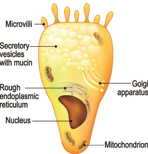
Ethmoid sinuses have many functions but their primary role is associated with our innate immunity. Mucus-producing cells called goblet cells are found in the epithelium of all the paranasal sinuses. This mucus runs into the nasal cavity, trapping bacteria that enter this early stage of the respiratory system.
Labyrinths lie next to the upper nasal conchae or turbinates. These swirled openings increase the volume of air and possibly help make the skull a little lighter; this theory is not always taken seriously. All of the nasal sinuses make a very slight difference to the total weight of the skull; on its own, the ethmoid bone structure makes very little difference.
Ethmoid air cells and superior and middle conchae increase air volume – inhaled air can be warmed and dampened before entering the lungs, further protecting the airway. Goblet cell mucus and nasal hairs also trap many pathogens.
Nasal dendritic cells protect the respiratory system and enable innate immunity in the form of phagocytes and acquired immunity as antigen-presenting cells. Dendritic cells are found in a multitude of tissues, including the epithelial lining of ethmoid air cells.
Nasal Nerve Pathways
The many grooves, perforations, and canals of the ethmoid bone allow important branches of two cranial nerves (olfactory nerves and nasociliary nerve) to reach their destination. The perforations through which these nerves pass are called foramina – the cribriform plate of the ethmoid bone has about twenty foramina.
The olfactory nerve (CN I) is a sensory nerve. The olfactory pathway begins with receptors in the nasal epithelium and ends in the brain. To pass from the nasal cavity to the brain the olfactory nerve must go through the ethmoid bone.
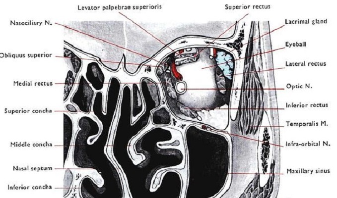
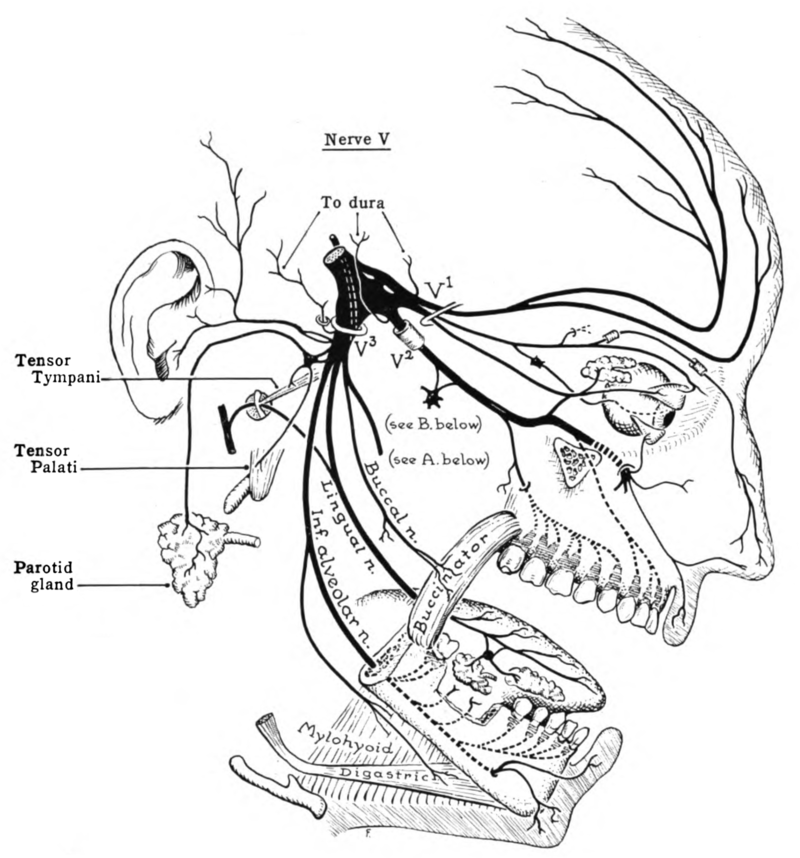
The nasociliary nerve is a smaller branch of the trigeminal nerve (CN V). In the ethmoid, it splits further to form the anterior ethmoidal nerve. This sensory nerve provides sensation in the nose and part of the eye. In the illustration, the V1 branch of the trigeminal nerve splits into further branches; the nasociliary nerve can be followed over the eye socket and down to the tip of the nose.
Quiz