Definition
The cranial bones of the skull are also referred to as the neurocranium. The neurocranium is a group of eight bones that form a cover for the brain and brainstem. The 8 cranial bones are the frontal, parietal, temporal, occipital, sphenoid, and ethmoid bones. Some of these are paired bones.
What Are Cranial Bones?
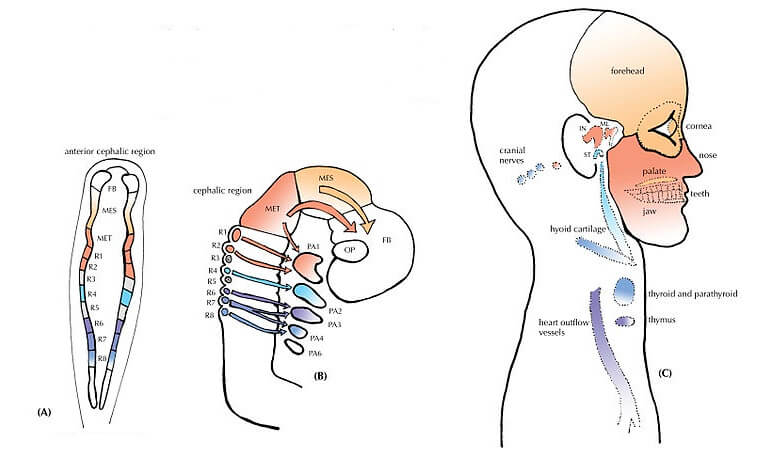
Cranial bone development starts in the early embryo from the neural crest and mesoderm cells. The cranial bones develop by way of intramembranous ossification and endochondral ossification. Endochondral ossification replaces cartilage structures with bone, while intramembranous ossification is the formation of bone tissue from mesenchymal connective tissue.
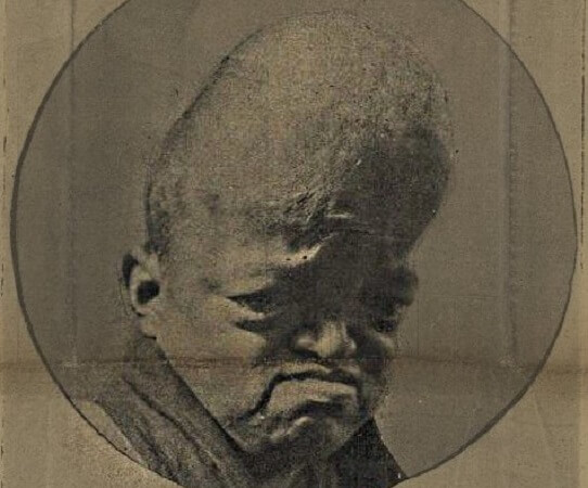
The cranial bones of the skull join together over time. They must be flexible as a baby passes through the narrow birth canal; they must also expand as the brain grows in size. The gaps between the neurocranium – before they fuse at different times – are called fontanelles.
Craniosynostosis is the result of the cranial bones fusing too early. Often, only one or two sutures are affected. This causes a misshapen head as the areas of the cranium that have not yet fused must expand even further to accommodate the growing brain.
The 8 cranial bones are the:
- Frontal bone
- Parietal bone (x 2)
- Temporal bone (x 2)
- Occipital bone
- Sphenoid bone
- Ethmoid bone
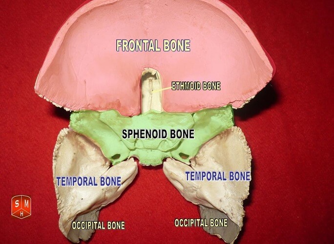
The sphenoid and ethmoid bones are sometimes categorized as part of the facial skeleton. This is because these bones contribute to both areas.
Cranial Bones: Anatomy
Cranial bone anatomy can be confusing when we consider the various terms used to describe different areas. The following words are often used incorrectly; this list gives their true meaning:
- Skull or cranium: all bones of the head, from the top of the head to the hyoid bone (tongue bone). The cranium is the sum of the cranial and facial bones, as well as the bony part of the larynx.
- Neurocranium: the top part of the skull that covers and protects the brain.
- Viscerocranium: the bottom part of the skull that makes up the face and lower jaw.
- Chondrocranium or cartilaginous neurocranium: so-called because this area of bone is formed from cartilage (endochondral ossification). More descriptive terms include skull base and cranial floor.
- Cranial vault, calvaria/calvarium, or skull-cap. Together, the cranial floor and cranial vault form the neurocranium

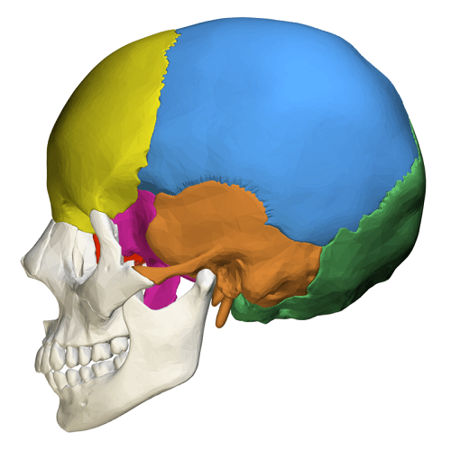
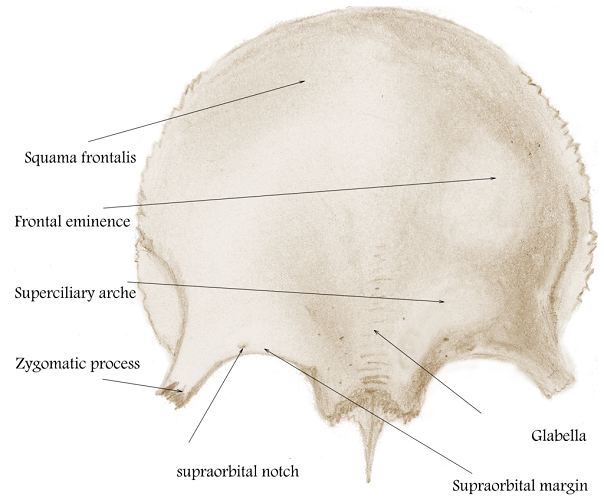
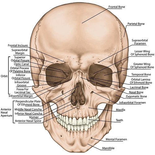
The front of the cranial vault is composed of the frontal bone. This bone forms the ridges of the brows and the area just above the bridge of the nose called the glabella. The frontal bone extends back over the curved line of the forehead and ends approximately one-third of the way along the top of the skull.
This single bone articulates (joins) with the nasal bones, some orbit bones, and the zygomatic bone. At the side of the head, it articulates with the parietal bones, the sphenoid bone, and the ethmoid bone.
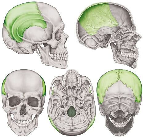
The two parietal bones continue the shape of the cranial vault; these are quadrilateral, smooth, and curved bony plates. They articulate with the frontal, sphenoid, temporal, and occipital bones, as well as with each other at the top of the head (see the final image in the five views below).
The sides of the neurocranium are formed by the parietal, temporal, and sphenoid bones. The temporal bone provides surfaces for both the cranial vault and the cranial floor. It articulates with the mandible by way of a synovial joint. Each temporal bone has sutures with a greater wing of the sphenoid bone and its neighboring parietal bone.
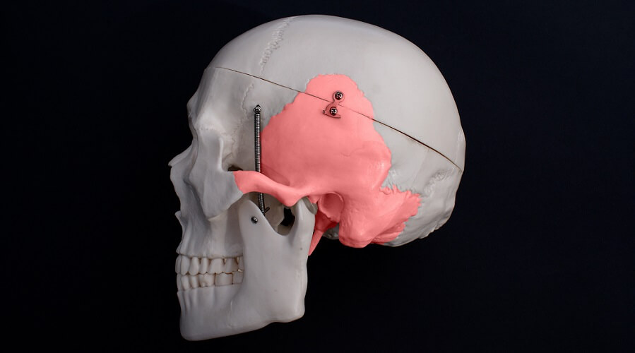
The sphenoid is occasionally listed as a bone of the viscerocranium. However, it also provides important structures at the side and base of the neurocranium. The irregularly-shaped sphenoid bone articulates with twelve cranial and facial bones.
The ethmoid bone, also sometimes attributed to the viscerocranium, separates the nasal cavity from the brain. Like the sphenoid, it is very irregular in shape. It articulates with fifteen cranial and facial bones.
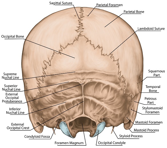
The final bone of the cranial vault is the occipital bone at the back of the head. The occipital bone – located at the skull base – features the foramen magnum. This is a large hole that allows the brain and brainstem to connect to the spine.
Cranial Bone Anatomy: Internal Surface
If you separate the cranial bones from the facial bones and first cervical vertebra and remove the brain, you would be able to view the internal surfaces of the neurocranium.
The midsagittal section below shows the difference between the relatively smooth upper surface and the bumpy, grooved lower surface. The picture also helps us to view the cranial vault in its natural position; the cranial floor is at a distinct angle, starting at the level of the frontal sinus and continuing at an angle to include the small pocket that contains the cerebellum. You can see this small indentation at the bottom of the neurocranium.
The inner surface of the vault is very smooth in comparison with the floor. It does feature a few bumps and grooves. For example, the frontal crest – a notch of bone just behind the frontal sinus. The frontal crest is an attachment point for a fold in the membranes covering the brain (falx cerebri).
Just above the occipital bone and close to the midline of the skull cap are the parietal foramina. Just as with all foramina, important blood vessels and nerves travel through them. They are not visible in the above image.
A vertical groove passes through the middle of the cranial vault – the sagittal groove or sulcus – that provides space for the superior sagittal sinus (part of the drainage mechanism for cerebrospinal fluid and blood). The raised edge of this groove is just visible to the left of the above image.
Below, the position of the various sinuses shows how adept the brain is at removing waste products and extra fluid from its extremely delicate tissues.
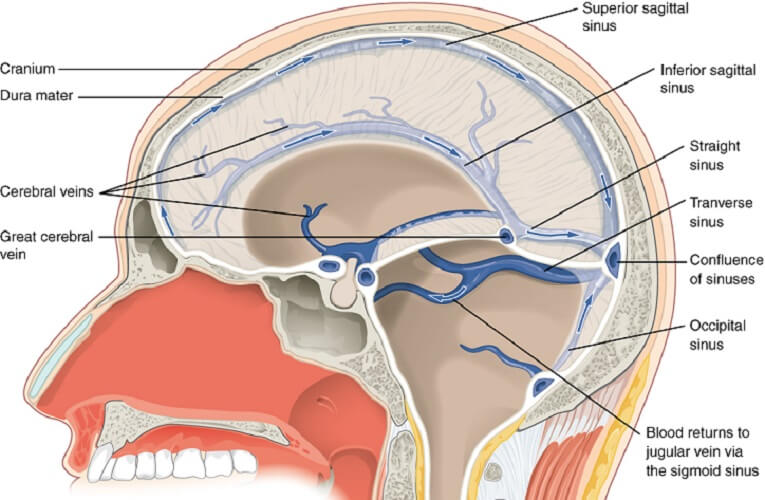
At the back of the skull cap is the transverse sulcus (for the transverse sinuses, as indicated above).
The cranial floor is much more complex than the vault.
Looking down onto the inner surface of the skull base, the first thing you notice is a series of divisions. These form indentations called the cranial fossae. In the cranial vault, there are three:
- Anterior cranial fossa: houses the frontal lobe, olfactory bulb, olfactory tract, and orbital gyri (intellectual and emotional expression)
- Middle cranial fossa: a butterfly-shaped indentation that houses the temporal lobes, features channels for ophthalmic structures, and separates the pituitary gland from the nasal cavity
- Posterior cranial fossa: contains the cerebellum, pons, and medulla oblongata; the point of access between the brain and spinal canal

The inner surface of the skull base also features various foramina. These include the foramen cecum, posterior ethmoidal foramen, optic foramen, foramen lacerum, foramen ovale, foramen spinosum, jugular foramen, condyloid foramen, and mastoid foramen. And let’s not forget the largest of them all – the foramen magnum. A separate Biology Dictionary article discusses the numerous cranial foramina.
Cranial floor grooves provide space for the cranial sinuses that drain blood and cerebrospinal fluid from the lower regions of the meninges (dura mater, arachnoid, and pia mater), the cerebrum, and the cerebellum.
Cranial Bone Sutures
The neurocranium has several sutures or articulations. The first four in the following list are the most important:
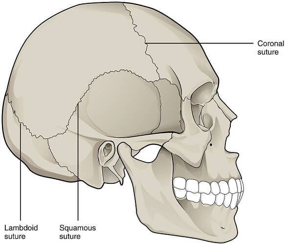
- Coronal suture: between the two parietal bones and the frontal bone
- Sagittal suture: between the left and right parietal bones
- Lambdoidal suture: between the top of the occipital bone and the back of the parietal bones
- Metopic suture: only found in newborns between the two halves of the frontal bone that, once fused (very early in life), become a single bone
- Squamous suture: between the temporal and parietal bones
- Sphenosquamous suture: vertical join between the greater wings of the sphenoid bone and the temporal bones.
- Frontoethmoidal suture: very short suture between the orbital projections of the frontal and ethmoid bones
- Petrosquamous suture: refers to the join between the petrous and squamous parts of the temporal bone, close to the middle ear and at the skull base
- Sphenoethmoidal suture: between the sphenoid and ethmoid bones
- Sphenopetrosal suture: joins the greater wing of the sphenoid bone with the petrous part of the temporal bone
Cranial and Facial Bones
Cranial and facial bones slightly overlap according to textbook sources. Some books include the ethmoid and sphenoid bones in both groups; some only in the cranial group; some only in the facial group.
None of these sources are wrong; these two bones contribute to both the neurocranium and the viscerocranium.

It is, therefore, perfectly acceptable to list them in both groups. This source does not include the ethmoid and sphenoid in both categories, but is also correct.
As we should now be very aware, the 8 cranial bones are the:
- Frontal bone (1)
- Parietal bones (2)
- Temporal bones (2)
- Occipital bone (1)
- Sphenoid bone (1)
- Ethmoid bone (1)
The facial bones are the:
- Sphenoid bone (1 – depending on the source)
- Ethmoid bone (1 – depending on the source)
- Maxillae (2 – sometimes considered to be 1 fused bone)
- Mandible (1 – sometimes considered to be 2 fused bones)
- Zygomatic bones (2)
- Nasal bones (2)
- Palatine bones (2)
- Inferior nasal conchae (2)
- Lacrimal bones (2)
- Vomer (1)
Cranial Bone Fractures
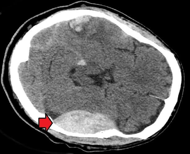
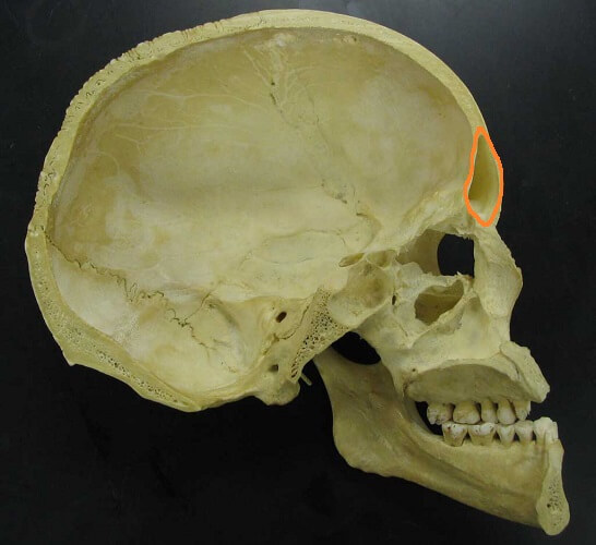
Neurocranium or cranial bone fractures are most likely to occur at a weak spot called the pterion. This refers to an almost H-shaped group of sutures that join the greater wing of the sphenoid bone, the temporal bone, the frontal bone, and the parietal bone at both sides of the head, close to the indentation behind the outer eye sockets.
As one of the meningeal arteries lies just under the pterion, a blow to the side of the head at this point often causes an epidural hematoma that exerts pressure on the affected side of the brain. This can occur in up to 85% of pterion fracture cases. Treatment often requires the placement of hollow tubes (drains) under the skull to allow this blood to drain away.