Definition
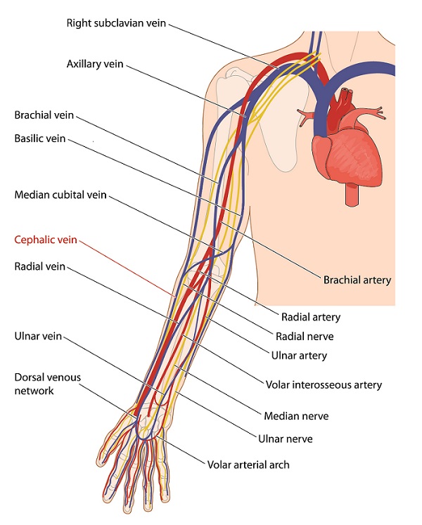
The cephalic vein is a superficial vein in the upper arm that is often visible through the skin. The name means head (likely coming from the Latin ‘cephalicus’ or the Greek ‘kephalikos’).
Veins form a complex network that allows all the tissues of the body to transport deoxygenated blood back to the heart. Lots of smaller veins in the lower limb pass blood into larger veins, like the cephalic vein. The direction of the blood flow of the cephalic vein is from the hand towards the shoulder (i.e., towards the heart).
Cephalic Vein Location and Anatomy
The cephalic vein is one of the two primary veins in the upper arm, the other being the basilic vein. Its path – from its formation from the lower limb to its drainage near the clavicle – is outlined below.
Hand and Lower Forearm
The cephalic vein forms from the dorsal venous network, which is the collection of veins on the back of the hand. This network is clearly visible on most people, due to the lack of subcutaneous fat in the area.
Specifically, the cephalic vein forms from the lateral end of the dorsal venous arch, which is a connection of the dorsal metacarpal veins. The dorsal venous arch drains into the basilic vein and the cephalic vein. The cephalic vein runs along the ‘thumb-side’ of the forearm, whereas the basilic vein runs along the ‘pinky-side.’
After it is formed at the back of the hand, the cephalic vein then crosses the roof of the anatomical snuff box, which is a small triangular deepening where the hand meets the wrist on the upper radial side (i.e., the thumb side). You can see it if you extend your thumb out as in the below image. It is named the anatomical snuffbox because it was a common location to place powdered tobacco (snuff) before sniffing it!

After crossing the anatomical snuffbox, the cephalic vein extends along the radial border of the forearm in front of the elbow.
Elbow
At the elbow, the cephalic vein communicates with the basilic vein (the other of the main veins in the arm) through the median cuboidal vein. Here, much of the blood from the cephalic vein is moved to the basilic vein, through the median cuboidal vein. You might be able to see it at the crease of your elbow joint if you are very lean!
Upper Arm
The cephalic vein then moves along the lateral border of the biceps brachii (commonly called the biceps). It runs superficially alongside the lateral cutaneous nerve of the forearm (sometimes referred to as the lateral antebrachial cutaneous nerve). This nerve is a sensory branch of the musculocutaneous nerve, which itself is a branch of the brachial plexus. It innervates the anterior muscles of the forearm.
Finally, the cephalic vein pierces the deep fascia at the lower border of the pectoralis major muscle (commonly called the ‘pecs’ or the chest muscle). It then runs along the deltopectoral groove, which is an indentation between the border of the pectoralis major muscle (the pec) and the deltoid muscle (the shoulder muscle). It travels along this groove until it reaches the infraclavicular fossa, which is an indentation right below the clavicle.
Drainage
At this groove (the infraclavicular fossa), the cephalic vein pierces the clavipectoral fascia, which is a structure of connective tissue buried under the pectoralis major muscle. It empties the traveling blood into the first part of the axillary vein. Thus, this is where the cephalic vein terminates. At this point, the axillary vein is now called the subclavian vein.
The subclavian vein then meets the internal jugular vein to form the brachiocephalic vein. The brachiocephalic vein is found in the upper chest, where it merges with its partner on the other side of the body (i.e., the veins from the left and right side come together). When they unite, the pair of brachiocephalic veins form the superior vena cava, which directly returns blood to the heart for oxygenation.
Accessory Cephalic Vein
The accessory cephalic vein joins the cephalic vein just below the elbow. The anatomy of this vein is variable. Sometimes it arises from the dorsal venous network; other times, it stems from a small network of veins on the back of the forearm.
Clinical Relevance of the Cephalic Vein
Medical Procedures
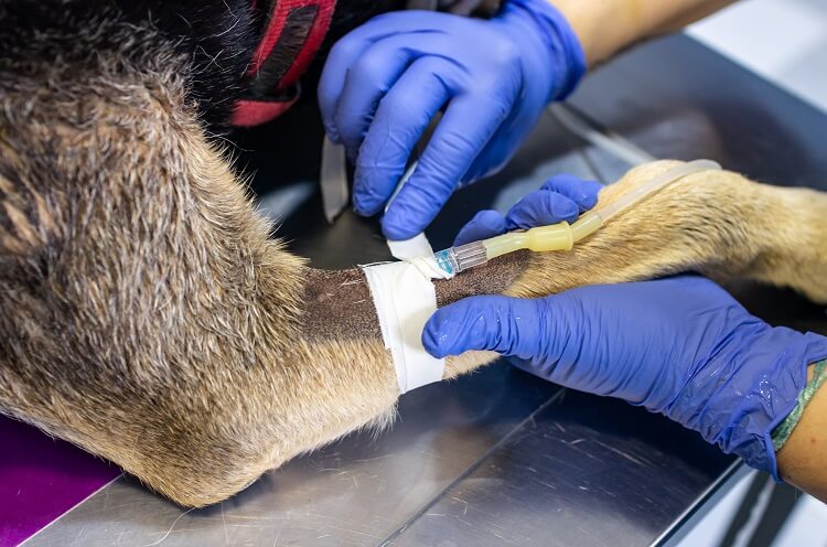
The cephalic vein is relatively large and often visible through the skin, and can be an attractive candidate for healthcare practitioners to draw blood through venipuncture. This is also the case for dogs! Collecting blood from the cephalic vein in canines is the most comfortable position for the animal during the procedure.
Additionally, the cephalic vein is often where the leads of permanent pacemakers are inserted to maintain an adequate heart rate. This is because, at the deltopectoral groove, the anatomy and location are highly consistent between individuals, so it is a reliable choice.
Superficial Vein Thrombosis
Superficial vein thrombosis (SVT) is a blood clot in a superficial vein (as opposed to deep vein thrombosis, which is a blood clot in a deeper vein system). SVT is far less dangerous than deep vein thrombosis, but it can lead to complications such as pulmonary embolism.
Symptoms of SVT include pain, redness, and swelling in the area. They usually involve the veins of the legs, but they can involve the arms, including the cephalic vein. Treatment includes compression, increased physical activity to reduce time spent sedentary, and anti-coagulant medication.
Quiz