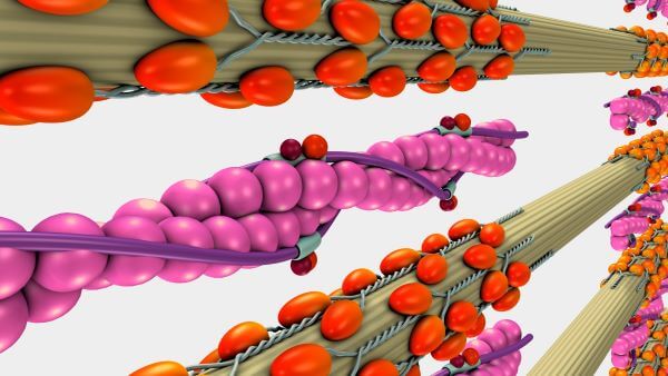
Actin and myosin are both proteins that are found in every type of muscle tissue. Thick myosin filaments and thin actin filaments work together to generate muscle contractions and movement. Myosin is a type of molecular motor and converts chemical energy released from ATP into mechanical energy. This mechanical energy is then used to pull the actin filaments along, causing muscle fibers to contract and, thus, generating movement.
Types of Muscle
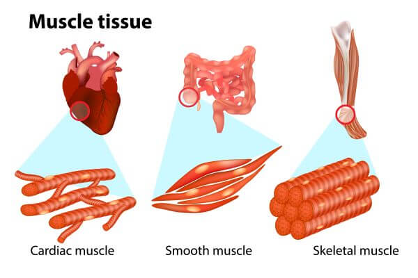
Muscle is a type of contractile tissue found in animals, and its main function is to facilitate movement. There are three types of muscle tissue found in the human body, and these are:
Smooth muscle – Smooth muscle contracts involuntarily and is found in the internal organs (except the heart) and blood vessels.
Cardiac muscle – Cardiac muscle is found only in the heart and also contracts involuntarily.
Skeletal muscle – Skeletal muscle is attached to the bones. It is the most abundant type of muscle tissue in the human body, and the only type of muscle that can be moved voluntarily. Skeletal muscle contractions pull on the bones of the skeleton and allow those bones (and the body structures they support) to move.
All three muscle types contain actin and myosin filaments, which work together to produce muscle contractions.
What Are Actin and Myosin?
Actin and myosin are both proteins that are found in all types of muscle tissue. Myosin forms thick filaments (15 nm in diameter) and actin forms thinner filaments (7nm in diameter). Actin and myosin filaments work together to generate force. This force produces the muscle cell contractions that facilitate the movement of the muscles and, therefore, of body structures.
The Structure of Muscles
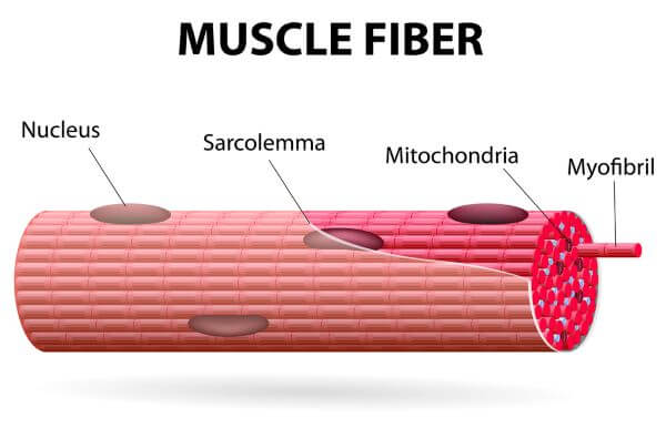
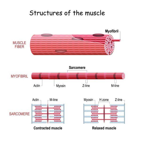
Muscle tissue is made up of bundles of muscle fibers. Muscle fibers are long, skinny cells that can be up to several inches long and, in the case of skeletal muscle, may contain several nuclei. The cytoplasm of muscle fibers contains long, thread-like structures called myofibrils, which are made up of bundles of thick, myosin filaments and thin actin filaments. Surrounding the actin and myosin filaments is a structure called the sarcoplasmic reticulum (SR); a network of tubules that store calcium ions. The SR also plays an important role in transmitting electrical signals. These electrical signals are delivered to the muscle cells by neurons.
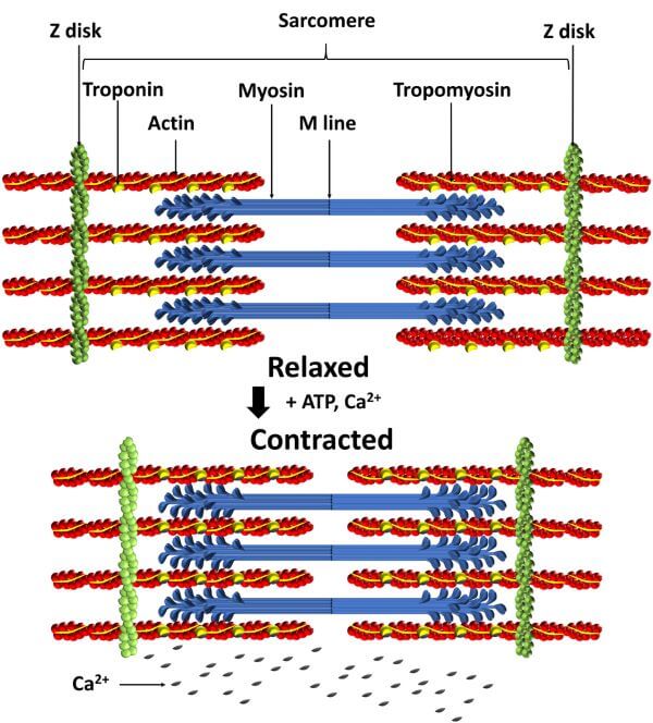
Each myofibril is made of a chain of repeating contractile units called sarcomeres. At the end of each sarcomere is a Z disc.
Sarcomeres contain two distinct types of bands. These are the dark-colored A bands, which contain thick, myosin filaments, and the I bands, which have a lighter color and contain only thin, actin filaments. The actin filaments are attached to the Z disc, whereas the myosin filaments are anchored to a region in the middle of the sarcomere called the M line.
How Do Actin and Myosin Work?
Actin and myosin work together to produce muscle contractions and, therefore, movement. First, a motor neuron delivers an electrical signal to the muscle cell from the brain. This triggers the release of a chemical called acetylcholine. Acetylcholine causes calcium ions to be released from the sarcoplasmic reticulum. Next, the calcium ions bind to a protein called troponin. Troponin is attached to another protein, called tropomyosin, and is found between the actin filaments in muscle tissue.
When calcium ions bind to troponin, the shape of troponin changes. This moves tropomyosin from the myosin-binding sites on the actin filament and ‘unblocks’ them, making it possible for the myosin heads to bind to the actin filament.
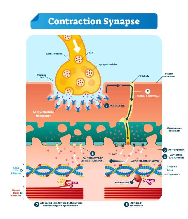
Once tropomyosin has moved out of the way, the myosin heads can bind to the exposed binding sites on the actin filaments. This forms actin-myosin cross-bridges and allows muscle contraction to begin.
A hydrolysis reaction releases energy from ATP, and the myosin works like a motor to convert this chemical energy into mechanical energy.
The myosin uses that mechanical energy to move its head groups towards the middle of the sarcomere. This movement pulls the actin filaments towards the center of the sarcomere, causing the sarcomere to shorten and contract. The contraction of the sarcomere causes the muscle fiber to contract and generates muscle movement
The mechanism of muscle contraction is explained by the Sliding Filament Model, which was first proposed in 1954.