Vertebral Column Definition
The vertebral column, also called the spine, is a series of bones known as vertebrae that are separated by intervertebral discs. The vertebral column is only found in vertebrates, or members of the subphylum Vertebrata, which is part of the phylum Chordata.
Vertebral Column Overview
The vertebral column consists of a variable number of bones which are separated by small, cartilaginous disks. The vertebral column typically runs from the skull through the end of the tail, although its exact features, density, length, and structure ultimately depends on the species. Sharks, for example, have a vertebral column made entirely of cartilage and connective tissues. Snakes, on the other hand, only have a a vertebral column and head.
While the column has been adapted in many ways in different groups of animals, its main purpose is to protect the spinal cord within the central nervous system. The spinal cord is a dorsally-located, hollow nerve cord made of many nerves which carries signals in both directions between the body and the brain. The vertebral column surrounds the spinal cord on all sides. Between each vertebrae, a system of connective fibers and cartilage also surround the spinal cord, followed by muscles on top. As such, the vertebral column protects the spinal cord well.
Vertebral Column Bones
In the human body, there are 33 vertebrae which make up the vertebral column. These bones are identified by the section of the vertebral column that they make up. Near the skull are the 7 cervical vertebrae, including the atlas and axis bones which connect the skull to the vertebral column.
Next are the thoracic vertebrae, which are 12 vertebrae that span the thorax. 5 lumbar vertebrae follow these bones. The final parts of the vertebral column are the 5 fused sacral vertebrae, finished by the 4 fused coccygeal vertebrae commonly called the coccyx. Other vertebrates can have more or fewer vertebrae, but the arrangement and various regions are fairly typical.
Vertebral Column Function
The spine is structured to not only provide support and places for muscle attachment but also to protect the spinal cord of the vertebrates. The vertebral column developed at a series of bony arches that surround the notochord from the top and bottom. The neural arches cover the spinal cord and extend to the hemal arches, which cover the bottom of the notochord and allow for rib attachments.
This simple arrangement can be seen in the most primitive vertebrates such as lampreys and gets slightly more complex through the fishes. As vertebrates evolved to move onto land, the demands for a stronger spine and more muscle attachment along the vertebral column drove the tetrapods to completely replace the notochord with vertebrae.
Vertebral Column in Animals
Members of the Chordata, or the chordates, share 4 derived characters which no other groups have. They have a dorsal hollow nerve cord, a muscular post-anal tail, an endostyle, and a notochord. The notochord is a strong, flexible rod seen in tunicates and cephalochordates, which are the only chordates without a vertebral column. The endostyle is a glandular groove which secretes mucus to trap food in primitive chordates. In the vertebrates, the notochord has evolved into the spine, while the endostyle has become the thyroid. The thyroid is an endocrine system gland which helps regulate the metabolism of an organism.
Within a Typical Tetrapod
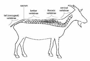
The modern tetrapod vertebral column consists of a series of interlocking vertebrae that extend from the skull through the tail of an organism. The vertebral column is divided into regions, which form similar functional regions in animals. The cervical vertebrae attach the skull to the rest of the body and create the structure for the neck of an animal. The thoracic vertebrae often have the most rib attachments and create a ribcage, which protects the heart and lungs. The lumbar vertebrae connect the top of the organisms to the sacrum, which is a series of fused vertebrae. This region allows for the connection of the pelvic bones and the tail or coccygeal vertebrae.
Altered Vertebral Columns
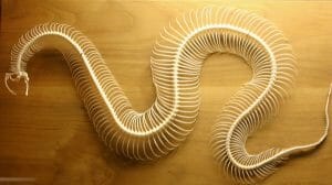
This basic pattern of the spine has been modified throughout the organisms within the Vertebrata. Some extreme examples include the snake, which has lost all appendages and relies solely on a vertebral column and ribcage for support. The ribs of the snake have extended throughout most of the body, which provides additional support. It also allows the snake to expand to several times its starting diameter, enabling snakes to swallow meals bigger than themselves. A snake skeleton can be seen below, with both the spine and ribs intact.
On the other hand, many animals have reduced the size of their vertebral column, including humans. As seen in the following picture of a human spine, the tail vertebrae are almost completely absent. As an organism that evolved to walk on two legs and usually on the ground, a tail became a hindrance. This can also be seen in our closest wild relatives: the great apes. The size increase from tailed monkey and the increased activity sitting in trees and on the ground reduced the need for a tail.
Eventually, many apes and humans have lost their tail, or only a small trace of it remains. The only bone left of the tail in humans is the coccyx and is considered a vestigial structure because it seems to serve no further purpose. In fact, people generally only find out they have a tail when they break their tailbone. While it is painful, the break is certainly not that harmful. A break anywhere else in the spine can cause a separation of the spinal cord, leading to paralysis.
Quiz


