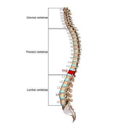
Thoracic Vertebrae Definition
The thoracic vertebrae are bones located between the cervical and lumbar vertebrae. There are 12 thoracic vertebrae in humans, and these bones increase in size as you move down the body. They are thicker and larger than the cervical vertebrae but smaller than the lumbar vertebral bones. The increase in size ensures they can support the weight of the body.
These vertebrae are readily identifiable because they articulate with the ribs and have facets on the sides of their bodies to allow this. They are generally labeled T1 through T12 in humans, with T1 being closest to the neck and T12 furthest down the body.
The vertebrae are illustrated in the below image:
Thoracic Vertebrae Function
The thoracic vertebrae function to support the back. Their articulations with the ribs allow them to provide a protective cage around the delicate organs of the thorax, including the heart and lungs. There is limited mobility in this section of the spine due to these joint articulations and their design. Unlike sections of the cervical vertebrae that allow nodding and shaking of the head, the thoracic vertebrae allow some twisting and bending motions, but little beyond that.
Thoracic Vertebrae Structure
The main portion of each of the thoracic vertebrae is the body or centrum, which is heart-shaped in the central vertebrae. In T1, it resembles the cervical vertebrae; they are similar to the lumbar vertebrae in the lower thoracic vertebrae. They contain ovoid surfaces known as facets on the transverse processes that allow the heads of the ribs to articulate on either side of the body. T1 is unique compared to the other vertebrae as it supports two pairs of ribs. It has a pair each of facets and demi-facets, which are facets shared between two vertebrae.
In the thoracic vertebrae, the spinous processes are long and triangular. These processes are also overlapping. They have pedicles that are angled backward and upward, with thick, broad laminae. Each vertebrae has two intervertebral foramina that allow nerve roots to exit the spinal cord. The vertebral foramen allows the spinal cord to pass through each of the vertebrae and is formed by the position of the vertebral arches projecting off each body.
Between each vertebral body is an intervertebral disc. These are made of fibrous connective tissues, and they hold the vertebrae in place while allowing some minor motion. There is a core of gel-like material known as the nucleus pulposus, which helps to provide a cushion and act as a shock absorber, especially for movements between the vertebrae.
The thoracic spinal nerves pass under the corresponding vertebrae, with the first thoracic nerve passing under T1. The fourth and fifth vertebrae are located at the sternal angle, a palpable landmark that is useful in clinical procedures. The xiphoid process of the sternum is located at the level of T9.
The number of thoracic vertebrae varies depending upon the species. Some aquatic mammals only have 9, while horses may have 18 to 20. This number can increase to 25 in certain sloth species.
Why is it Important
The thoracic vertebrae are prone to problems such as stress and strain, especially given their lack of a range of motion. They are used constantly, and people may face issues caused by poor posture, as well as traumatic fractures. Some injuries, including disc herniation, may put pressure on the spinal cord or spinal nerves, causing symptoms aligning with the lesion.
There are two long muscles associated with the thoracic vertebrae, the spinalis and the longissimus. These run along the back and help with posture and movements. Overuse or trauma can lead to muscle strains and pain in the back.
Herniated discs are another common injury involving the thoracic vertebrae. The intervertebral discs are situated between each vertebrae and provide a cushion to pad the joints. When there is a herniation, the disc material puts pressure on the spinal cord, which can result in pain and nerve abnormalities.
Other injuries can occur around the thoracic vertebrae, including vertebral fractures. Osteoarthritis may develop in conjunction with degenerative joint disease, as can spondylolisthesis, a degenerative disc disease. Although less common, infections can also develop around the spinal cord, affecting the bones, discs, or nerves.
Quiz