Definition
Spindle fibers are microscopic protein structures that help divide genetic material during cell division and organize cellular components. The spindle fibers form out of the centrosome, also known as the microtubule-organizing center, or MTOC.
Overview
Spindle fibers are formed from microtubules with many accessory proteins which help guide the process of genetic division. Each spindle fiber forms during cellular division near the poles of the dividing cell. As they extend across the cell, they search for the centromere of each chromosome.
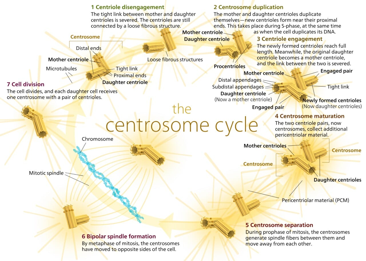
Once attached, the spindle fiber is pulled back. With each fiber comes the chromosome it is attached to, which separates the chromosomes into each daughter cell. The process can be seen in the image above. The spindle fibers can be seen extending in all directions from the centrosomes in step 6. Each spindle fiber is formed from several microtubules. The spindle fibers act like small machines during cell division. They carefully assemble and divide the chromosomes, and have been doing so for billions of years. But how does this complex process take place?
Structure of Spindle Fibers
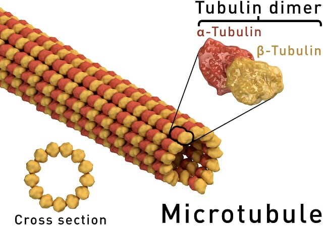
The centrosome, or MTOC, always has some microtubules preassembled. On the surface of the MTOC are small proteins, responsible for lengthening or shortening the microtubules. These proteins respond to signals from the cell, and when it is time for cell division, they begin lengthening the spindle fibers. To do this, they must add subunits of alpha-tubulin and beta-tubulin. Together, these two small proteins form the structure of a microtubule. Many individual microtubules together are called spindle fibers. A single microtubule can be seen in the graphic below.
Functions of Spindle Fibers
Shrinkage and Growth
The main feature of microtubules, and therefore of larger fibers, is that the proteins which control them can extend or contract the microtubule by adding or removing tubulin dimers. At first, the MTOCs must add many of these dimers to the microtubule, to extend it across the cell. As the microtubule travels, it eventually reaches a chromosome. Special proteins within the centromere of the chromosome can attach to the microtubule. Here, there are also proteins which can shorten and extend the spindle fibers.
This is one of the main ways that the chromosomes get aligned on the metaphase plate, a hypothetical middle of the cell. It is also the main way they are separated during anaphase of mitosis or meiosis. While the addition and subtraction of dimers is one of the main ways that spindle fibers help carry chromosomes about the cell, there are two other primary methods.
Spindle Fibers: Sliding
When two fibers from opposite poles of the cell meet, they are bound together by a special protein. Instead of grabbing onto a chromosome, they more or less attach to each other via the protein. This protein is a specialized motor protein, which reacts to signals from the cell. At the appropriate time during cell division, the motor protein will begin crawling along each microtubule it is attached to. This “sliding action” causes pressure to be exerted against the poles and helps drive the poles apart. This action of the spindle fibers is what forces the cell apart and allows for it to be divided in half during telophase.
Spindle Fibers: Anchors
The final action carried out by some spindle fibers is that of anchoring to the cell surface. On the inside surface of the cell membrane, specialized proteins are placed to anchor the microtubules. While these anchors cannot assemble dimers into the microtubule, they can bind onto it. Then, when the MTOC starts removing microtubule dimers, the whole spindle fiber shortens. In this way, it pulls the cell membrane toward the MTOC and starts to define the area of the newly forming cell.
Quiz
1. Which of the following is NOT caused by the actions of spindle fibers?
A. The movement of chromosomes
B. The change in the shape of the cell
C. The structure of the cell when not dividing
2. Microtubules form in a peculiar fashion. While the entire structure is just repeated units of the small tubulin dimer, the structure has polarity to it. That is, each side of the microtubule is different. On one side the beta-tubulin is more exposed, while on the other side the alpha-tubulin is more exposed. How must the proteins in the MTOC and the proteins on chromosomes be different in order to work?
A. They must be the same
B. They must be able to add dimers from opposite sides
C. They are completely different processes, therefore they are completely different proteins
3. Often, when products of an organelle are exported, they are contained within vesicles. These small compartments of the cell membrane are then attached to a microtubule via a small motor protein. The protein works its way down the microtubule, like in the sliding example above. It carries the vesicle to another organelle or the cell surface. Here it can be expelled or absorbed. Are these microtubules considered spindle fibers?
A. No
B. Yes
C. Maybe