Definition
The sphenoid bone of the skull base is one of the most complex bones of the body. It is an unpaired bone with many foramina (holes) and grooves that allow nerves and blood vessels to pass through or along it. The form of the sphenoid bone is often referred to as bat-shaped, wasp-shaped, or butterfly-shaped. It forms part of the orbits (eye sockets), paranasal sinuses, and cranium; it articulates (joins) with twelve other cranial or craniofacial bones.
Sphenoid Bone Location
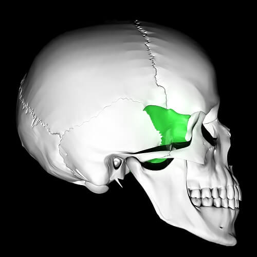
The location of the sphenoid bone lies behind the top of the nasal cavity and stretches from the left side of the skull to the right. Each end meets the outer surface of the skull in front of the left and right parietal bones and under the frontal bone.
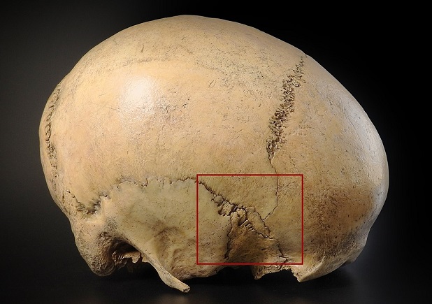
The place where frontal, sphenoid, parietal, and temporal bones meet is called the pterion.
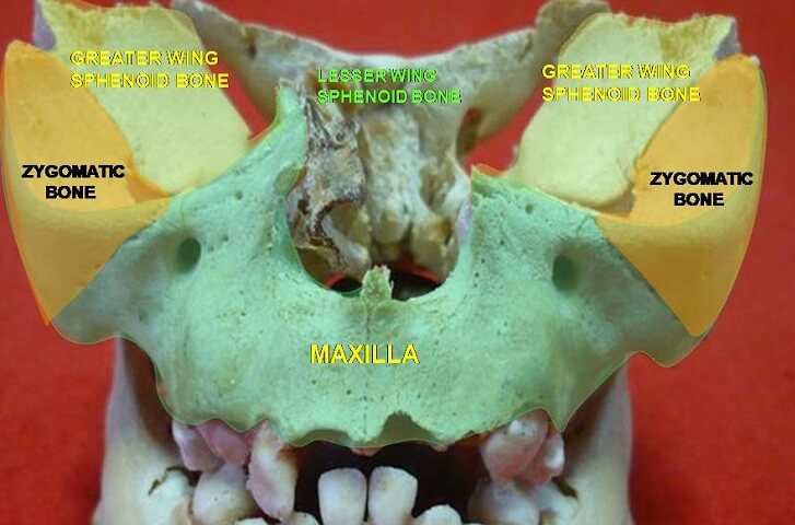
The greater wing of the sphenoid bone can be seen in the below image as the oval shape in the center of the red rectangle.
The sphenoid bone body sits behind the nasal cavity with various protrusions that play important roles in the construction of other bony structures.
A sphenoid bone fracture – trans-sphenoidal basilar skull fracture – is often life-threatening; the bone lies close to the soft tissues of the pituitary gland, frontal and temporal lobes and meninges of the brain, internal carotid arteries, and facial nerves.
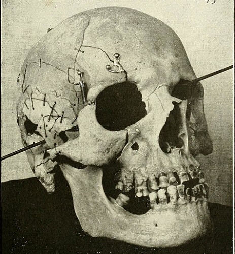
Patients with healed sphenoid bone fractures may need months or years to reverse or partially reverse associated nerve damage. The trajectory of the gunshot wound below caused multiple fractures to the cranium and face and also shattered the sphenoid bone.
Sphenoid Bone Anatomy
Sphenoid bone anatomy is complex, not only because of its strange shape but also because of its many articulations with other bones. It is much easier to split this topic into four subheadings, each heading describing both the shape and position of a particular part.
Body of the Sphenoid Bone
The (central) body of the sphenoid bone (corpus) contributes to multiple facial structures.
At the front it helps to construct the nasal cavity; the lateral (side) surfaces play a role in the structure of the optic canal.
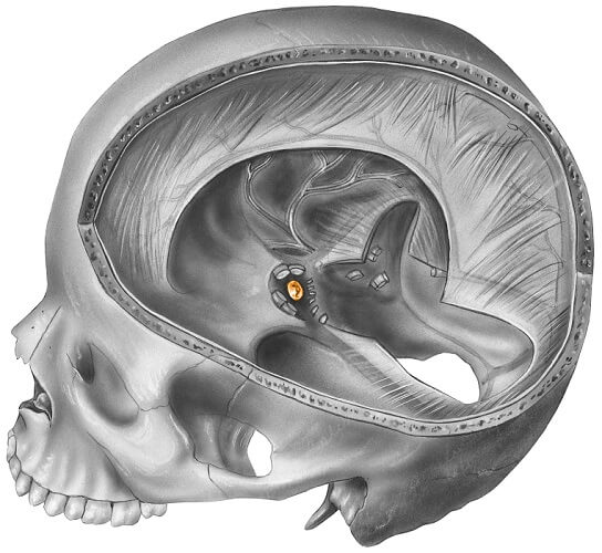
The sphenoid bone sella turcica (tuberculum sellae), hypophyseal fossa, and dorsum sellae at the back of the body.
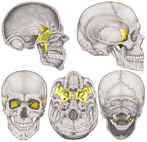
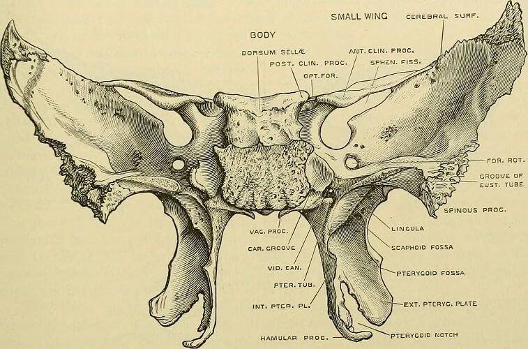
The labeled sphenoid bone views below depict various substructures.

If we look at a picture of the sphenoid bone, it is easy to see the position of the body. The ‘antennae’ of the lesser wings, the ‘wings’ of the greater wings, and the ‘legs’ of the pterygoid process show us why the shape of this bone is often compared to butterflies, wasps, and bats.
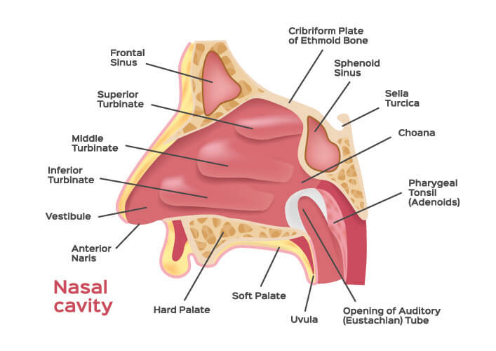
The corpus is almost cube-shaped and the front is partially hollow. The hollow area is divided into two to form the sphenoidal sinus. This sinus is one of four paranasal sinuses that allow air to be warmed and humidified and cleared of pathogens before it travels to the lungs. Each sinus lies at the back of the nasal cavity, at its apex (top).
The bony walls of the sphenoid sinus separate it from the optic canal, dura mater, pituitary gland, and cavernous sinus; an infection in this sinus can spread to cause extremely serious consequences.
The back of the sphenoid body consists of two sellae (seats). The first is the sella turcica, shaped like a saddle. The dip of this saddle – the hypophyseal fossa – leaves space for the pituitary gland.
Empty sella syndrome refers to a shrunken pituitary gland that has been damaged by inflammation of the cerebral membranes, high intracranial pressure, tumors, surgery, and trauma.
The dorsum sellae forms the boundary of the sella turcica and part of the clivus (slope) that supports the pons of the brainstem.
Greater Wings of the Sphenoid Bone
The two greater wings of the sphenoid bone (alae majores) are positioned laterally (to the side) of the sphenoid body. The greater wings contribute to the structure of the left and right orbital bone, optic canal, and the sides of the skull.
The orbital surface of the sphenoid bone provides space for the lacrimal artery and an attachment point for the lateral rectus muscle – one of the muscles that control eye movement.
The edges of the greater wings contribute to the construction of the superior and inferior orbital fissures. The superior orbital fissure separates the greater and lesser wings and allows motor nerves of the extraocular muscles to pass through. The inferior orbital fissure, formed by the greater wings and maxilla bones, is a passageway for the maxillary nerve.
The greater wings also feature four foramina – the foramen ovale, foramen spinosum, foramen Vesalli, and foramen rotundum.
- The foramen ovale is a gateway for the mandibular and lesser petrosal nerves, the meningeal artery that feeds the membranes surrounding the brain, and the emissary vein.
- The hole of the foramen spinosum allows meningeal blood vessels and the meningeal branch of the mandibular nerve to pass through.
- The foramen rotundum connects the middle cranial fossa and the pterygopalatine fossa, allowing multiple facial nerves and blood vessels to pass through from the brain to various tissues of the face.
- The foramen Vesalii is not always present. Only a small vein of the cavernous sinus passes through.
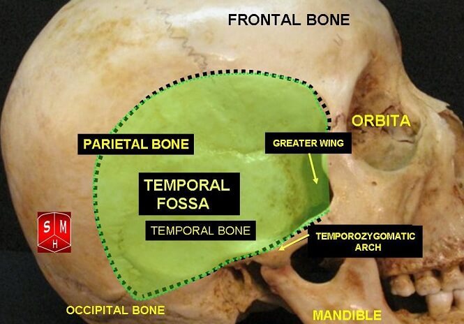
The tips of the greater wings articulate with the temporal and parietal bones to create part of the cranium. They meet with the parietal bones at a weak spot called the pterion.
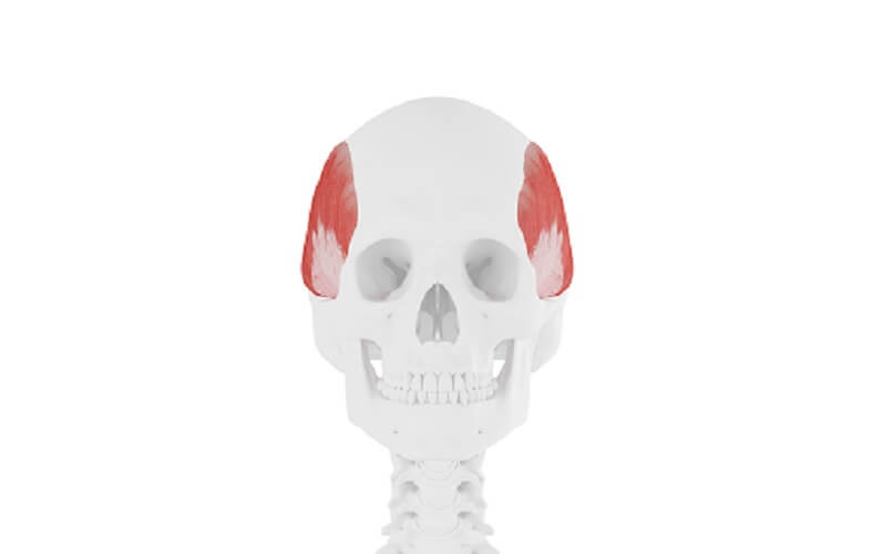
A crest runs through the sides of the greater wings of the sphenoid bone that divides them into two anatomical parts. The temporal part is part of the temporal fossa – a dip in the side of the skull (see below) and provides a point of attachment for the temporalis muscle that helps us to chew.
The infratemporal part constructs part of the infratemporal fossa and provides a point of attachment for the pterygoideus externus muscle, another masticatory muscle.
A further feature of the greater wings is the spina angularis – another muscle attachment point – this time for the tensor veli palatini that help us to swallow and the sphenomandibular ligament that stops the lower jaw from dropping.
Lesser Wings of the Sphenoid Bone
The two lesser wings of the sphenoid bone (alae minores) are positioned anteriorly (at the front of) the sphenoid body. You can imagine them as the antennae of a butterfly. Just like the body, the lesser wings also aid in the construction of the optic canal (where the optic nerve runs through to the eye) and orbit; the superior surfaces are involved in the construction of the cranial cavity.
Pterygoid Process of the Sphenoid Bone
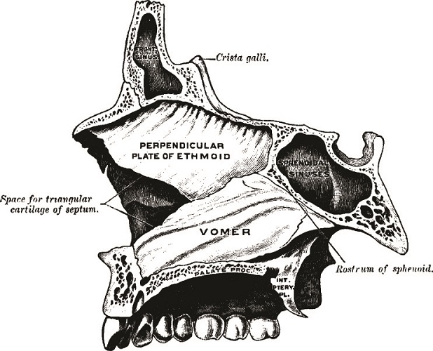
The pterygoid process of the sphenoid bone points downward from the body, trailing behind it like the legs of an insect in flight. It contains two important channels (canals) that protect two types of nerve fibers. These are the petrosal nerves (taste and parasympathetic innervation) that run through the pterygoid canal, and the pharyngeal nerve (motor nerve of the pharynx) that travels along the palatovaginal canal.
Each process is constructed from two bony plates – the medial and lateral pterygoid plates labeled respectively as the interior (int.) and exterior (ext.) plates in the above image. The lateral plate is where the pterygoid muscles that help us to move our jaw from side to side originate. The medial plate is an attachment point for the superior pharyngeal constrictor muscle.
Both plates are separated by the pterygoid notch that fits snugly with the corner of the palatine bone (pyramidal process). Between the open ends of the plate is the pterygoid fossa. The pterygoid fossa houses deep masticatory muscles – the medial pterygoid and tensor veli palatini muscles.

Sphenoid Bone Articulations
The sphenoid bone has articulations with twelve other paired and unpaired bones – this makes it the most complex bone in the human body. While the sphenoid bone is unpaired, it stretches from one side of the skull to the other. Its articulations are with the:
- Temporal bones (paired)
- Parietal bones (paired)
- Zygomatic bones (paired)
- Palatine bones (paired)
- Occipital bone (unpaired)
- Vomer (unpaired)
- Ethmoid bone (unpaired)
- Frontal bone (unpaired)
Sphenoid Bone Function
The sphenoid bone has many functions. As we have already seen, each part has more than one task to fulfill, either in cooperation with other bones of the cranium and face, or alone.
Structural Functions
Several bony structures involve the sphenoid bone.
- Cranium – at the skull base and pterion
- Sphenoid bone sinus
- Nasal cavity
- Orbital bone floor (eye sockets)
- Clivus (slope) of the skull base
- Temporal, infratemporal, hypophyseal, and pterygoid fossae
- Optic canal (containing the optic nerve)
- Pterygoid and palatovaginal canals
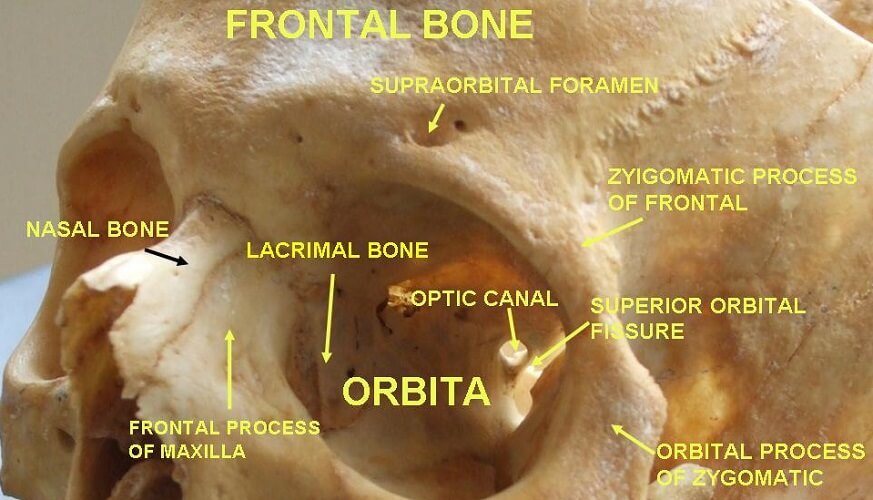
- Superior and inferior orbital fissures
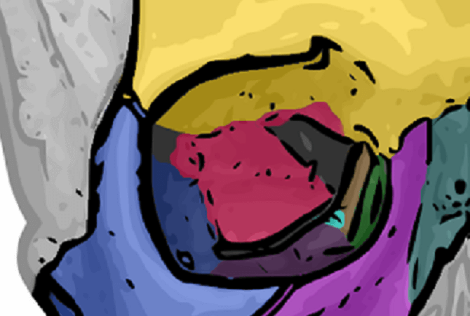
In the above image, the frontal bone is colored yellow, the maxilla in purple, zygomatic bone in lilac, lacrimal bone in green, nasal bone (not part of the orbit) in gray-green, sphenoid in deep pink, ethmoid in brown, and the small bright blue area indicates the position of the orbital process of the palatine bone.
Muscular Attachment Function
The sphenoid bone is an attachment point for multiple muscles:
- Lateral and medial pterygoid muscles
- Lateral rectus muscle
- Pterygoideus externus muscle
- Tensor veli palatini muscle
- Sphenomandibular ligament
- Temporalis muscle
- Superior pharyngeal constrictor muscle
Innate Immunity
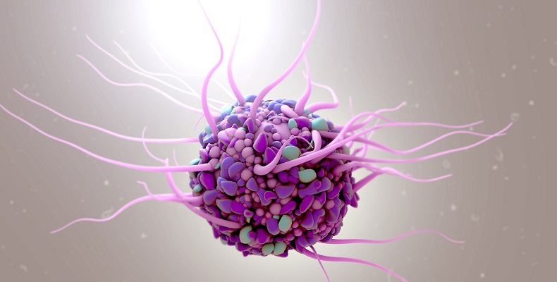
As the sphenoid bone is the source of the sphenoid sinus, this small, complex structure also contributes to our innate immunity. The mucous linings of the sinuses trap potential pathogens and also contain dendritic cells and other phagocytes. Phagocytes recognize proteins on the cell membranes of foreign cells and tell the acquired immune system to produce more antibodies.
Nervous and Circulatory System Function
Finally, the sphenoid bones contain important foramina that allow nerves to pass from brain to face and vice versa. These are both motor and sensory nerves that play crucial roles in our senses of touch, sight, smell, and taste, and the actions of facial expression, chewing, and speaking.

Quiz