Somatic Nervous System Definition
The somatic nervous system (SoNS), also known as the voluntary nervous system, is a part of the peripheral nervous system (PNS). It consists of neurons that are associated with skeletal or striated muscle fibers and influence voluntary movements of the body.
The peripheral nervous system is made up of all the neurons that exist outside the brain and spinal cord. It acts as an intermediary between the central nervous system (CNS) and muscles, skin and sensory organs. The nerves of the PNS send electrochemical signals back and forth between the CNS, and the rest of the body. A large part of the PNS is composed of 12 pairs of cranial nerves and 31 pairs of spinal nerves. Some of the neurons in these nerves have sensory function and others have a motor function. The motor neurons that innervate striated muscles form the somatic nervous system.
Functions of the Somatic Nervous System
The SoNS contains both afferent nerves traveling towards the CNS and efferent nerves responsible for sending signals from the CNS towards the rest of the body. The brain and spinal cord process the input from a variety of sources and integrate them before devising a response. This response determines the location and strength of muscle contraction across different parts of the body. Therefore, the primary function of the somatic nervous system is to connect the CNS with organs and striated muscles in order to enable complex movements and behavior.
Additionally, the SoNS also mediates a subset of involuntary muscle responses called reflex arcs. A reflex arc results in an extremely quick muscle contraction in response to a stimulus, with minimal intervention from the brain. While the impulse for most voluntary muscle contraction originates in the brain or brainstem, a reflex action can be brought about with just a single sensory and motor neuron that synapse in the spinal cord. The motor response is practically ‘hard-wired’ for a particular stimulus. The knee-jerk response to the stimulation of the patellar ligament in the knee is an example of a reflex response. Other examples include the immediate withdrawal of a hand on touching a hot stove or a quick change in posture when the foot is placed on a sharp stone.
Examples of the Somatic Nervous System Response
The somatic nervous system is intricately linked to the central nervous system with the sensory and motor neurons of the SoNS communicating with the brain and spinal cord. Striated skeletal muscles under voluntary control receive signals to contract on the basis of stimuli relayed to the CNS. For instance, while walking in a tropical forest, you watch the forest floor for fallen twigs, insects or undergrowth. As the CNS constantly receives visual input, it sends messages to the peripheral nervous system, particularly the SoNS, to alter posture and contractility of skeletal muscle, and accommodate changes to the surface of the forest floor. At the same time, if a leech is stuck to your calf muscle, sensory neurons indicate the presence of a persistent damp feeling on your leg. Skeletal muscles act to alter your position so that the area can be visually inspected. On finding a leech, the CNS, through memory and learning, directs skeletal muscles of the arms and fingers through the SoNS to reach for some salt. Gross and fine motor skills are used to sprinkle a pinch of salt on the leech to ensure that it falls off.
Similar events are occurring within the nervous system in vastly varied activities. For example, a dancer on stage is integrating her memory of the music and choreography in the CNS to direct the movement of her skeletal muscles through the SoNS. From the still readiness of her body before the music begins till the last bow and smile, the neurons of the SoNS signal every large and small striated muscle group in the body based on the directions of the CNS.
Motor Neurons
The neural pathway that results in skeletal muscle contraction can be functionally divided into two main types of neurons – the upper motor neurons in the central nervous system and the lower motor neurons of the somatic nervous system. Lower motor neurons can be a part of cranial or spinal nerves. They innervate muscle fibers and directly cause their contraction.
Upper motor neurons have their cell bodies in the precentral gyrus of the brain. This region is located towards the posterior end of the frontal lobe in the cerebral cortex and is associated with the primary motor cortex. The axons of upper motor neurons related to voluntary muscle movement travel along the CNS in two pathways – the corticospinal and corticobulbar tracts. Neurons whose axons travel along the corticobulbar tract synapse with lower motor neurons in the brain stem. The axons of these lower motor neurons form cranial nerves such as the oculomotor, trochlear or trigeminal nerves that are involved with the contraction of skeletal muscles in the face, neck, jaw and tongue.
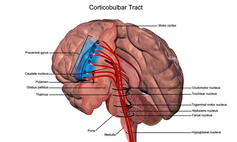
The image shows upper motor neurons emerging from the precentral gyrus and traveling along the corticobulbar tract towards the brainstem.
The axons of other upper motor neurons travel along the corticospinal tract, passing the medulla oblongata and reaching the ventral horns of the spinal cord.
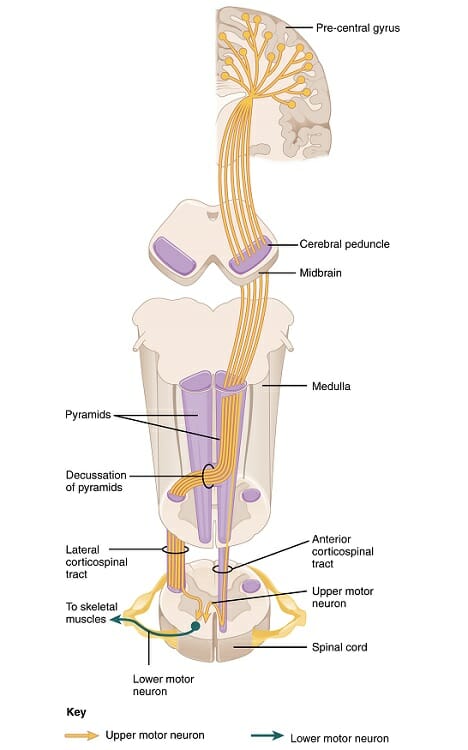
The image shows the origin of upper motor neurons from the precentral gyrus, moving through the midbrain and medulla to form the lateral and anterior corticospinal tracts. The primary function of these neurons is to connect the brain with the spinal cord. At the spinal cord, upper motor neurons form synapses with lower motor neurons, and release glutamate into the synaptic cleft. The depolymerization of the lower motor neuron results in the transmission of the action potential towards skeletal muscles.
There are three types of lower motor neurons – alpha, beta, and gamma. Alpha motor neurons are thick, myelinated, multipolar nerve fibers that are involved in innervating most skeletal muscle fibers and causing their contraction. Gamma motor neurons support the activity of alpha motor neurons by keeping muscle spindles taut. Alpha motor neurons can receive signals from upper motor neurons for voluntary muscle movement. At the same time, they can receive input from sensory and inter neurons as well, in order to initiate reflex actions. The number of alpha motor neurons innervating a single muscle depends on the extent of fine motor control required at the site. Therefore, the muscles of a finger will have substantially more alpha motor neurons associated with them than the muscles of the thigh or upper arm.
Neuromuscular Junction
The axon terminus of an alpha motor neuron forms a neuromuscular junction with striated muscle fibers, where acetylcholine is released as the neurotransmitter. When an action potential reaches the axon terminus of the alpha motor neuron, a voltage-gated ion channel allows the entry of calcium ions into the neuron. These ions induce the fusion of synaptic vesicles with the plasma membrane resulting in the release of acetylcholine into the neuromuscular junction. Acetylcholine then binds to the nicotinic receptors on muscle cells. These receptors are ion channels that open upon ligand binding, which then leads to a cascade of ions within the muscle fiber, leading to muscle contraction.
Two potent toxins that affect the neuromuscular junction are botulinum toxin and tetanus toxin. Both chemicals are produced by bacteria – the former by a bacterium called Clostridium botulinum and the latter by Clostridium tetani. Botulism can affect humans through inhalation or ingestion of the toxin or through ingestion of bacterial spores from contaminated food. This is particularly true for improperly prepared canned food, since the warm, moist, and anaerobic environment within food containers can provide a fertile environment for the growth of the bacteria. The toxin interferes with the fusion of synaptic vesicles with the neuronal plasma membrane and thus prevents the release of acetylcholine into the neuromuscular junction. Therefore, it leads to paralysis, initially of facial muscles and in severe cases, even the smooth muscles of the diaphragm. It is among the most potent neurotoxins known, with a lethal dosage of 1 microgram for an adult. The only other toxin of this potency is the tetanus toxin, and it functions in a similar manner. When the tetanus toxin enters the presynaptic nerve terminus, it prevents the release of neurotransmitters into the neuromuscular junction. While botulinum toxin produces a flaccid paralysis, tetanus toxin produces a spastic or rigid paralysis.
Sensory Neurons
Afferent sensory neurons of the somatic nervous system provide information to the CNS about joint angle, muscle length, muscle tension, and the presence of noxious stimuli.
Proprioceptors
In addition to the typical extrafusal muscle fibers, the body of a muscle also contains muscle spindles. These small sensory organs contain specialized muscle fibers that have a central non-contractile segment. Afferent neurons of the somatic nervous system have their sensory dendrites in this area. These dendrites contain ion channels that open in response to mechanical forces on the cell. When the muscle spindle is stretched, the opening of ion channels generates an action potential in these sensory neurons. The presence of mechanically gated ion channels allows these neurons to carry detailed information about the condition of the muscle, and its contractile activity.
Nociceptors
Nociceptors are pain receptors found across the body, and are an essential part of injury prevention, especially in muscle fibers. These neurons are activated in response to potentially damaging stimuli, such as heat, cold, or extreme forces. The presence of nociceptors prevents us from hyperextending joints, overstretching muscles and protects us from a wide range of injuries.
Related Biology Terms
- Alpha Motor Neurons – Lower motor neurons of the brainstem and spinal cord associated with striated muscle fibers and directly responsible for their contraction.
- Extrafusal Muscle Fiber – Skeletal striated muscle fibers innervated by alpha motor neurons, and attached to bone through tendons. Primary function is the generation of skeletal movement through contractile tension. Constitute a large proportion of striated muscles within the human body.
- Lower Motor Neurons – Effector neurons of voluntary muscle contraction, usually synapsing at neuromuscular junction with striated muscle fibers. Cell body is located either in the medullary pyramids of the brainstem or in the anterior horn of the spinal cord.
- Upper Motor Neurons – Neurons that originate in the brainstem or the motor cortex and carry information to lower motor neurons in the spinal cord or medullary pyramids.
Quiz
1. Which of these statements about motor neurons is NOT true?
A. Upper motor neurons have their cell bodies in the prefrontal cortex
B. Acetylcholine is the only neurotransmitter released in the synapse between upper and lower motor neurons
C. Axons of upper motor neurons descend towards the spinal cord in the corticobulbar tract
D. All of the above
2. Which of these lethal toxins is used in cosmetic procedures?
A. Botulinum toxin
B. Tetanus toxin
C. Curare
D. All of the above
3. Which of these are features of alpha motor neurons?
A. Small and pyramidal shaped
B. Myelinated
C. Long and thin
D. All of the above