Definition
The optic nerve is a nerve made up of a bundle of Ganglionic cells located in the back of the eye which connects the eye to the brain. Its function is to carry visual information in the form of electrical impulses from the retina to the areas of the brain which deal with vision, allowing you to see these impulses as images in your head as your brain processes and translates them.
Optic Nerve Structure
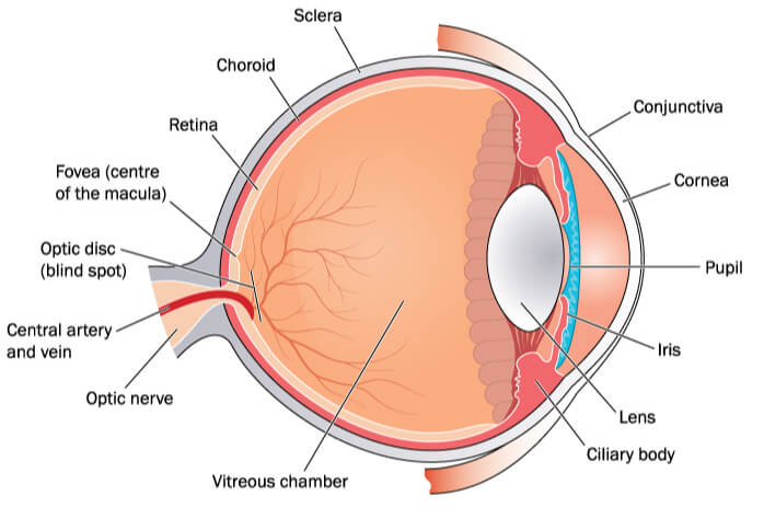
The optic nerve is made up of a bundle of more than a million fibers of nerve cells called Ganglionic cells which converge at the papilla (also known as the optic disc). The optic nerve is, in fact, a constituent of our central nervous system (as opposed to part of the peripheral nervous system).
The optic nerve, funnily enough, is partially responsible for the blind spot we have in our eyes – this blind spot is caused by an absence of photosensitive cells (cells that are sensitive to light) right in the area of the retina where the optic nerve leaves the eye (the papilla).
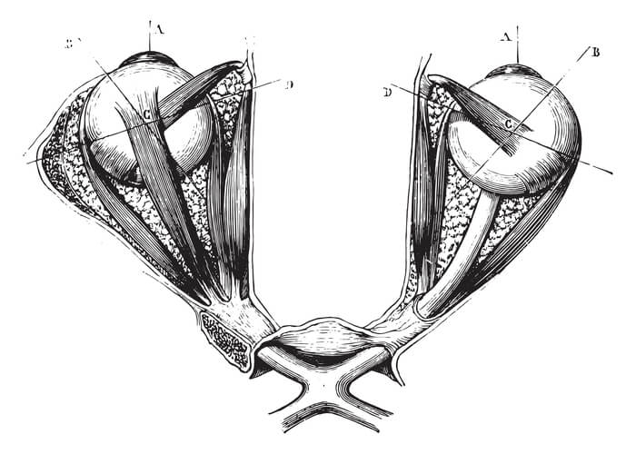
The optic nerve is divided into sub-sections known as fascicles by connective tissue, and is also surrounded by cerebrospinal fluid as well as all three meningeal layers (the dura mater, the arachnoid, and the pia mater). The optic nerve can also be said to have four distinct parts:
- The optic head – where the optic nerve begins in the eyeball
- The orbital part – the part within the orbit
- The intracanicular part – the part within a canal in the bone known as the optic canal
- The cranial part – the part of the optic nerve that’s within the cranial cavity
The optic nerve is also known as the second cranial nerve (there are twelve cranial nerves) and is actually the only tract in the central nervous system to leave the cranial cavity. In addition to this, the head of the optic nerve can be observed through an ophthalmoscope, meaning it is the only part of your brain that can be seen clinically from outside the body!
Optic Nerve Function
The optic nerve’s main function is to transfer visual information from the retina in your eye to the visual centers of your brain in the form of impulses, allowing your brain to translate these impulses as images in your head.
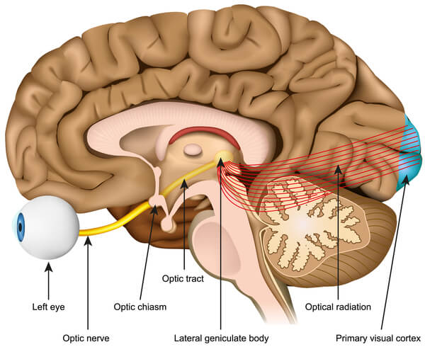
Visual information is picked up by the rod and cone cells found in the retina, which pick up on different wavelengths of light and trigger the cells to send electrical impulses to the optic nerve via the retinal ganglia. These impulses travel all the way up the optic nerve up until the optic chiasm of the brain. The optic nerve connected to the right eye carries impulses to the left cerebral hemisphere, whilst impulses from the left eye are carried to the right cerebral hemisphere. As a result, these optic nerve fibers cross.
Not only does the optic nerve carry visual information to the brain such as visual acuity and brightness and color perception, but it also conducts the impulses in control of the light reflex and the accommodation reflex.

Light Reflex
The light reflex (also known as the pupillary light reflex) refers to the eye’s reaction to light intensity – this reaction is observed through the changing in diameter of the pupil in different light levels. For instance, if a light is shone into your eye, your pupil will constrict so that less light enters your eye.
On the other hand, if the intensity of light is lower, the pupil will dilate in order to allow more light to enter your eye. This is an adaptation to help you see better in different light conditions – if you’ve ever had your pupil dilated by a doctor during an eye exam and then had to go out into the sun, you can understand why the pupil needs to react to high light intensity the way it does!
Accommodation Reflex
The accommodation reflex (also known as the accommodation-convergence reflex or near response) is the eye’s reaction to focusing on a near object, and then looking at a distant object, and vice versa. It allows objects that are nearby to be brought into focus, which is accomplished through constriction of the pupils, thickening of the lens and eye convergence (inward rotation of the eyes). The brief moment where your vision is blurry when having to suddenly focus on an object in front of you is an example of the transition period where your eyes need to adjust and carry out these changes.
Optic Nerve Damage
Unfortunately, as is the case with all parts of the body, the optic nerve can get damaged and diseased, too. This will cause a wide range of symptoms such as pain with eye movement and visual impairments such as vision loss in one eye or loss of color vision, as well as an abnormal pupillary reflex.
Generally, damage to the different parts of the optic nerve will cause distinct symptoms. For example:
- Damage to the optic chiasm – lateral vision loss
- Damage to anterior optic nerve – areas of vision loss
- Damage to the optic tract – loss of the entire visual field from the side opposite that of the damage
There are many different ailments that can negatively affect the functioning or structure of your optic nerve; some of the more common types of optic nerve damage and disease are as follows:
Optic Nerve Hypoplasia
Optic nerve hypoplasia is a congenital disorder (a disorder that is present from birth) which results in the underdevelopment (AKA hypoplasia) of the optic nerves. This disorder can often arise from maternal factors such as a premature birth, maternal diabetes or fetal alcohol syndrome, and can cause moderate to severe vision loss in one or both eyes in children. It can often be characterized by involuntary, rapid eye movements (nystagmus) observed in an infant or child
Optic nerve hypoplasia can also be associated with several other disorders such as cerebral palsy and developmental delay, as well as abnormalities in ventricles or in white- or gray-matter development. The hypothalamus in patients with optic nerve hypoplasia can also be abnormal. However, not all of these complications arise in children affected by optic nerve hypoplasia.

Optic Nerve Glaucoma
Optic nerve glaucoma is a condition that damages the optic eye, often through a build-up of pressure in the eye (intraocular pressure). This build-up of intraocular pressure is caused by the blockage of the channel through which aqueous humor (the fluid inside your eye) flows, causing the liquid to gradually build up.
There are other factors other than a build-up of intraocular pressure that can cause glaucoma however, though these are less common. Such causes include chemical or blunt injury to the eye, blockage of the blood vessels inside the eye, severe eye infection and inflammation. Eye surgery can also cause glaucoma in the optic nerve, though this is also incredibly rare.
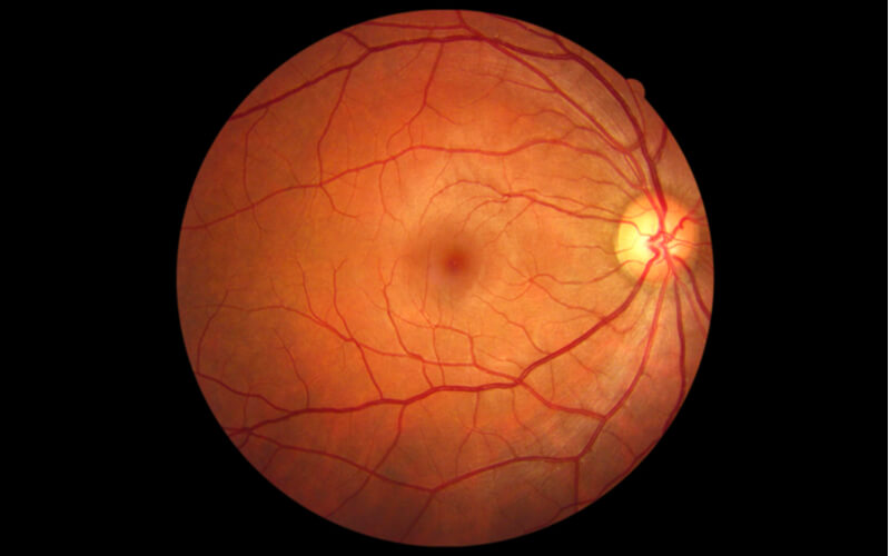
In those who have sustained glaucoma damage to the optic nerve, a process called “optic nerve cupping” can begin to occur. This is where the center portion (“cup”) of the optic disc begins to progressively grow larger as a result of the increased intraocular pressure, as the structure that supported it is not there due to death of nerve fibers in the eye.
Though it does not cause much harm to the eye itself, optic nerve cupping is a sign of glaucoma which, if left untreated, can cause severe damage to your vision.
Optic Neuritis
Optic neuritis refers to optic nerve swelling (AKA optic nerve inflammation), which often causes damage to the optic nerve. This inflammation, though not extremely harmful in most cases (as long as it is caught early), can progressively worsen over time if left untreated, causing vision loss in one eye, visual field loss, and loss of color vision.
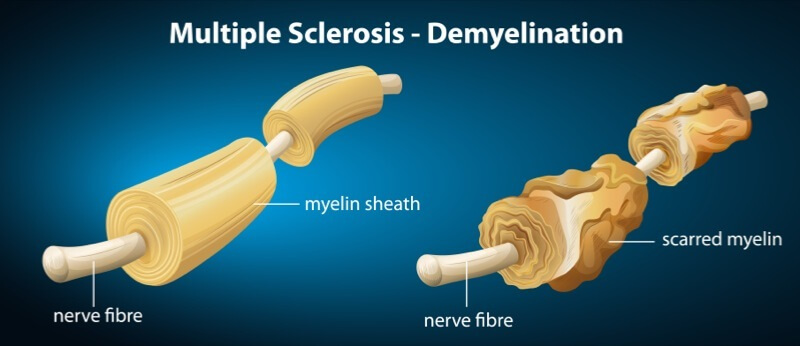
Other symptoms include pain that is worsened by eye movement and flashing lights seen with eye movements. Optic neuritis can also be associated with a number of other diseases, one of which being multiple sclerosis (a disease which affects the myelin sheath of nerve fibers, eventually causing severe nerve damage).
Optic Nerve Atrophy
Optic nerve atrophy is the general term used to describe the degradation and death of tissues in the optic nerve caused by damage to it. This damage leads to a loss of vision, and can be caused by several factors:
- Brain tumor putting pressure on the nerve or blood vessels, or cutting off the supply of blood, oxygen, and nutrients.
- Glaucoma
- Cranial or temporal arteritis (inflammation of the arteries which cuts off blood, leading to tissue damage)
- Multiple sclerosis
- Stroke
Another disorder that can cause optic nerve atrophy is Leber’s hereditary optic neuropathy, which is a disorder inherited from mother to child (mitochondrial inheritance) that results in the degeneration of the retinal ganglion cells and their axons. The symptoms begin with vision loss in one eye and then, unfortunately, this ultimately leads to sub-acute or acute loss of vision in both eyes.