Definition
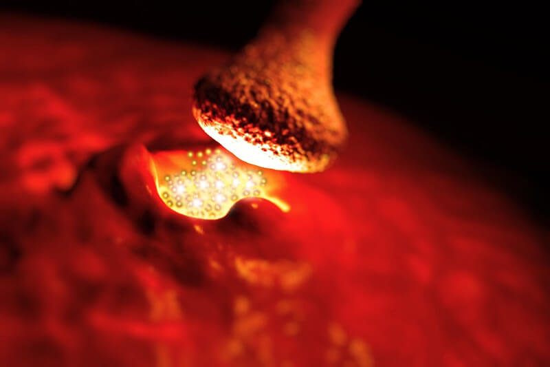
A neuromuscular junction (NMJ), also called a myoneural junction, is the connection between a motor neurons and a muscle fibers. These neurons are the site at which the neuron transmits a signal from the brain to the muscle fiber, causing it to contract.
Therefore, neuromuscular junctions represent the channel of communication between the nervous system and muscle cells. Their function is to allow the nervous system to control the contraction of muscles, and hence they represent an important structure in the regulation of much of our biological functions.
Structure of a Neuromuscular Junction
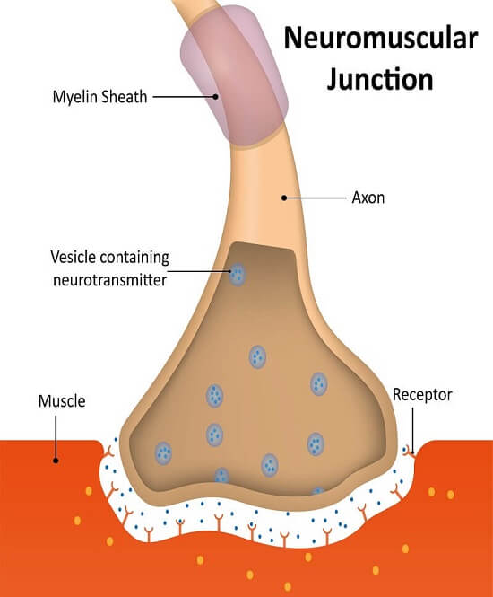
The anatomy of a neuromuscular junction can be divided into three parts:
- the presynaptic terminal (i.e. the motor neuron)
- the synaptic cleft
- the postsynaptic membrane (i.e. the membrane of the muscle cell).
Presynaptic Terminal
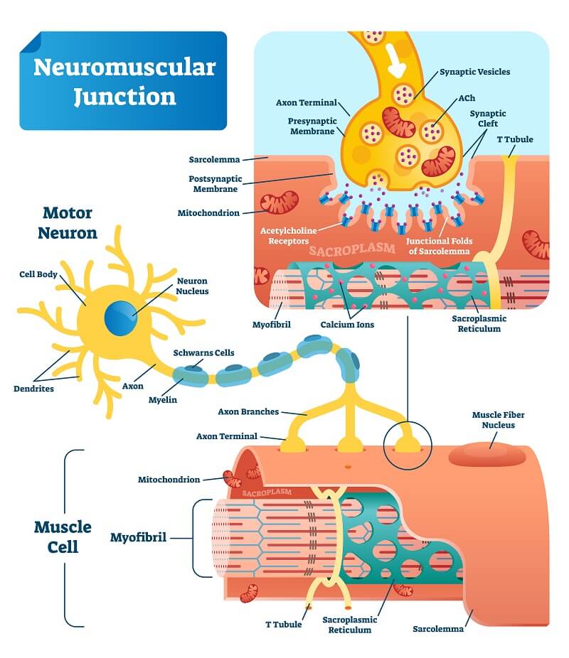
A motor neuron has a dendritic end and an axonal end. The dendrites receive the signals from adjacent neurons, whereas the axon is where the signal is passed on to the next neuron or cell.
The presynaptic terminal of a neuromuscular junction refers to the axonal terminal of a motor neuron. Motor neurons are the neurons that directly control effector organs, in this case, muscle cells. This axon terminal end is the presynaptic terminal of a neuromuscular junction.
Importantly, there are synaptic vesicles present in the presynaptic terminal. These vesicles are small pockets that are separated from the rest of the cell. These vesicles contain neurotransmitters, which are chemical messengers that are responsible for the transmission of the message. In the case of the neuromuscular junction, the neurotransmitter is acetylcholine (Ach).
Synaptic Cleft
The synaptic cleft, also sometimes referred to as the synaptic gap, is the approximately 20 nm space between the presynaptic terminal (the axonal terminal) and the postsynaptic membrane (the muscle cell that will receive the signal). This gap is important for the control of the concentration of neurotransmitter that communicates the signal to the muscle cell.
Postsynaptic Membrane
The postsynaptic membrane is the membrane of the muscle fiber cells to which the signal is travelling. This membrane has many indents that increase the surface area of the membrane, which is important for the transmission of the signal from the motor neuron.
Additionally, the muscle cells have a specialized cell membrane, called the sarcolemma, that aids in the transmission of signals throughout the fiber.
Steps of Signalling at Neuromuscular Junctions
The events involved in the transmission of a signal at a neuromuscular junction are summarized in the six steps below.
- Firstly, the signal from the axon terminal of the previous neuron travels down the motor neuron to the presynaptic axon terminal. This causes the activation and opening of calcium channels in the membrane, allowing calcium ions to enter the neuron.
- The axonal terminal contains neurotransmitters (specifically, acetylcholine) that are packaged into vesicles. When calcium floods into the neuron, it binds the proteins on the surface of these vesicles, called SNARE proteins. These SNARE proteins mediate vesicle fusion, prompting the vesicles to fuse with the cell membrane.
- Once they have fused to the membrane, the vesicles can release their contents (acetylcholine) outside the cell, through the process of exocytosis.
- As a result, acetylcholine floods the synaptic cleft, where it can reach the postsynaptic membrane by diffusion.
- Acetylcholine binds to acetylcholine receptors, also called nicotinic acetylcholine receptors. These are present in the many folds of the postsynaptic membrane (the sarcolemma). Thus, the increased surface ara that is generated by these folds serves to maximize the number of receptors to which acetylcholine can bind on the membrane.
- The binding of acetylcholine to its receptors causes ion channels to open, allowing sodium and potassium ions to flood the cell. This causes depolarization, permitting calcium ions to enter the cell. It is the calcium ions that carry out muscle contractions.
Calcium ions can propagate the signal to contract to other muscle cells by moving between cells through structures called gap junctions, which link the muscle cells, allowing them to behave in sync.
There is also a reservoir of calcium ions present in the muscle cell, in an organelle called the sarcoplasmic reticulum. The signaling at the neuromuscular junction also causes this organelle to release its calcium ions, contributing to muscle cell contraction.
Disorders of Neuromuscular Junctions
Neuromuscular junctions perform an important role by bridging the gap between the nervous system and the muscular system. If any of the signalling steps or structures are compromised, diseases can occur. Two examples of such diseases are Myasthenia Gravis and Lambert–Eaton myasthenic syndrome.
Myasthenia Gravis
Myasthenia Gravis is an autoimmune disease in which the immune system attacks the acetylcholine receptors present in the postsynaptic terminals of neuromuscular junctions. This causes muscle weakness because the junction can no longer initiate the signals required to contract skeletal muscles.
It affects 1 in 10,000 individuals, and primarily affects the muscles of the eyes and face, resulting in the eyelids drooping, double vision, and facial weakness. It can also cause people to have trouble walking and talking.
Lambert–Eaton Myasthenic Syndrome
Lambert–Eaton myasthenic syndrome is another autoimmune disease, but in this case, the immune system attacks the calcium channels (and likely other proteins) in the presynaptic terminal.
Interestingly, this disease is often associated with cancer, with around half of the affected individuals developing the disease after a diagnosis of small cell lung cancer. Lambert–Eaton myasthenic syndrome primarily causes muscle weakness in the arms and legs, particularly the muscles closest to the torso.
Quiz