Definition
Elastic cartilage is one of three types of cartilage found in the human body. A form of connective tissue, elastic cartilage is also recognized by its ability to snap back into an original form – or resting form – due to the addition of elastin fibers to the extracellular matrix. This fiber type distinguishes elastic cartilage from all other forms.
Overview
Elastic cartilage is sometimes confusingly referred to as fibroelastic cartilage (this is confusing because there is another type of cartilage called fibrocartilage). It is usually recognizable by its dull yellow color. The other two types of cartilage – hyaline and fibrous – are white in appearance.
Elastic cartilage looks very similar to hyaline cartilage under a microscope, and special stains must be used to show the otherwise invisible elastic fibers that give this particular cartilage type its name. Like hyaline, elastic cartilage also has single or multiple chondrocytes housed within spaces called lacunae. The extracellular matrix of elastic cartilage contains higher levels of type II collagen. Chondrocytes and ECM are contained within an outer layer known as a perichondrium. The perichondrium is made up of two differently functioning layers: the inner layer contains chondroblasts (chondrocytes which are not yet fixed within the extracellular matrix, and which continue to produce the components of this thick gel-like substance). The outer layer houses fibroblasts, which differentiate into chondrocytes as they progress towards the inner layer. During this move and accompanying differentiation, fibroblasts stop secreting type I collagen and hyaluronic acid and start to secrete type II collagen and chondroitin sulfate as they change into chondroblasts.
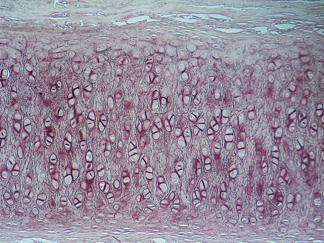
The above image, multiplied in size by 100, clearly shows one, two, and very rarely three chondrocytes within a single lacuna inside the elastic cartilage. These chondrocytes are surrounded by the extracellular matrix crisscrossed by a darker, fibrous network of collagen and elastin fibers. On either side of the cartilage are the paler perichondrial layers.
Elastic Cartilage Function
Elastic cartilage function is two-fold: to change cartilage shape in response to tension, compression, and bending before returning to an at-rest state, and to provide a strong but flexible structure.
In Outer Ear
The outer ear is relatively unprotected and sticks out from the skull. The aneural and avascular properties of cartilage mean that it is unlikely that one is woken up if the ear is folded when being slept on. A change of position means the ear returns to its original shape.
As an external organ, the ear must be able to withstand trauma. If the outer ear were made of bone, ear fractures would be regular occurrences. If the outer ear was fleshy, the folds of the pinna which modify sound waves before funneling them through to the middle ear would not keep their form and work less efficiently.
In Epiglottis and Larynx
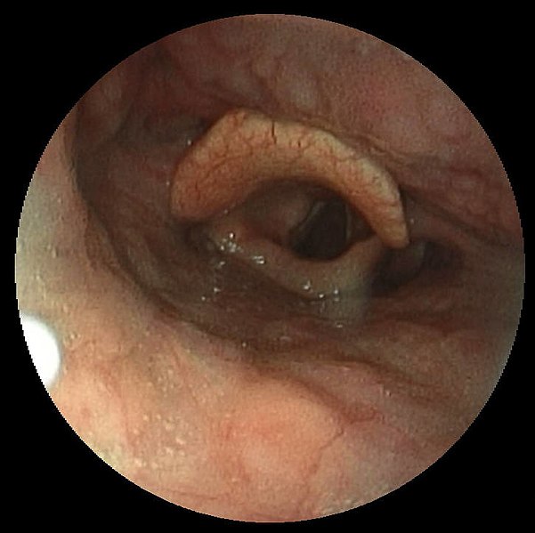
At rest, the epiglottis is upright. In this position air can pass through the larynx during inspiration and expiration. Should a person swallow, the epiglottis is pulled back and covers the opening into the larynx which lies in front of the opening into the esophagus. This action prevents saliva and food from entering the airway, where choking and aspiration pneumonia may be the result. Elastic cartilage in the epiglottis is important as it maintains the form of this valve-like appendage and always springs back into the at-rest position, allowing for the body’s most important process – breathing. In this case, elastic cartilage has a protective and structural function.
The image below shows an epiglottis at rest. The white vocal chords of the larynx can be seen at the edges of the dark hole of the trachea. The upper flap of the epiglottis covers the entrance to the esophagus, which lays posterior to the trachea. When swallowing, the flap of the epiglottis moves forward, closing the entrance to the airway and revealing that of the esophagus.
Elastic cartilage is also found in the corniculate and cuneiform cartilages of the larynx, where the joints they create allow for the movement of the vocal chords and play a role in voice quality and pitch.
In Eustachian Tube
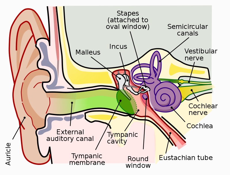
The Eustachian tube (pharyngotympanic tube/auditory tube) is a narrow tube of approximately 3mm wide. Its main function is the equalization of middle ear pressure to the ambient atmospheric pressure. The effect of an imbalance in middle ear and atmospheric pressure is most readily experienced during the take-off and landing of an airplane. Swallowing during take-off or landing often causes the ears to ‘pop’. This effect can also be achieved through nose blowing or yawning, as during these actions the Eustachian tube opens and allows air to enter into the middle ear, reducing the difference between middle ear and atmospheric pressures.
Elastic cartilage has a structural and functional purpose in this case, as the narrow tube must return to its original, at-rest form (closed) when free from the tensile forces of the tensor veli palatini muscle. The function of elastic cartilage, in this case, is therefore to spring back into a closed form upon the relaxation of this muscle, as such a narrow but important feature must be stable enough to retain its shape, and must be elastic enough to change it. The image below shows the position of the Eustachian tube colored in red.
Where Is Elastic Cartilage Found
Elastic cartilage locations are varied. As a springy and highly flexible connective tissue, elastic cartilage is found in specific places, primarily in the pinnae (or auricles) of the outer ear, shaping the folds which efficiently channel sound waves towards the inner ear. A clear distinction can be felt in the ear lobes, which have no cartilage, in comparison to the cartilaginous pinnae.
The other prominent anatomical sites composed of elastic cartilage are the epiglottis, Eustachian tube, and corniculate and cuneiform cartilages of the larynx.
The Extracellular Matrix of Elastic Cartilage
The ECM of elastic cartilage contains elastin, fibrillin, glycoproteins, collagen types II, IX, X and XI, and (predominantly) the proteoglycan Aggrecan. These components are produced by the chondroblasts at the inner edges of the perichondrium, and are located in an environment that is gel-like. Chondrocytes within the cartilage only make up to 2% of total tissue volume.
The extracellular matrix of all types of cartilage binds with water (osmotic effect) due to the action of Aggrecan, the predominant proteoglycan of cartilage. This osmotic effect produces a flexible but firm gel. The chains of glycosaminoglycan (GAG) that are part of the Aggrecan structure attract water and produce the lubricating and shock-absorbing qualities of cartilage. Other noncollagenous proteins contain GAGs that bind with cytokines or help organize the extracellular matrix.
The elastic fibers within the ECM of elastic cartilage are this tissue’s defining characteristic. Elastic or yellow fibers are composed of elastin proteins which co-polymerize with another protein type – fibrillin – forming fiber-like elastic chains. When relaxed, these chains are disorganized. Stretching them creates uniformity which is once again lost when the tensile pressure is released. This allows the cartilage to spring back into the original position (into a relaxed state).
The collagen fibers similarly form a network which provides strength and a structural framework for other molecules within the ECM. Collagen fibers are found in all types of cartilage, unlike elastic fibers, and are found in high quantities. Collagen fibers are also found within the perichondrium; the outer layer is more collagen-rich.
Quiz