Chorion Definition
The chorion is one of the membranes that surround the fetus while it is still being formed. In mammals, the fetus lies in the amniotic sac, which is formed by the chorion and the amnion and separates the embryo from the mother’s endometrium. During development, the embryo grows inside, and beside, four extraembryonic membranes that protect and nurture it. These membranes are, from closest to the embryo (innermost) to furthest (outermost): the umbilical vesicle (called yolk sac in reptiles and birds), the allantois, the amnion and the chorion. The two innermost membranes—the umbilical vesicle and the allantois—do not surround the embryo but rather sit beside it; the outermost membranes—the amnion and the chorion—surround the embryo. These four membranes sit in the endometrium of the female while the embryo is developing, and are discharged once the embryo is born.
The chorion in turn comprises two layers: a double layer of trophoblasts on the outer side and the mesoderm on the inner side, in contact with the amnion. The outer layer of the chorion is made of trophoblasts (also known as the trophoblast), which are the first cells to differentiate once the mammalian egg has been fertilized. They first form the outer layer of the blastocyst and eventually develop into most extraembryonic tissues, including a part of the chorion referred to as the chorion trophoblast cells, also known as the extraembryonic ectoderm. The inner layer of the chorion is the mesoderm, which is one of the first layers to develop in the embryo and lies between the endoderm and the ectoderm. The mesoderm forming the allantois (one of the other extraembryonic membranes) fuses with the chorion and ends up forming the chorionic villi (see below).
Chorion Function
The chorion has two main functions: protect the embryo and nurture the embryo.
To protect the embryo, the chorion produces a fluid known as chorionic fluid. The chorionic fluid lies in the chorionic cavity, which is the space between the chorion and the amnion. The chorionic fluid protects the embryo by absorbing shock originating from forces such as movement.
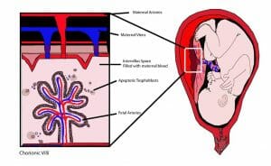
To nurture the embryo, the chorion grows chorionic villi, which are extensions of the chorion that pass through the uterine decidua (endometrium) and eventually connect with the mother’s blood vessels. An image of chorionic villi can be seen here:

On the left side of this figure you can see an amplification of the maternal-fetus interface. At the top are the mother’s veins and arteries, and at the bottom is a structure that contacts with the interviillius space filled with maternal blood. This structure is a chorionic villus, which extends from the chorion, contains fetal blood vessels, and is the site where nutrients and oxygen are delivered to the fetus and waste products are taken by the mother to be excreted afterwards. Chorionic villi enable the maximum contact between embryo and mother due to their tree-like shape granting a very large area of contact.
Chorion Development
Chorionic villi develop in three stages. In the primary stage, chorionic villi are non-vascular, i.e., they do not have blood vessels for the blood exchange to take place between mother and embryo, and they are formed exclusively of trophoblast. In the secondary stage, chorionic villi become larger, with more ramifications, and the mesoderm starts growing into them; at this point, they are constituted by trophoblast and mesoderm. In the tertiary stage, chorionic villi become vascularized because blood vessels begin growing into the mesoderm; chorionic villi are thus at this stage made of trophoblast, mesoderm and umbilical arteries and veins (fetal blood vessels).
The chorion interacts with other membranes and tissues, such as the allantois and the decidua basalis, to develop into the placenta, the function of which is to exchange substances and protect the embryo. Another part of the chorion, which is in contact with the decidua capsularis, will atrophy and the chorionic villi will end up disappearing.
Chorion in Monotremes and in Non-Mammals
Monotremes (mammals that lay eggs), reptiles and birds also have a chorion around the embryo. In this case, however, the albumin—the egg white—surrounds the chorion. In insects, the chorion fuses with the allantois (one of the four extraembryonic membranes) and this fusion, called the chorioallantoic membrane, facilitates the exchange of oxygen and carbon dioxide.
Quiz
1. Where is the chorion located?
A. Between the amnion and the allantois.
B. Between the allantois and the umbilical vesicle.
C. Between the amnion and the maternal interface.
D. Between the amnion and the umbilical vesicle.
2. What are the functions of the chorion?
A. To protect the embryo from shock.
B. To provide the embryo with nutrients.
C. To provide the embryo with oxygen.
D. To get rid of waste products.
E. All of the above.
3. What is the name of the structures that extend from the chorion to the fetus-maternal interface and allow the exchange of maternal and fetal blood?
A. Amnions
B. Chorionic villi
C. Chorionic cavities
D. Trophoblasts
E. Umbilical vesicles
References
- Hemberger, M. & Cross, J.C. (2001). Genes governing placental development. Trends in Endocrinology and Metabolism, 12, 4: 162-168.
