Definition
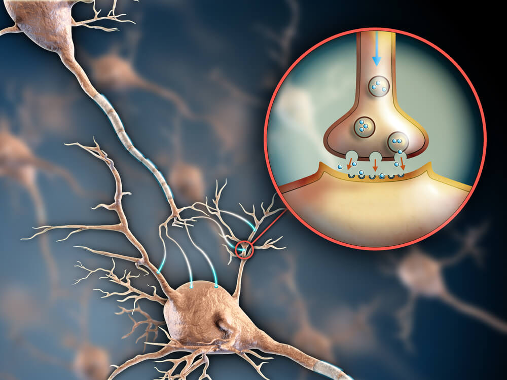
The axon terminal, also known as the synaptic bouton and terminal bouton, is the most distal portion of a neuron’s axon and is critical for neural communication. When action potentials reach the axon terminal, calcium floods the neuron, allowing synaptic vesicles to fuse with the membrane and release stored neurotransmitters to target cells. This results in communication between stimulated neurons and target cells.
Review of the Neuron
The nervous system is made up of single cells called neurons. Neurons are able to communicate with each other by passing along electrochemical signals at speeds of up to 120 meters per second. This speed is critical for the rapid response of the nervous system, which allows incoming stimuli to be processed to the brain and back almost instantly.
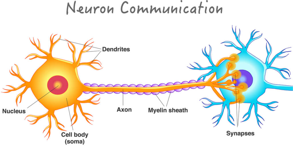
Neurons are made up of three main parts:
- Dendrites- which are short extensions on the cell body
- The cell body (or soma)- which contains the nucleus and organelles
- The axon- which is the long branch that extends from the cell body
Dendrites receive incoming signals, which are then converted from chemical to electrical. The cell body – otherwise known as the soma- integrates the signal through the nucleus. The axon then passes the signal along the length of the neuron.
The Resting Membrane Potential
What Is It?
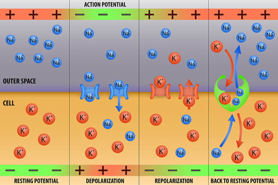
Neurons are considered to be at rest when they are not actively communicating with another cell. This resting phase is determined by the relative number of ions on either side of the cell membrane, and thus by the resulting relative charge. Therefore, this phase is known as the resting membrane potential.
The resting membrane potential is defined by a lower concentration of sodium ions (Na⁺) and chloride ions (Cl⁻) inside the neuron, with a higher concentration of these ions outside the cell in the extracellular fluid. Additionally, there is a higher concentration of potassium ions (K⁺) and several organic anions- which are ions that carry a negative charge- found inside the neuron, with a lower concentration of these ions outside the cell.
Ion Movement in the Resting Membrane Potential
While the neuron may be in a “resting” state, ions still move across the membrane. To maintain the ion ratios on either side of the membrane however, this movement does not occur via simple diffusion. Instead, this movement can occur in three ways:
- Primary active transport: Transport across the membrane against the concentration gradient that directly uses energy from adenosine triphosphate (ATP)
- Secondary active transport: Transport across the membrane against the concentration gradient that uses stored energy gained from electrochemical gradients
- Facilitated diffusion via ion channels: Transport across the membrane following the concentration gradient by use of a facilitator protein
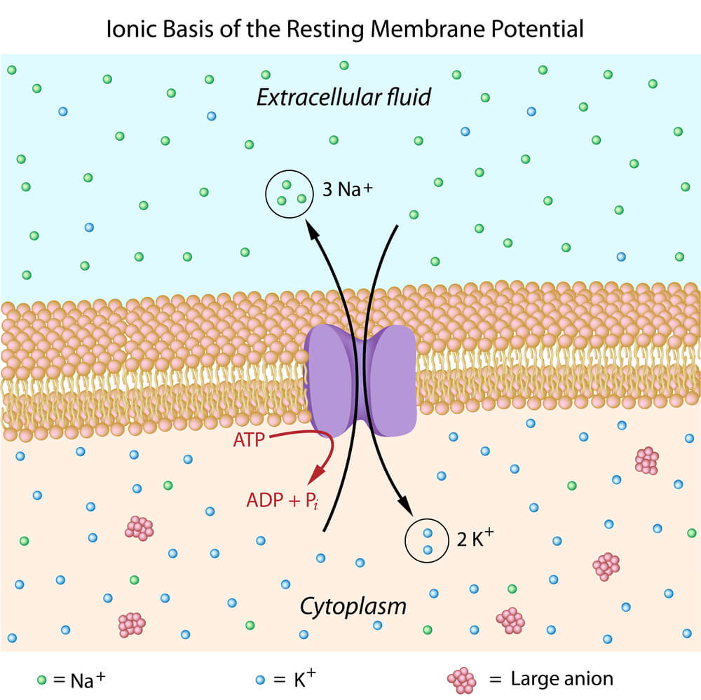
During the resting membrane potential, neurons use a specific primary active transport protein known as the sodium-potassium pump (or Na⁺/K⁺- ATPase) to control ion movement across the membrane. As indicated by the three parts in the name, the sodium- potassium pump uses the energy gained when it receives a phosphate from ATP to move three Na⁺ ions out of the neuron and two K⁺ ions into the neuron. Because there are three positive charges leaving the neuron and only two coming in, the outside of the neuron is considered more positive compared to the inside. Alternatively, the inside of the neuron is considered negative while at the resting membrane potential.
Action Potentials
Definition and Stages
Neurons must cease their resting membrane potential in order to create a signal that can be passed along and used to communicate with target cells (where target cells can include additional neurons, muscle cells, and glandular cells). Neurons begin this communication by creating an action potential, which is a very quick and temporary change within the resting membrane potential.
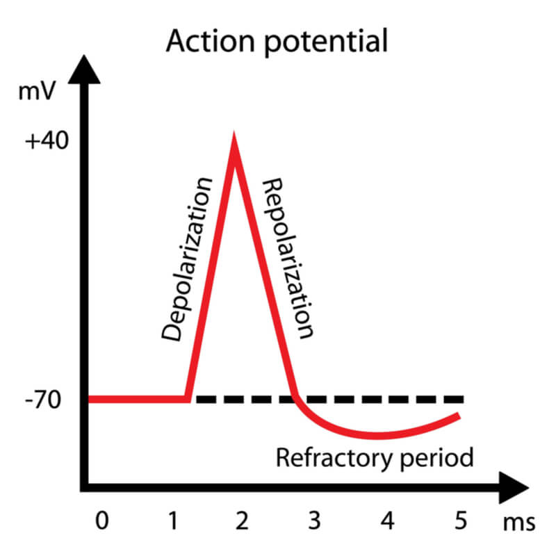
Action potentials consists of three stages:
- Depolarization
- Repolarization
- Refractory period (also known as hyperpolarization)
Polarization is a state where two sides of a neuron’s membrane consist of different charges. As mentioned previously, the inside of the neuron at the resting state is more negative compared to the outside. If a neuron is depolarized, then the two sides become less polarized. Therefore, there must be an increase of charge to one of the sides. In neurons, this is achieved as Na⁺ ions flux into the cell through voltage- gated sodium channels (an example of facilitated diffusion), thus making the inside of the cell more positive. This increase in positivity only lasts for a split millisecond before the cell repolarizes.
During repolarization, the inside of the cell increases in negativity. This is achieved as K⁺ ions flux out of the cell through their own voltage-gated channels. The opening of the voltage- gated potassium channels (another example of facilitated diffusion) slightly lags behind the opening of the voltage-gated sodium channels. This lag differentiates the steps between depolarization and repolarization.
Repolarization is quickly followed by a refractory period- known as hyperpolarization – where the inside of the cell becomes even more negative than typical while at the resting membrane potential. The neuron then stabilizes back to the resting membrane potential conditions.
Action Potentials Use Positive Feedback
Action potentials are an “all or nothing” event, meaning it is not possible to have only a partial action potential. When an action potential is started, it will continue down the length of the axon. This is due to the positive feedback of the incoming Na⁺ ions. The more Na⁺ ions that enter the cell, the more depolarized the cell becomes, and therefore the more Na⁺ channels that open sequentially down the axon. Because Na⁺ channels go through a refractory stage after they close where they are unlikely to open again for a short time period, the action potential can only continue in the direction of the axon terminal and synapse.
The Synapse
Neurons are not in direct contact with each other. There are extremely small spaces between each neuron known as synapses. Because there is not a direct connection, neurons must communicate with each other indirectly through the use of neurotransmitters. This communication is facilitated by the conversion of the electrical signals within the neuron to the chemical signals within the synapse.
Neurotransmitters
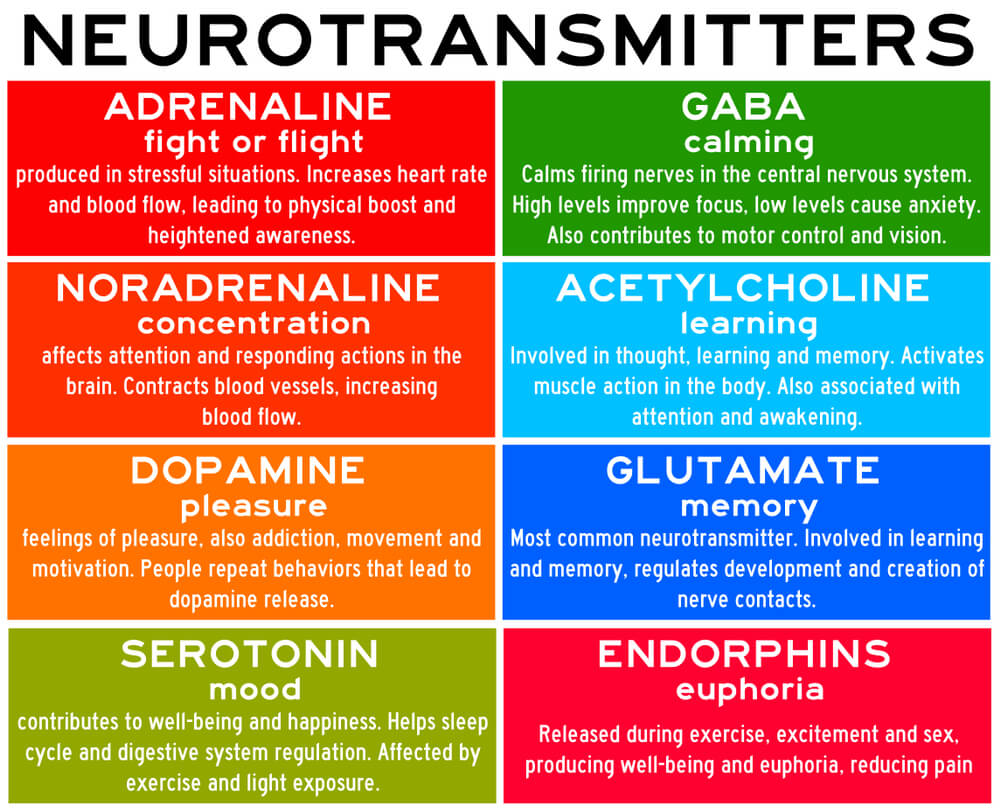
Neurotransmitters are small chemical messengers that are stored in synaptic vesicles at the end of each axon. Examples of neurotransmitters and some of their common functions are listed in the chart below, where a more detailed overview can be found in this chart.
The neuron that has the synaptic vesicles is referred to as the presynaptic neuron, while the target cell receiving the neurotransmitters are referred to as the postsynaptic cell.
Communicating Across the Synapse
Once action potentials arrive at the axon terminal of the presynaptic neuron, the membrane depolarizes, which allows voltage- gated calcium (Ca2+) channels to open. This then allows Ca2+ ions to flux into the neuron. This influx of Ca2+ causes the synaptic vesicles containing the neurotransmitters to fuse with the neuron’s membrane, allowing the neurotransmitters to exit the neuron and bind to corresponding receptors on the postsynaptic cell. Once bound, the resting membrane potential of the postsynaptic cell is altered, which results in a change within the cell.
The change within the postsynaptic cell is specific to the neurotransmitter received. If the neurotransmitter is excitatory- such as with acetylcholine (typically) or glutamate- then an excitatory postsynaptic potential (EPSP) will result. In this case, the reception of the neurotransmitter is more likely to induce an action potential in the postsynaptic cell. If the neurotransmitter is inhibitory- such as with GABA- then an inhibitory postsynaptic potential (IPSP) will result. In this case, it is not as likely that the postsynaptic cell will produce an action potential. Some neurotransmitters- such as with norepinephrine- can be both excitatory and inhibitory depending on the situation and localization.
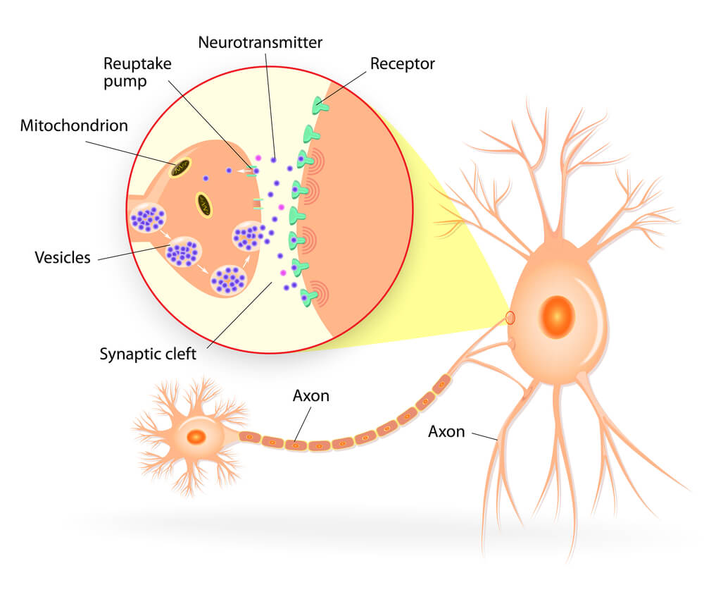
Once the communication is delivered to the postsynaptic cell, the bound neurotransmitters are released from the receptors. They can either be broken down and recycled or reused by the presynaptic neuron.
Disruptions at the Synapse
Drugs for Non-Medical Purposes
Interferences in communication at the synapse can lead to both temporary and long-term disruptions. These disruptions can be a direct result of chemical imbalances as well as physical harm to the structural components.
In a properly performing axon terminal and synapse, the use of drugs can directly interfere with neurotransmitters and their associated receptors. This can occur when chemicals within the drug bind to the receptor in place of the neurotransmitter, thus artificially inducing or inhibiting a reaction in the body. Alternatively, drugs can increase or decrease the presence of the neurotransmitter, also leading to the artificial stimulation or inhibition of a reaction.
Example 1: Drugs that block neurotransmitter reception – Adenosine is a neurotransmitter that leads to drowsiness and indicates when it is time for an individual to go to sleep. Caffeine- which is a drug found in coffee and certain teas- blocks reception of this neurotransmitter by binding to the adenosine receptors in the brain. As a result, the individual is less likely to feel tired. Blocking this receptor can further lead to a cascade of events, as levels of acetylcholine and dopamine can be eventually increased. While an increase in acetylcholine can lead to increased jitters, the increase in dopamine can result in mood changes and altered emotional regulation.
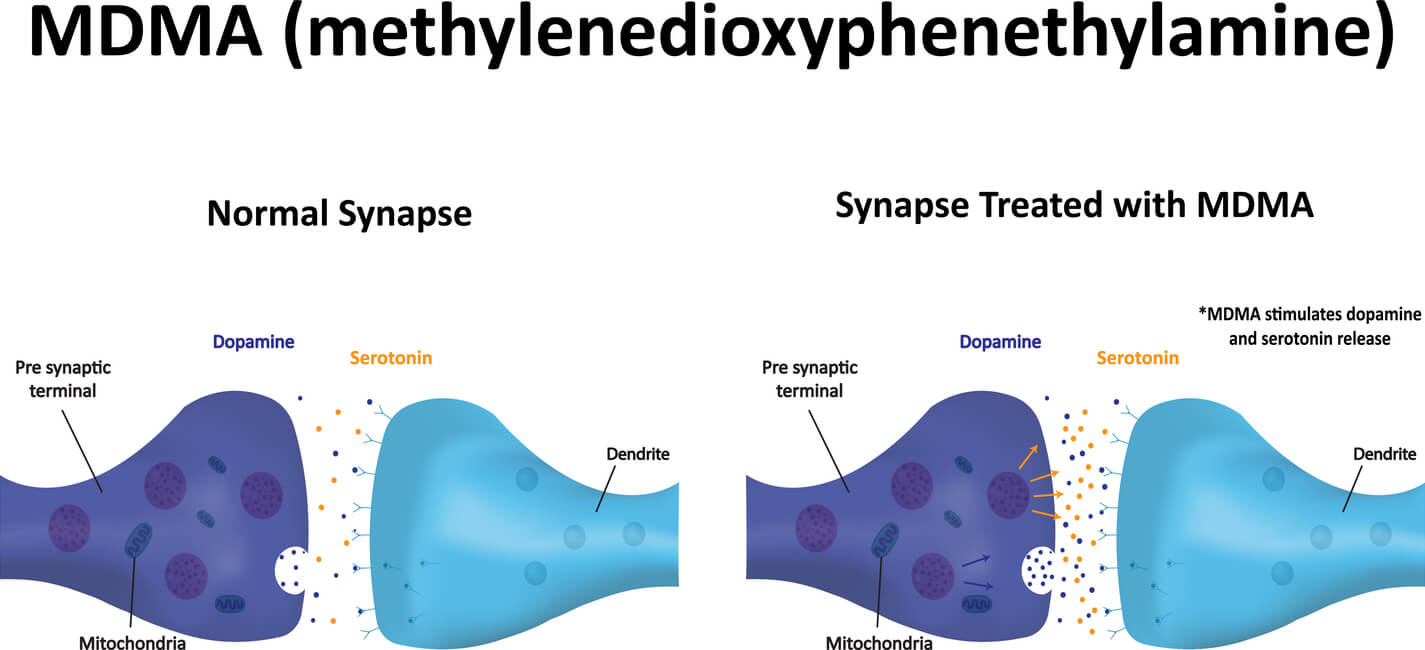
Example 2: Drugs that increase neurotransmitter presence – Drugs such as MDMA (otherwise known as ecstasy) can increase serotonin levels in a short time period, leading to heightened feelings of self-wellness. Having drastically increased levels in a very short time period then reduces the available levels of serotonin for several days to weeks, which can result in short term depression. If used consistently, the axons that release serotonin can become severely damaged, leading to chronic depression as the serotonin cannot be properly utilized in the body.
Drugs for Medical Treatments
While drugs can be dangerous when not used properly, they can also offer themselves as potential treatments to pre-existing conditions if administered correctly by a medical professional.
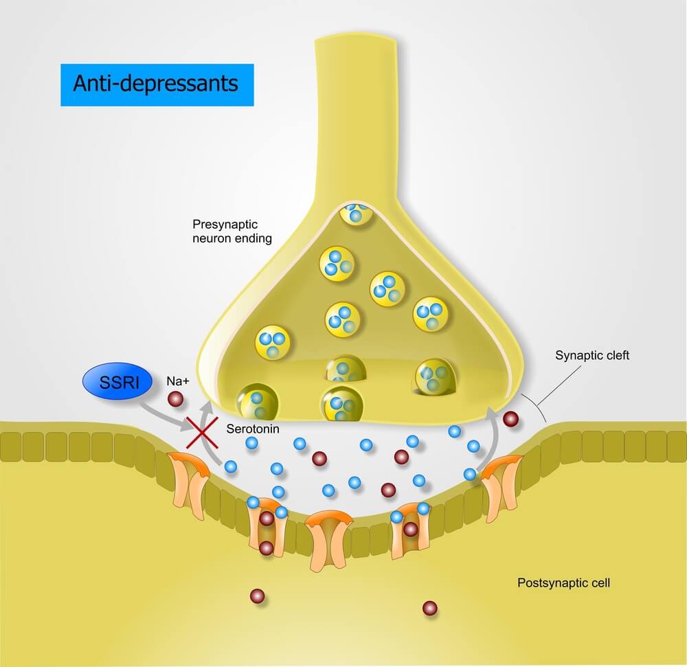
Example 1: Drugs that prevent reuptake of neurotransmitters – Where decreased levels of serotonin is common in individuals expressing depression, many anti-depressant drugs have been discovered that inhibit the reuptake of serotonin back into the presynaptic neuron. By stopping this reuptake, serotonin levels are expected to remain high and thus improve depression over time. These selective serotonin reuptake inhibitors (SSRIs) are very common anti-depressant drugs. However, as SSRIs typically take several weeks for individuals to see improvement in their conditions, research has been expanding to identify alternate factors that correlate with depression. As a result, additional neurotransmitters have been linked to depression- such as norepinephrine, dopamine, and GABA. There are now multiple classes of drugs containing reuptake inhibitors to control neurotransmitter levels in the synapse.
Additionally, as professionals expand to personalize medical practices, new research seeks to find alternative patterns in the brain that could indicate early on how effective certain treatments may be on individuals.
Example 2: Drugs that prevent release of neurotransmitters – Acetylcholine stimulates muscle contractions. Botulism toxin is a chemical that can prevent the release of acetylcholine, thus resulting in weakened and/or paralyzed muscles. When administered appropriately by professionals, this chemical – found in the drug Botox- is capable of alleviating medical issues, such as cervical dystonia (a disorder that results in severe muscle contractions in the neck and shoulder) and blepharospasm (a disorder that causes uncontrollable blinking).
Conclusion
Axon terminals make up the most distal portion of the axon within a neuron and contain structures that are critical for neural communication. Because neurons are separated by synapses, communication is achieved by converting the electrical signals created within the neurons into chemical signals by use of neurotransmitters. The neurotransmitters are stored in synaptic vesicles located at the axon terminals, which are released when the synaptic vesicles fuse with the neural membrane following an influx of Ca2+ ions. The neurotransmitters then bind with receptors on the postsynaptic cell to carry out the communication. This region is commonly studied for psychological and pathological diseases, where further research is needed to find more effective drugs to aid treatment for chemical imbalances.
Quiz