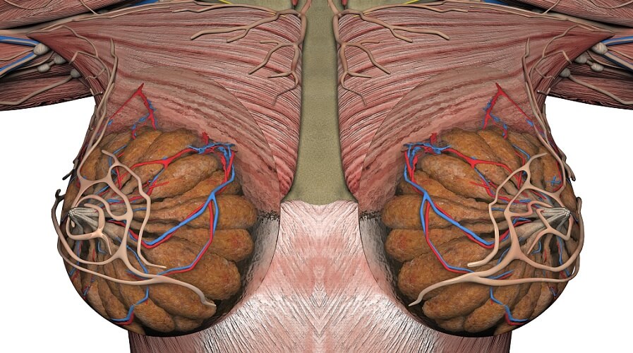
Definition
Mammary glands (mammae) produce and secrete milk and are found in developed and undeveloped forms in females and males respectively. Mammae are apocrine glands, found between the second and seventh ribs in humans. Mammary glands are present at birth but develop in females during puberty. Hormones control milk production during pregnancy and when breastfeeding.
What are Mammary Glands?
When asked what are mammary glands, it is normal to associate them with the female sex. Even so, both males and females have mammary glands or breasts. Both also have systemic and local control systems that cause mammary glands to produce and secrete milk. The only difference is that male mammary glands are not developed enough to respond to these systems.
The reason mammals are called mammals is because they have mammary glands – this is a distinguishable feature of the mammalian class. Monotremes are an order of mammals that lay eggs (the platypus and echidna). Instead of having nipples, monotremes lactate via mammary gland tubules that have a similar structure to sweat glands. It is understood that all mammary glands are modified sweat glands.

Marsupials (kangaroos, koalas, opossums, and wombats) give birth to embryos that further develop in the mother’s protective pouch. Their mammary glands are located at the lower abdomen.
The majority of mammals have mammary glands at the rib and abdominal levels. How many glands a species has and where these are located depends on that species’ form. Humans, whales, and primates have two mammary glands, in dogs and cats eight (thoracic, abdominal, and inguinal) are the norm; rats have twelve and mice have ten. Each gland is paired – rats have six paired mammae, for example.
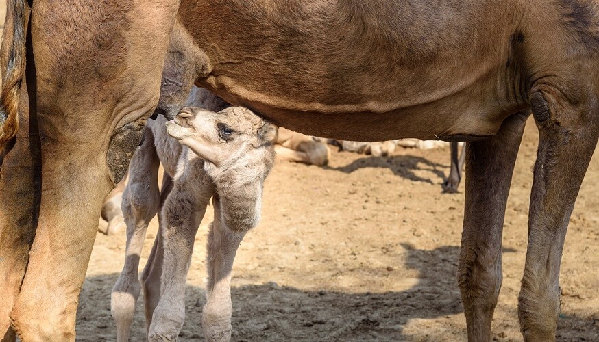
In ruminants, mammary glands take the form of udders. Udders allow much greater storage space. Cattle usually have two pairs of mammary glands; deer, sheep, and goats just two. Camels have six pairs of mammae.
Mammary Gland Anatomy
Mammary gland anatomy is similar in both sexes, although size and level of development differs. Each gland is made up of lobules that are divided into areas of fat and stroma. Within each lobule lies an alveoli network – lots of sacs surrounded by alveolar cells or glandular epithelia. Alveolar cells produce milk and milk is stored in the alveoli. These sacs have additional layers of cells (myoepithelium) that make them contract. When the myoepithelium contracts, the result is milk let-down or milk ejection.
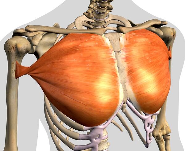
Human mammary glands are positioned at a level that begins at the second rib and ends at the sixth or seventh rib. They are supported by the pectoralis major muscle of the chest and the pectoral fascia.
Adipose tissue surrounds a network of ducts and lobules. Men have milk ducts but only a few, scattered lobules. Suspensory ligaments called Cooper’s ligaments support the breast; regular exercise without a good supporting bra or overly heavy mammae can thin and weaken these ligaments, leading to sagging breasts.
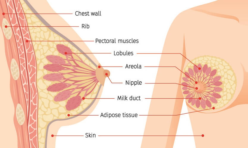
The nipple of males and females is where the lactiferous ducts secrete milk out of the body. Nipples can also be referred to as papillae and teats. They are highly innervated. The nipple is surrounded by the areola – a specialized tissue that helps the skin to withstand suckling; the areola also becomes a visually-conditioned feeding cue for a hungry baby.
The areola contains Montgomery glands that secrete a fatty fluid, helping to keep the nipple lubricated. Bumps on the areola indicate the position of the Montgomery glands that share common ostia (exits) with the lactiferous ducts.
All lactiferous ducts or milk ducts merge into between five and ten main ducts that open at the nipple and secrete milk. These ducts connect the nipple to the mammary gland lobules and are surrounded by epithelium; this epithelium also features myoepithelial cells that can contract.
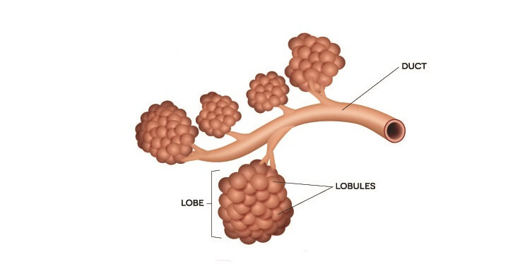
Each lactiferous duct branch begins at a lobe. Lobes are commonly referred to as terminal ductal lobular units or TDLU. This is the functional unit of the mammary gland.
A TDLU has an extralobular terminal duct that connects a lobule to its lactiferous duct branch, an intralobular terminal duct that runs deep into the lobule, and many clusters of acini. Acini or alveoli are tiny sacs. It is in the intralobular terminal ducts and acini that milk is produced and stored.
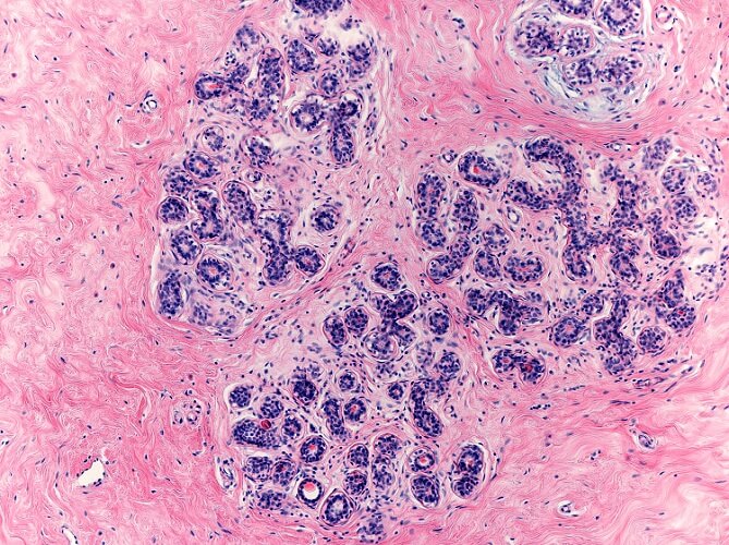
Up to 100 acini make up a lobule. Each sac is composed of a hollow area surrounded by luminal (inner) epithelium and basal (outer) myoepithelium.
The interlobular stroma is a dense connective tissue that surrounds the larger ducts and TDLUs. This part of the breast is responsible for breast size. The intralobular stroma surrounds the acini. It contains many immune cells that can filter into breastmilk and so provide a means of passive immunity to the feeding baby.

Mammary Gland Function
Mammary gland function revolves around the production, storage, and secretion of milk. Even though different mammals have different numbers, locations, and structures of mammae pairs, how they work is essentially the same.
Mammary glands develop at puberty under the influence of hormones; however, the structure for milk production is complete by birth and milk production in human babies (witch’s milk) can occur under the influence of the mother’s hormones.
Mammary gland fat pads grow during puberty when estrogen (ovary), growth hormone (pituitary gland), and insulin-like growth factor-1 (liver) work together to make these cells proliferate. The tree-like network of ducts also produces extra branches during puberty in response to high growth hormone and insulin-like growth factor levels.
During pregnancy, the mammary glands mature. In early pregnancy, more ductal branches form. These allow the mammae to develop even more alveoli. Increased numbers of blood vessels form to oxygenate and feed this additional tissue.
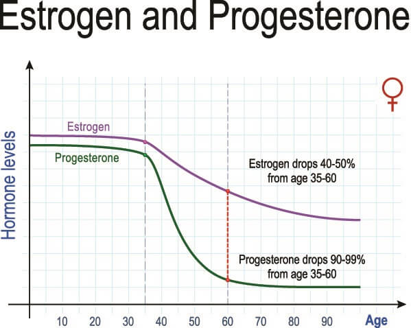
The development of alveoli is called alveologenesis and relies upon the hormone progesterone (ovary). Together with prolactin (pituitary gland) the newer structures form and the breasts become ready to produce milk. Only after birth, when progesterone and estrogen levels fall at a rapid rate, does prolactin encourage milk production and secretion.
During suckling, prolactin levels increase and stimulate further milk production and release; as prolactin rises to its highest levels at night, night-time breastfeeding is important to keep up production.
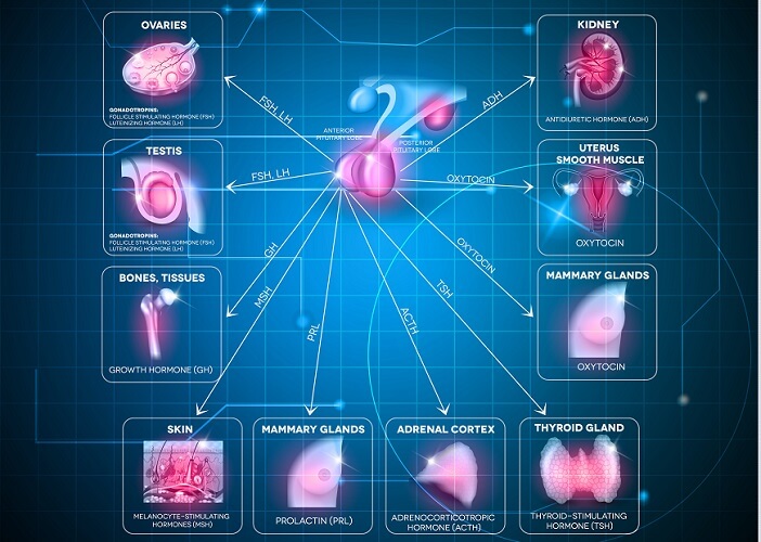
The myoepithelial cells around the alveoli contract in response to oxytocin. The oxytocin reflex is also known as the letdown or ejection reflex. This reflex is an example of a conditioned response – eventually, seeing, smelling, and even thinking about her baby can cause the breasts to secrete milk. With oxytocin inhibition due to stress and/or pain this reflex may not occur.

Do Men Have Mammary Glands?
Mammary glands or breasts are milk-producing structures found in both women and men. Only one male species of mammal can produce milk (lactate) spontaneously – the Dayak fruit bat. However, as the duct and lobe structures are immature in other mammalian males, it is not possible for them to lactate.

Lactation should not be confused with galactorrhea – nipple discharge that is not related to milk production.
While male nipples are said by many to be evolutionary remains that are neither advantageous nor harmful (and so left as is), men do produce the hormones necessary to produce breast milk but do not have the mature structures to make this happen. The lower levels of female hormones during puberty also mean that the duct network and alveolar tissue do not increase in volume and this tissue is unable to specialize. Male lactation has only been found in two species of fruit bat – whether this might be a future option for the human race is still a source of discussion.
Mammary Gland Pathology
Mammary gland pathology includes pregnancy-related disorders such as mastitis or infection of the milk ducts but most commonly concerns breast cancer. Swollen mammary glands can be the result of hormonal changes (premenstrual syndrome) or, in the case of small, hard lumps, an indication of tumor growth.
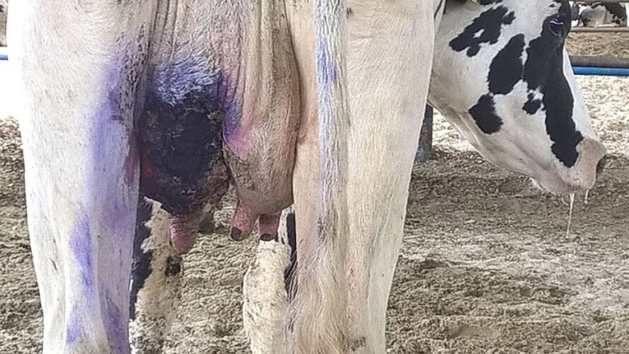
Breast cancer is the most common malignancy in women. Most breast tumors, however, are not malignant but the result of breast tissue cysts (fibrocystic disease).
Around 40,000 women die as a result of breast cancer every year in the US, most of these women over the age of 50 who express the BRCA1 or BRCA2 gene. In men, breast cancer is almost always fatal. It is supposed that most malignant breast cancers are the result of increased receptors for estrogens – one of the reasons why anti-estrogen therapies are recommended. Other treatments are surgery, irradiation, and chemotherapy.
There are multiple types of mammary gland cancer. These can be named according to how the cancerous tissue is distributed (nodular or diffuse), cell type (adenocarcinoma, for example), and the area where the cancer is (such as Paget’s disease of the nipple or intraductal cancer along the milk ducts).
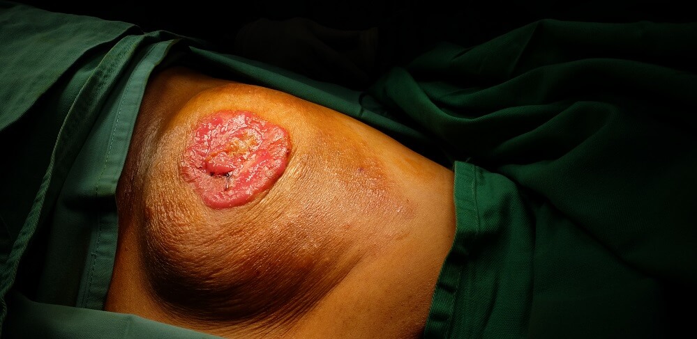
Because the breasts are mammary glands and close to the subclavian, parasternal, perithoracic, and axillary lymph nodes, metastasis is common. It is said that breast cancer patients should not be considered fully recovered until up to 20 years have passed.

