Skeletal Muscle Definition
Skeletal muscle is a specialized contractile tissue found in animals which functions to move an organism’s body. Skeletal muscle is comprised from a series of bundles of muscle fibers, surrounded by protective membranes. This arrangement allows skeletal muscle to contract quickly and release quickly without subjecting the individual fibers to too much friction. Skeletal muscle tissue can be found across the animal kingdom, in most multi-cellular forms of life.
Skeletal Muscle Structure
Skeletal muscle is comprised of a series of muscle fibers made of muscle cells. These muscle cells are long and multinucleated. At the ends of each skeletal muscle a tendon connects the muscle to bone. This tendon connects directly to the epimysium, or collagenous outer covering of skeletal muscle. Underneath the epimysium, muscle fibers are grouped into bundles called fascicles. These fascicles are surrounded by another protective covering formed from collagen. The perimysium, as it is called, allows nerve and blood vessels to make their way through the muscle. These structures can be located in the image below.
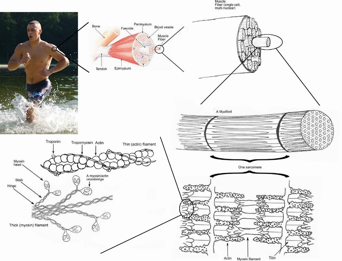
Each fascicle is formed from tens to hundreds of bundled muscle fibers. Each muscle fiber is formed from a chain of multinucleated muscle cells. These fibers are then protected by another layer called the endomysium as they are bundled into fascicles. Each muscle cell has distinct regions when viewed under a microscope. These are known as sarcomeres, and give skeletal muscle a banded or striated appearance. Each sarcomere is a complex of proteins, which operates to contract the muscle.
Sarcomeres are formed from actin and myosin, as well as a number of associated helper proteins. The filaments seen between the dark bands are actin and myosin filaments. Actin, as seen in the image above, is composed of many units of actin and takes the form of a twisting filament. Actin is accompanied by a number of proteins which help stabilize it and provide a pathway for muscle contraction. The two most important are troponin and tropomyosin. Tropomyosin surrounds the actin filament, and stops the heads of myosin from attaching. Troponin locks tropomyosin in place until receiving the signal to contract. Myosin is a fiber composed of many interlaced tails of individual myosin units. The heads of the units stick above the fiber and are attracted to the actin filament.
Function of Skeletal Muscle
When you want to move your arm, your brain sends a nervous signal through your nerves. The simple act of raising your arm requires many muscles, so the signal is sent down many nerves to many muscles. Each skeletal muscle receives the nervous impulse at neuromuscular junctions. These are places where nerves can stimulate an impulse in a muscle cell. The impulse travels down channels in the sarcolemma, the plasma membrane of skeletal muscle cells. At certain places in the membrane, there are channels that lead inside the cell. These transverse tubules carry the nervous impulse inside the cell. The impulse releases calcium ions from a specialized endoplasmic reticulum, the sarcoplasmic reticulum. These calcium ions active troponin to release from tropomyosin. Tropomyosin can then shift position, allowing the myosin heads to attach to the actin filament.
Once the myosin heads are attached, the ATP available will be used to contract the filament. This is done by each pair of myosin heads slowly crawling down the filament. Energy from ATP is used to move one head, while the other is attached. When many hundreds or thousands of heads are involved, this quickly contracts the sarcomere up to 70% of its original length. As the nervous impulse hits each muscle fiber and muscle at the same time, the arm can lift in a fluid motion. As an added feedback measure, every skeletal muscle has special sensory cells which send feedback to the brain. These cells, called muscle spindles, have specialized proteins which can sense tension. When tension is received by the cell, the cell starts a nervous impulse and sends the signal through neurons to the brain.
By piecing together this complicated framework of inputs and outputs, the brain can sense where the body is in space. The somatic nervous system controls these actions, and allows us to move our body in a coordinated manner. Skeletal muscle is controlled almost exclusively by the somatic nervous system, while cardiac and smooth muscle is controlled by the autonomous nervous system. This system can be easily demonstrated. Close your eyes, then clap your hands together several times. Did your hands meet? This is because your brain has been training in coordination since birth, and recognizes the specific tensions on each muscle as you swing your hands. As you clap, these inputs are monitored and adjustments are made to ensure your hands continue to make contact with each other. The same system is responsible for balance, coordination, and most physical movements.
Skeletal Muscle Location
Skeletal muscle, as the name implies, is any muscles that connects to and controls the motions of the skeleton. In all there are somewhere between 600 and 900 muscles in the human body, but an exact number is hard. Many muscles are obscurely small or are sometimes grouped together with similar muscles. Skeletal muscle is found between bones, and uses tendons to connect the epimysium to the periosteum, or outer covering, of bone.
Skeletal muscle is adapted and shaped in many different ways, which give rise to complex movements. Skeletons are not always internal as they are in humans. Even animals with exoskeletons, like crabs and mussels, have skeletal muscle. While the muscle might be adapted differently depending on the animal, skeletal muscle is defined by its striations and connections to skeleton. Everything from the flapping of a bird’s wings to the crawling of a beetle are carried out by skeletal muscle.
Quiz
1. A very bad doctor is trying to test a patient who is unconscious from a head injury. . The doctor tells the patient to raise his arm. The patient is unable to do so, and the doctor records the response. Why was this an unnecessary test at this point in the patient’s recovery?
A. Unconscious patients are notoriously bad listeners
B. To control the somatic nervous system, a conscious patient is needed
C. No test is a waste of time!
2. Clams have an interesting adaptation to help them survive. Their adductor muscle, which holds their shell shut, is composed of two smaller muscle. One of the muscles is a skeletal muscle, while the other one is a smooth muscle. Why would this be beneficial to a clam?
A. Double the muscle, double the power!
B. Skeletal muscle can act fast, while smooth muscle can maintain a contraction
C. The smooth muscle is used to open the shell, and the skeletal to close it
3. Stretching before and after a workout is often recommended by trainers. Why is this?
A. Stretching helps extend the muscle fibers
B. Stretching helps keep tendons elastic
C. Both of the above!
References
- Brusca, R. C., & Brusca, G. J. (2003). Invertebrates. Sunderland, MA: Sinauer Associates, Inc.